Keratocentesis and Vitreous Biopsy


Keratocentesis and Vitreous Biopsy


Michael S. Tsipursky

Gholam A. Peyman

Joseph B. Michelson


The dazzling panorama of new diagnostic laboratory techniques allows for the identification and characterization of cells, proteins, and histopathologic specimens and even for ultrastructural analysis of very small samples obtained by paracentesis. Diagnostic paracentesis of the eye (keratocentesis of the anterior chamber fluid) and vitreous biopsy (paracentesis of the vitreous fluid in the posterior segment of the eye) have definite value in the following scenarios:


  • Diagnosing the presence of specific microbial pathogens that are the likely cause of infectious disease in the eye.


  • Detecting a predominance of certain cell types (e.g., eosinophils, macrophages, epithelial ingrowth, ghost erythrocytes, phacolytic cells) that may provide a clue as to the etiology of an inflammatory disease, which may be autoimmune or allergic in nature.


  • Identifying:


    • Specific antibodies in the aqueous humor or vitreous aspirate that are suggestive of infection (e.g., Toxocara, Toxoplasma, herpesvirus, syphilis).


    • Proteins (e.g., lens proteins, angiotensin-converting enzyme) that are suggestive of granulomatous inflammation, such as sarcoidosis.

Immune complexes and antibodies associated with Behçet’s disease may be found. Polymerase chain reaction (PCR) analysis has suggested the presence of DNA from the infection. Tumor cells may be identified when a malignant infiltration of the eye (e.g., large cell lymphoma, leukemia, retinoblastoma, malignant melanoma) masquerades as a uveitis, or by the presence of tumor cell enzymes and antigens (Table 35.1).1,2

Although keratocentesis had been advocated historically as a treatment for active uveitis, it lost the attention of ophthalmologists until 1919, when Bruckner3 first examined the aqueous humor for diagnostic purposes. Laboratory techniques were revolutionized in the 20th century in areas such as: (1) evaluating very small aliquots of fluid (0.2 to 0.3 mL of aqueous or vitreous), and (2) identifying specific microbial organisms and the predominance of other cell types, antibodies, and proteins in these fluids (Figs. 35.1, 35.2, 35.3, 35.4, 35.5, 35.6). These advancements have led to the development of diagnostic paracentesis for sight-threatening ocular inflammations that are difficult to diagnose. Witmer4 and O’Connor5 have provided strong evidence that samples of the aqueous humor reflect the antibodyproducing capabilities of the iris and ciliary body, particularly when more specific antibody per unit of gamma globulin can be found on the aqueous humor than in the blood of the same patient.6,7,8 These determinations may be highly significant when one considers the fact that diseased tissue is being bathed in an antibody-containing fluid that is elaborated locally. For instance, in the case shown in Figure 35.1, the immunofluorescent antibody titer to toxoplasmosis is four times greater in the vitreous aspirate at the time of vitrectomy for repair of retinal detachment than in the plasma. These same considerations have long been recognized in syphilis of the central nervous system, wherein specific antibodies may be present in the cerebrospinal fluid but not in the blood. This is also the case with an unusual presentation of ocular coccidioidomycosis
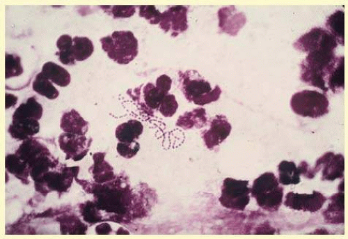
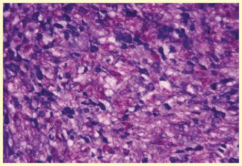
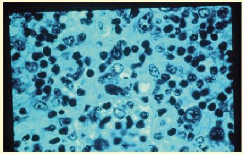
9 or toxocariasis. Many forms of uveitis are characterized by specific types of inflammatory cells. Usually, however, one encounters mixtures of cell types in any given specimen, with the relative percentages of lymphocytes and polymorphonuclear leukocytes varying. There may be unusual number of eosinophils, or macrophages laden with lens material. Thus, an enumeration of the cells and a careful analysis of their structure can be useful as a diagnostic aid (Figs. 35.7, 35.8, 35.9, 35.10, 35.11, 35.12, 35.13, 35.14, 35.15, 35.16, 35.17, 35.18, 35.19 and 35.20). 
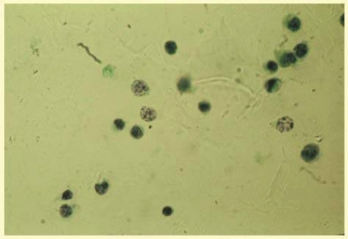
Figure 35.15 demonstrates eosinophils that were aspirated from the anterior chamber of a patient with Toxocara canis endophthalmitis. 
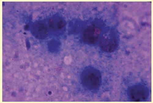
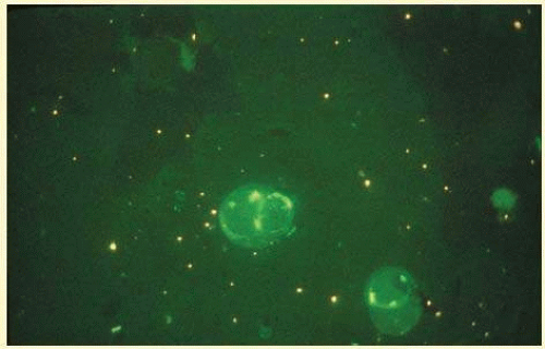
Figure 35.12 demonstrates malignant cell infiltrate from the vitreous, showing the stained presence of monoclonal light chains being elaborated in the cytoplasm. Interleukin-10, detectable in the vitreous of intraocular lymphoma patients, is also directly indicative of both the clinical activity and the number of malignant cells as observed by cytopathology.


TABLE 35-1 Diagnostic Paracentesis


Finding


Condition or Disease Indicated


Aqueous


Bacteria


Endophthalmitis


Fungi


Candida, Aspergillus, etc.


Tumor cells


Retinoblastoma, malignant melanoma, reticulum cell sarcoma, leukemia, metastatic cancer


Eosinophils


Toxocara canis


Macrophages


Phacolytic glaucoma


Antibodies (ELISA)


Toxoplasma gondii, T. canis, reticular cell sarcoma, Behçet’s disease, syphilis


Immune complexes


Behçet’s disease


Other proteins


Angiotensin-converting enzymes


Sarcoid


Lactate dehydrogenase isoenzymes


Retinoblastoma


Lens fragments


Phagocytolytic glaucoma


Ghost erythrocytes


Hemorrhagic glaucoma


Metastatic cancer cells


Metastatic cancer


JXG


JXG


Mesenchymal fibrous cells


PHPV


Amyloid


Amyloid


Epithelial cells


Epithelial ingrowth


Vitreous


Bacteria


Endophthalmitis


Fungi


Candida, Aspergillus sp., etc. (e.g., cryptovirus)


Tumor cells


Retinoblastoma, malignant melanoma, reticular cell sarcoma, leukemia, metastatic cancer


Eosinophils


T. canis


Antibodies


T. gondii, reticular cell sarcoma, Behçet’s disease, syphilis (and immune complexes)


Macrophages


Sympathetic ophthalmia, severe retinitis


Amyloid


Amyloid


Calcium soaps


Asteroid hyalosis


Polymerase chain reaction of viral DNA


CMV retinitis1,2


Interleukin-10 (ELISA)


RCS3


Monoclonal antibodies to Behçet’s disease (long)


Behçet’s disease (clinical activity, numbers of cells)4


HLA DR and DRY


VKH (equally prevalent among Hispanics and Japanese)5


CMV, cytomegalovirus; ELISA, enzyme-linked immunosorbent assay; HLA, human leukocyte antigen.


FIG. 35.1 Fundus photograph of a patient with toxoplasmosis who, after vitrectomy and scleral buckling, demonstrates the etiologic chorioretinal lesion at the 11:30 position on the buckle, which is now inactive. The patient’s serum indirect fluorescent-antibody toxoplasmosis titer was 1:1,024 in the plasma; the vitrectomy fluid yielded a titer of 1:4,096.


FIG. 35.2 Typical gram-positive cocci occurring in clusters as isolated from the aqueous of a patient with staphylococcal endophthalmitis.

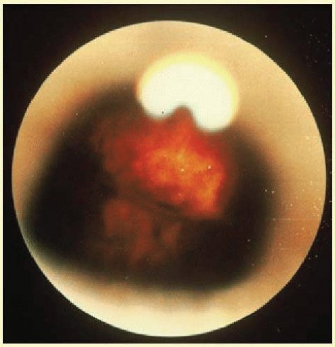
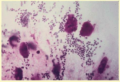
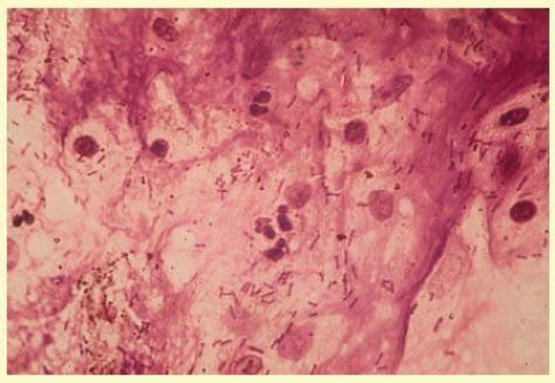
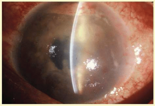
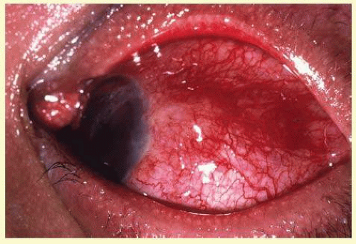
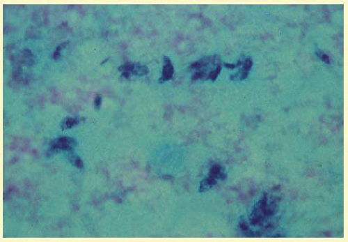
Precise identification and culture of bacterial and fungal pathogens from both the aqueous humor and the vitreous fluid can be obtained. Gram’s stain and Giemsa’s stain smears of centrifuged specimens from the aqueous humor and the vitreous humor frequently demonstrate the bacterial or fungal causative agent. Attempts to isolate bacteria and fungi and to identify them on Gram’s stain or Giemsa’s stain smears have been most rewarding in the following cases: (1) postoperative endophthalmitis, (2) infection after a penetrating injury of the eye, (3) drug abuse patients with endogenous endophthalmitis (Figs. 35.21, 35.22, 35.23, 35.24 and 35.25), (4) patients receiving hyperalimentation, and (5) patients who are immunocompromised as a result of exogenous immunosuppressive agents.


FIG. 35.3 Chains of gram-positive cocci obtained from aqueous fluid in a patient with streptococcal endophthalmitis.


FIG. 35.4 Gram-negative rods obtained from the vitreous of a patient with endophthalmitis.


FIG. 35.5 Aqueous specimen demonstrating acid-fast bacilli of acute lepromatous leprosy in the aqueous aspiration of a patient with a profound anterior segment inflammation that presented as a diagnostic dilemma.


FIG. 35.6 Scleral nodule specimen showing a profuse number of acid-fast bacilli from a patient with lepromatous uveitis.


FIG. 35.7 Anterior segment photograph showing a petechiaespeckled hypopyon in a patient with profound anterior segment inflammation resulting from acute lepromatous leprosy.


FIG. 35.8 Inflamed scleral nodule removed from the same patient as in Figure 35.7, who had acute lepromatous uveitis.


FIG. 35.9 Trophozoites of toxoplasmosis in an aspirate from mouse peritoneum, which incubated acute material from a patient with acute acquired systemic toxoplasmosis with retinitis.


FIG. 35.10 Axillary lymph node of a patient with acquired systemic toxoplasmosis demonstrating trophozoites and a toxoplasma cyst.


FIG. 35.11 Vitreous aspirate demonstrating large cell lymphoma infiltration in a patient with vitreitis and masquerade uveitis of reticulum cell sarcoma or large cell lymphoma.


FIG. 35.12 Large cell lymphoma aspirate of vitreous. Immunofluorescence demonstrates a monoclonal infiltrate of lambda light chains on the B cells.

Only gold members can continue reading. Log In or Register to continue

Stay updated, free articles. Join our Telegram channel

Jul 11, 2016 | Posted by in OPHTHALMOLOGY | Comments Off on Keratocentesis and Vitreous Biopsy

Full access? Get Clinical Tree

Get Clinical Tree app for offline access