Introduction to Otolaryngic Genetics


Introduction to Otolaryngic Genetics


Selena E. Heman-Ackah

Anil K. Lalwani


Human genetics forms the foundation of existence and the backbone of basic human formation and function. Since the turn of the century, the body of knowledge and understanding of human genetics has grown tremendously. The decoding of the human genome has unlocked the door to understanding gene function. Further investigation and unveiling of the genetic code will facilitate the identification of fundamental genes, critical genetic pathways, and deleterious genetic disorders. Current studies are now aiming to decipher the complex interaction between environmental factors and genetics in the manifestation and evolution of genetic disorders. As the body of knowledge continues to grow, human genetics will increasingly be an integral component in the diagnosis, prevention, and ultimately treatment of genetic disorders. This will further aid in identifying disease susceptibility to treatments and targeted therapies to improve individualized drug response. The utilization of genetics in diagnosis and treatment planning may dramatically revolutionize the management of chronic diseases that have plagued mankind for centuries.

Consequently, a keen understanding of human genetics, both the current triumphs and future endeavors, is essential to the practice of otolaryngology—head and neck surgery. Numerous genetic disorders are encountered in otolaryngology and its subspecialties. These genetic disorders may occur as a result of sporadic chromosomal anomalies and mutations or may be transmitted from generation to generation. The purpose of this chapter is to review the basic principles of human genetics and to review genetics as it pertains to the practice of otolaryngology. This chapter provides an overview of normal human genetics, patterns of genetic transmission, genetic disorders in otolaryngology, and molecular therapies in genetics.


MOLECULAR BASIS OF INHERITANCE

The human genetic code provides the basis upon which information encoded within genetic material is translated into proteins vital to cellular function and the sustenance of life. This process starts with deoxyribonucleic acid (DNA), which is transcribed as ribonucleic acid (RNA) and ultimately translated into proteins and polypeptides (Fig. 9.1). This is a highly specific and regulated process by which the building blocks of life are created.


DNA and Chromosomes

DNA provides the basis for all genetic material and is the cornerstone of the human genomic code. DNA provides the code from which all human proteins are derived. The majority of DNA is housed within the cell’s nucleus with a relative minor proportion being contained within the mitochondria. Nuclear DNA is referred to as autosomal DNA, whereas DNA derived from the mitochondria is referred to as mitochondrial DNA. Roughly half of all autosomal DNA is derived from each parent respectively. All mitochondrial DNA is maternally derived. The structure of autosomal and mitochondrial DNA differs substantially.

DNA is a double-stranded helix comprised of two paired nucleotides with a phosphate-deoxyribose backbone joined by ester bonds. Within DNA, there are four nucleotides with specific binding patterns: adenine (A) pairs with thymine (T) and cytosine (C) pairs with guanine (G). The entire human autosomal DNA genome is comprised of 3 × 10⁹ base pairs measuring approximately 2 m in length, all of which is condensed and housed within a nucleus that is only 0.06 µm in diameter. Less than 5% of the entire human genome codes for the fewer than 30,000 genes that are translated into proteins. A trinucleotide sequence,
a codon, corresponds to a specific amino acid. Each DNA molecule is comprised of a sense or coding strand (5′ to 3′) that is paired with a complementary antisense or template strand (3′ to 5′). Segments of the DNA containing genetic information that is transcribed into corresponding RNA, proteins, and polypeptides are called genes.


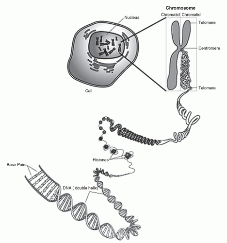
Figure 9.1 Structure and components of the chromosome.

Within the nucleus, DNA is organized into chromosomes following DNA replication and preceding cellular division. Chromosomes are comprised of dense DNA-protein complexes known as chromatin. The human genome contains 23 paired chromosomes of maternal and paternal origin for a total of 46 chromosomes: female (46, XX) and male (46, XY) (Fig. 9.2).

Mitochondrial DNA is also comprised of a doublestranded helix. However, instead of the linear organization, mitochondrial DNA is a circular molecule with 16,569 base pairs (Fig. 9.3). Each mitochondrion contains multiple copies—from 100 to 10,000—of this circular DNA (1). The mitochondrial genome consists of 37 genes coding for 2 rRNAs, 22 tRNAs, and 13 polypeptides. Mitochondrial DNA is essential in its contribution to enzyme complexes critical for oxidative phosphorylation (2). During fertilization, the nucleus of the sperm and oocyte fuse, allowing for transmission of both maternal and paternal autosomal DNA into the developing offspring. Interestingly, shortly following fertilization, the mitochondria derived from the
sperm undergo programmed destruction, leaving only the maternally mitochondria within the developing offspring (3). For this reason, mitochondrial DNA is solely of maternal origin.


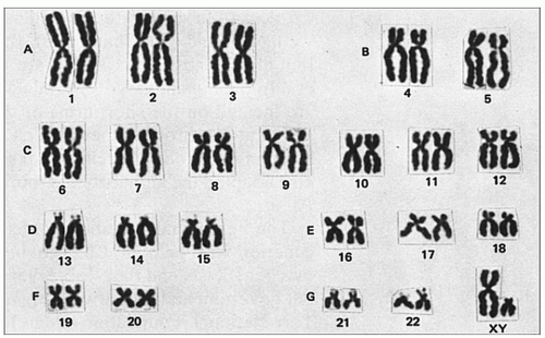
Figure 9.2 Human karyotype.


RNA and Proteins

Information coded within the genome is translated into the vital proteins by RNAs. Similar to DNA, RNA is comprised of a sugar-phosphate backbone with a nucleotide base. However, unlike the double-stranded configuration of the DNA molecule, RNA is configured as a singlestranded molecule. RNA is comprised of three of the same nucleotide base pairs encountered in DNA: adenine (A), guanine (G), and cytosine (C). The thymine (T) base associated with DNA is replaced by uracil (U) in the RNA molecule. Similarly, during the transcription process, adenine pairs with guanine, and uracil replaces thymine in pairing with cytosine. There are multiple types of RNA, which are critical to the cellular function (Table 9.1).


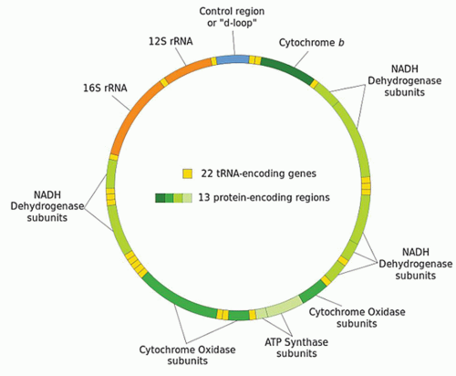
Figure 9.3 The mitochondrial genome.

RNA functions to aid in the transcription of the DNA code and the translation of the genetic code into protein
and polypeptide products (Fig. 9.4). In short, during the process of transcription, messenger RNA (mRNA) pairs with DNA, utilizing the sense strand as a template. Therefore, mRNA is configured in a 3′ to 5′ orientation and is identical to the antisense strand of DNA with the exception of the thymine to uracil substitutions. Transcription takes place completely within the cell nucleus. Once this process is complete, the mRNA exits the nucleus where, with the assistance of transfer RNA (tRNA) and ribosomal RNA (rRNA), the code is translated on a ribosome (a complex of protein and rRNA) into protein. Because the mRNA is similar to the antisense DNA strand, the nucleotide bases of the tRNA correspond directly to that of the sense DNA strand. For translation to occur, tRNAs specific for individual amino acid, with the amino acid covalently attached to it, are required. Each nucleotide triplet represents a codon, of which there are 64, corresponding to a specific amino acid or a stop signal (Fig. 9.5). The peptide produced by the tRNA ultimately forms proteins and polypeptides, which are critical to virtually every cellular function.


TABLE 9.1 TYPES OF RIBONUCLEIC ACIDS


Types of RNA


Abbreviation


Function


Messenger RNA


mRNA


Codes for protein sequence


Essential to transcription


Micro RNA


miRNA


Posttranscriptional regulators


Translational repression and gene silencing


Ribosomal RNA


rRNA


Decoding mRNA


Interact with tRNA during translation


Small interfering RNA (inhibitory RNA, silencing RNA, short interfering RNA)


siRNA


Interferes with specific gene expression


Antiviral mechanism


Shaping chromatin structure of the genome


Small nuclear RNA


snRNA


RNA splicing


Regulation of transcription factors


Maintenance of telomeres


Signal recognition particle RNA


SRP RNA


Translational translocation


Membrane integration


Posttranslational transport


Transfer RNA


tRNA


Translation


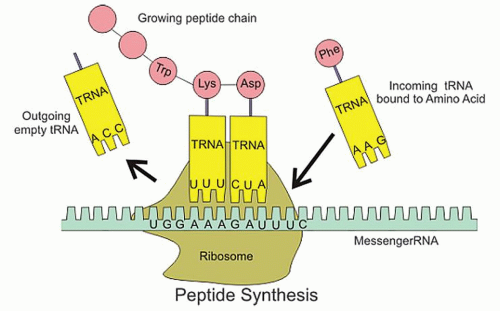
Figure 9.4 RNA translation.


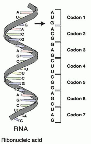
Figure 9.5 Nucleotide triplets.


Proteins are essential for cellular structure and life function. Proteins form the structural components of cells, the foundational material of tissue, and the enzymes that catalyze chemical reactions. Although every cell contains the entire genome, only specific genes are expressed to enable the production of the precise proteins required for that specific cell’s function. The entire process of gene expression to protein production, therefore, requires precise timing and orderly expression of genes specific to carry out functions within divergent cells and organ systems.


MENDELIAN GENETICS

The concepts of Mendelian genetics were first introduced by Gregor Mendel in his work Experiments in Plant Hybridization in 1865 presented on February 8 and March 8 before the Naturforschender Verein (Natural Science Association) in Brunn (4, 5). Using the pea plant for artificial insemination studies, he was able to identify various patterns of inheritance based upon transmission of specific physical characteristics in the plant’s offspring such as color variations and seed form. The works of Mendel were largely ignored for a period of 35 years until their rediscovery in 1900 independently by Correns et al. (6). Since then, Mendel’s model of inheritance has become the cornerstone of human genetics. The main forms of Mendelian inheritance include autosomal dominant, autosomal recessive, and sex-linked inheritance, all of which are discussed in detail below. Table 9.2 provides definitions of basic terminology upon which Mendelian genetics is based.


Autosomal Dominant Inheritance

Each individual possesses two copies or alleles of a specific gene, one maternally derived and the other paternally derived. Autosomal dominant inheritance is a form of inheritance wherein only a single mutated allele is required for the transmission of disease. Therefore, an individual with the disease phenotype need only inherit the mutated allele from one parent while inheriting a normal allele from the other parent. Figure 9.6 depicts a prototypical pedigree of a family with autosomal dominant inheritance. If one parent possesses one diseased allele (heterozygous dominant) and the other parent possesses two normal alleles, their offspring have a 50% chance of inheriting the disorder. Alternatively, in extremely rare cases, if one parent possesses two diseased alleles (homozygous dominant) and the other parent possess two normal alleles, the offspring have a 100% chance of inheriting the disorder. There is no sexual predilection in the pattern of inheritance with autosomal dominant disorders. Additionally, there is no carrier state with autosomal dominant disorders. Therefore, inheritance of one disease allele confers the phenotype of disease unless there is “incomplete penetrance.” Inherited diseases may demonstrate complete penetrance (every individual with the disease allele has the disease phenotype) or incomplete penetrance (not all individuals with the disease allele have the disease phenotype). Because of the nature of transmission, autosomal dominant disorders are rarely associated with fatal mutations. This is because the presentation of fatal mutations with autosomal dominant inheritance would preclude its transmission.


TABLE 9.2 BASIC GENETIC TERMINOLOGY


Terminology


Definition


Genotype


Genetic constitution of a particular genetic locus


Phenotype


Observable characteristics of a cell or organism that are coded by a particular genetic locus


Alleles


The particular form of a gene on each chromosome


Homozygous


Individual who carries two identical alleles of a specific gene loci


Heterozygous


Individual who carries two different alleles of a specific gene loci


There are a number disease in otolaryngology with known autosomal dominant inheritance including achondroplasia, Charcot-Marie-Tooth, Ehlers-Danlos syndrome, Marfan syndrome, neurofibromatosis type I, neurofibromatosis type II, osteogenesis imperfecta, Pfeiffer syndrome, von Hippel-Lindau syndrome, Treacher-Collins syndrome, Noonan syndrome, Osler-Weber-Rendu syndrome, Gorlin syndrome, branchio-oto-renal (BOR) syndrome, DiGeorge syndrome, Gardner syndrome, Pallister-Hall syndrome, Stickler syndrome, van der Woude syndrome, and otosclerosis. These disorders are discussed later in the chapter.


Autosomal Recessive Inheritance

With autosomal recessive inheritance, two diseased alleles are required for the phenotypical presentation of disease. Figure 9.7 depicts a prototypical pedigree of a family with cystic fibrosis, an autosomal recessive disorder. Cystic fibrosis is caused by various mutations within the CFTR gene (7). If an individual possesses two mutated CFTR genes (homozygous recessive), they phenotypically present with cystic fibrosis. However, if an individual possesses only one mutated CFTR gene and possesses one normal copy (heterozygous recessive), this individual is considered a carrier. Carriers typically do not phenotypically present with the disease process, but possess the ability to pass the abnormal gene to their offspring. If two carriers procreate, 25% of their offspring will have the disease process (homozygous recessive), 25% of their offspring will have two normal alleles, and 50% of their offspring will be carriers (heterozygous recessive). Additionally, if one parent is a carrier and one parent is normal, half of their children will be carriers and half will be normal.


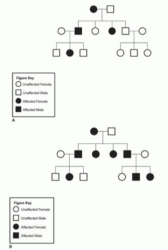
Figure 9.6 A: Pedigree of family with neurofibromatosis (heterozygous dominant parent inheritance). B: Pedigree of family with neurofibromatosis (homozygous dominant parent inheritance).

Like autosomal dominant disorders, there are numerous examples of autosomal recessive diseases within otolaryngology. These disorders include Usher syndrome, Refsum disease, Pendred syndrome, Jervell Lange-Nielsen syndrome, xeroderma pigmentosa, Fanconi anemia, Friedreich ataxia, and Letterer-Siwe disease. Unlike autosomal dominant disorders, genetic disorders with lethal phenotypes are more likely to be autosomal recessive in
nature owing to the normal phenotypical presentation of the carrier individuals at childbearing age. Autosomal recessive disorders with lethal phenotypes, such as Tay-Sachs disease and Bowen-Conradi syndrome, most often occur in patient with no prior family history of disease. The lethal phenotypes tend to occur more frequently among consanguineous families in which the parents are genetically related. Many autosomal recessive disorders tend to have a racial or ethnic predilection (i.e., sickle cell anemia, β-thalassemia, and Gaucher disease) believed to be a function of the founder effect. The founder effect describes the expression of a mutation initially occurring in one or few founding members of a group that has been propagated by the tendency for marriages to occur within the same group. As with autosomal dominant disorders, there is no sexual predilection associated with autosomal recessive disorders.


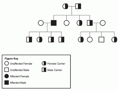
Figure 9.7 Pedigree of family with cystic fibrosis (autosomal recessive inheritance).


Sex-Linked Inheritance

Sex-linked inheritance refers to genes expressed on the X or Y chromosome. Among humans, most sex-linked genetic disorders are carried on the X chromosome. As the daughter inherits an X chromosome from the mother and the father, therefore, with X-linked disorders, a female may inherit a mutated gene allele from either the mother or the father. However, as the male inherits the X chromosome from the mother and the Y chromosome from the father, thus, sons can only inherit the diseased allele from the mother. X-linked disorders are further subdivided into X-linked dominant and X-linked recessive disorders.

X-linked dominant disorders are exceedingly rare. As with autosomal dominant disorder, X-linked dominant disorders only require one diseased allele for the phenotypical expression of disease. Therefore, both males and females may be affected. Figure 9.8 depicts a pedigree of a family with Aicardi syndrome, an X-linked dominant disorder characterized by partial or complete absence of the corpus callosum, retinal abnormalities, and seizures (8). All females born to a male with an X-linked dominant disorder will possess the disease mutation and express the disease phenotype as the father has only the single affected X chromosome. Paternal possession of X-linked dominant disorders has no influence on the genotype or phenotype of male offspring as the father only passes the Y chromosome to the son. Half of males born to a female with an X-linked dominant disorder will inherit the disease genotype and phenotype. In certain X-linked dominant disorders (i.e., focal dermal hypoplasia), expression of the genetic mutation within males is highly or uniformly fatal. There are few examples of X-linked dominant disorders including X-linked hypophosphatemia, CHILD syndrome, Lujan-Fryns syndrome, and incontinentia pigmenti.

The vast majority of X-linked disorders are recessive in nature, meaning that one functional form of the gene precludes the phenotypical presentation of disease. Figure 9.9 depicts a pedigree of a family with Kallmann syndrome, an X-linked disorder. With X-linked recessive disorders, females possessing one copy of the disease allele are carriers and typically do not have the disease. Females must possess two copies of the diseased allele for expression of disease, which is extremely uncommon. Females may inherit this diseased X-linked allele from either or both parents. However, because male offspring inherit their only X chromosome from their mother, males may only
inherit X-linked recessive disorders from their mother. Also, because males lack a second X chromosome, the disease allele represents the only functional X chromosome allele leading to the phenotypical expression of disease. There are a number of X-linked recessive diseases encountered in otolaryngology including X-linked stapes gusher syndrome, X-linked Alport syndrome, idiopathic hypoparathyroidism, X-linked agammaglobulinemia of Bruton, Lesch-Nyhan syndrome, severe combined immunodeficiency disease, Wiskott-Aldrich syndrome, Norrie disease, X-linked ichthyosis, Keipert syndrome (nasodigitoacoustic syndrome), and fragile X syndrome.


Figure 9.8 Pedigree of family with Aicardi syndrome (X-linked dominant inheritance).


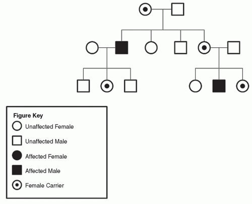
Figure 9.9 Pedigree of family with Kallmann syndrome (X-linked recessive inheritance).


Penetrance and Expressivity

When discussing Mendelian genetics, it is also important to understand the concepts of penetrance and expressivity. Penetrance describes whether individuals carrying a particular gene mutation also express an associated trait or phenotype. Penetrance may be complete or incomplete, that is, among individuals harboring a mutated gene, all or some have the disease, respectively. Expressivity describes the variation in phenotype among individuals carrying a particular genotype. For example, a mutation may be associated five different findings; in patients with a disease characterized by variable expression, they may have one to all five of the features.

Neurofibromatosis type I (NF1), an autosomal dominant disorder, is an excellent example to illustrate the concepts of penetrance and expressivity as it is a genetic disorder characterized by incomplete penetrance with variable expressivity. Individuals with the NF1 genotype may phenotypically appear normal representing the phenomenon of nonpenetrance. Nonpenetrance describes the lack of phenotypical presentation of a genetic disorder in an individual known to possess the genotype associated with disease. Additionally, the expression of NF1 varies greatly among affected individuals. Approximately, 97% of patients with the NF1 genotype have five or more café-aulait spots, 60% cutaneous neurofibromas by age 20 years, 17% scoliosis, and 13% optic gliomas. The variability in presentation of these components of the disease process is a reflection of the variable expressivity associated with NF1.


NON-MENDELIAN GENETICS

Mendel’s description of patterns of inheritance formed the foundation upon which the current level of knowledge and understanding of genetics was built. In addition to the Mendelian forms of genetic inheritance, further investigation has revealed a number of non-Mendelian processes by which genetic mutations can be inherited conferring the phenotype of a disease. These include mitochondrial inheritance, genetic imprinting, epigenetic influences, digenic inheritance, complex genetics, and chromosomal anomalies.


Mitochondrial Inheritance and Mutations

As described above, mitochondrial DNA is instrumental in the production of proteins and enzymes fundamental to oxidative phosphorylation. Mitochondrial DNA is solely maternally inherited. As with autosomal DNA, mutations within the mitochondrial genome can produce the phenotype of disease. These mutations may be inherited or acquired.

Because the mitochondrial genome in maternally transmitted, all inherited mitochondrial genetic disorders are maternally derived (9). Figure 9.10 depicts two pedigrees of families with mitochondrially inherited nonsyndromic hearing loss (MINSHL). The first family depicts the case of a family derived from the maternal unit with MINSHL, and the second depicts the case of a family derived from the paternal unit with MINSHL. Mitochondrially inherited disorders are more rarely encountered in otolaryngology than autosomal inherited disorders and range in severity from asymptomatic to fatal. Because the mitochondrial genome is essential to cellular energy production, tissues with high energy demand are preferentially affected including the nervous system, muscle, heart, and endocrine systems (10). Examples of mitochondrially inherited genetic disorders include aminoglycoside induce ototoxicity, NARP (neuropathy, ataxia, and retinitis pigmentosa), Leber hereditary neuropathy, MELAS (mitochondrial encephalomyopathy, lactic acidosis, and stroke-like episodes), and MINSHL. Certain forms of male infertility are also proposed to be secondary to inherited mitochondrial genomic mutations.

Mutations within the mitochondrial genome may also be acquired. Acquired mitochondrial mutations are proposed to be associated with aging in many organ systems including the inner ear, skin, and retina. Oxidative damage due to increased reactive oxygen species and a decrease in endogenous antioxidants has been identified as a major contributor to acquired mitochondrial genomic mutations (11, 12).

Various cancers have also been associated with mitochondrial genomic mutations. These mutations have been found to be germline related and de novo. In addition to head and neck tumors, renal adenocarcinoma, colon cancer, astrocytic tumors, thyroid tumors, breast tumors, and prostate tumors have been associated with mitochondrial genomic mutations (13, 14).


Genetic Imprinting

Genetic or genomic imprinting is an epigenetic process not described or explained by the traditional Mendelian patterns of inheritance. It describes a genetic process by which certain genes are expressed in a parent-of-origin specific manner. The process of genetic imprinting silences an allele derived from a specific parent such that only the genes from the nonimprinted allele are expressed. Imprinting may occur on either the maternally derived or paternally derived allele. Genetic imprinting is accomplished via methylation of cytosine within DNA and histone acetylation, which produces monoallelic gene expression without alteration of the genetic sequence. The markers for genetic imprinting are established through the germline and are maintained throughout all somatic cells. Imprinted genes are typically grouped within clusters, which allow them to share common regulatory units including noncoding RNA and differentially methylated regions, which together comprise the imprinting control regions (15).

There are a number of genes that have been identified that lead the disease phenotype as a function of genetic
imprinting. Beckwith-Wiedemann syndrome is a disorder of genetic imprinting coincidentally described by Hans-Rudolf Wiedemann in Germany and J. Bruce Beckwith of Loma Linda University, California in the 1960s. Beckwith-Wiedemann syndrome is the most common overgrowth syndrome. It is characterized by gigantism, macroglossia, anterior abdominal wall defects (most commonly congenital exomphalos), neonatal hypoglycemia,
organomegaly, and the development of multiple tumors during childhood, most commonly Wilms tumors (16, 17, 18). H19, also known as BWS, has been found to be associated with Beckwith-Wiedemann syndrome. The paternal allele of the H19 gene is always silenced, and only the maternal allele is expressed. Interestingly, variability in H19 or BWS has been associated with tumorigenicity and variable degree of aggressive behavior of certain tumors (19, 20, 21). Similarly, the cyclin-dependent kinase inhibitor 1C gene, also known as CDKN1C or p57KIP2, has been implicated in Beckwith-Wiedemann syndrome (22). CDKN1C, like H19 undergoes the process of imprinting and only the maternal allele is expressed. CDKN1C is proposed to be a tumor suppressor gene that, when mutated, leads to the formation of various tumors (22, 23). Insulin-like growth factor 2 gene (IF2) has also been implicated in the presentation of Beckwith-Wiedemann syndrome (24). In the case of IF2, in the normal situation, only the paternal copy of the allele is expressed. In tumors derived from patients with Beckwith-Wiedemann syndrome, a lack of imprinting has been demonstrated with both alleles being expressed in association with the disease.


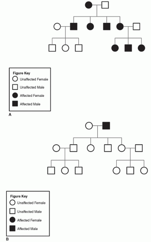
Figure 9.10 A: Pedigree of family with MINSHL (maternal inheritance). B: Pedigree of family with MINSHL (paternal inheritance).

Additional syndromes associated with genetic imprinting include Angelman syndrome, Prader-Willi syndrome, and Silver-Russell syndrome. Angelman syndrome is characterized by developmental delay, sleep disturbance, seizures, movement and balance disorders, microcephaly, and an unusually happy demeanor. Aberrations in UBE3A expression that codes for ubiquitin ligase have been implicated in Angelman syndrome. Angelman syndrome is caused by paternal imprinting of a normal UBE3A gene located on chromosome 15q11-13 with associated maternally inherited deletion or inactivation of the same genes (25). Converse to Angelman syndrome, Prader-Willi syndrome is caused by maternal imprinting of a normal UBE3A gene located on chromosome 15q11-13 with associated paternally inherited deletion or inactivation of the same genes (26). Prader-Willi syndrome is characterized by hypotonia, short stature, hyperphagia, obesity, behavioral issues, hypogonadism, and mental retardation. Silver-Russell syndrome is characterized by intrauterine growth restriction, dwarfism, hypoglycemia, failure to thrive, blue sclera, hemihypertrophy, craniofacial dysostoses, clinodactyly, hypotonia, precocious puberty, and cardiac defects (27, 28). Silver-Russell syndrome has been associated with anomalies in chromosome 7 and 11 (29). Hypomethylation of the imprinting control region 1 (ICR1) in 11p15.5 has been demonstrated to affect the expression of IGF2 and H19 in association with Silver-Russell syndrome (24, 29, 30).


Additional Epigenetic Influences

Inheritable changes in phenotype or gene expression caused by mechanisms other than changes in the underlying DNA sequence refers to epigenetics. Gene silencing is a major component of epigenetic phenomenon. Gene silencing is a regulatory process by which certain genes are inactivated by mechanisms other than genetic modifications. This process typically involves DNA methylation or histone acetylation. Genetic imprinting as described above is an example of gene silencing. Genetic imprinting as described above is one of the most common forms of epigenetics by which DNA methylation and histone acetylation lead to the silencing of a specific gene allele based upon it pattern of inheritance. Additional forms of genetic silencing include transposon silencing, transgene silencing, transcriptional gene silencing, and RNA-directed DNA methylation.

Bookmarking is an epigenetic mechanism by which cellular memory is passed to subsequent cellular generations. Patterns of gene expression in the cell are transmitted through cellular memory by bookmarking though mitosis (31, 32

Only gold members can continue reading. Log In or Register to continue

Stay updated, free articles. Join our Telegram channel

May 24, 2016 | Posted by in OTOLARYNGOLOGY | Comments Off on Introduction to Otolaryngic Genetics

Full access? Get Clinical Tree

Get Clinical Tree app for offline access