Purpose
To compare postoperative visual performance after implantable Collamer lenses with and without a central hole (Hole ICL and conventional ICL) are implanted to correct moderate to high myopia.
Design
Prospective intraindividual comparative study.
Patients and Methods
This study evaluated 58 eyes of 29 patients with spherical equivalents of −7.55 ± 2.09 diopters (D) [mean ± standard deviation] who underwent Hole ICL implantation in one eye and conventional ICL implantation in the other eye by randomization assignment. Ocular higher-order aberrations (HOAs) and contrast sensitivity (CS) function were measured by Hartmann-Shack aberrometry and a contrast sensitivity unit before and 3 months after surgery, respectively. From the contrast sensitivity, the area under the log CS function was calculated.
Results
For 4-mm and 6-mm pupils, the changes after Hole ICL implantation in coma-like aberrations, spherical-like aberrations, and total HOAs are similar to those after conventional ICL implantation ( P > .05, Wilcoxon signed rank test). The postoperative area under the log CS function was significantly increased after Hole ICL implantation ( P < .05), and was equivalent to that after conventional ICL implantation under photopic, mesopic, or mesopic with glare conditions. Subjective symptoms such as glare or halo were also essentially equivalent after Hole ICL and conventional ICL implantation.
Conclusions
A newly developed Hole ICL implantation appears to be equivalent in the induction of HOAs and CS function to conventional ICL implantation for the correction of moderate to high myopia, suggesting its viability as a surgical option for the treatment of such eyes, because it does not require additional peripheral iridotomies and may also reduce the risk of cataract formation.
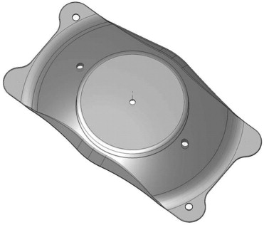
The Visian Implantable Collamer Lens (ICL; STAAR Surgical, Nidau, Switzerland), a posterior chamber phakic intraocular lens (IOL), has been reported to be effective for the correction of moderate to high ametropia. In addition, this surgical procedure is largely reversible and the lens exchangeable with another lens, unlike laser in situ keratomileusis (LASIK), even when unexpected refractive changes occur after surgery. Recently, toric ICLs have also been demonstrated to be effective for the correction of high myopic astigmatism. However, in order to prevent the occurrence of pupillary block, this surgical technique unavoidably requires preoperative laser iridotomy, which frequently involves some pain, especially in younger subjects, or intraoperative peripheral iridectomy, which is sometimes accompanied with iris hemorrhage, causing difficulties in the surgical procedure. Even after these additional procedures, the onset of pupillary block has occurred, possibly because of the closure of previously patent iridotomies attributable to chronic inflammatory responses. Moreover, there are still ongoing concerns about the possible risk of cataract formation, presumably resulting from direct physical contact between the ICL and the crystalline lens or else from malnutrition attributable to poor circulation of the aqueous humor. We recently developed a new ICL with a central artificial hole (Hole ICL) ( Figure 1 ) in order to rectify such disadvantages. It has been demonstrated that the modulation transfer function of an ICL with a 1.00-mm central hole in the optic region was similar to that of an unperforated ICL, and that the in vitro optical performance of an ICL with a 0.36-mm central hole at various IOL powers fulfills the International Organization for Standardization (ISO) criteria for modulation transfer function, which require a contrast above 28% with a 3.0-mm effective aperture diameter. We have already demonstrated in another study that Hole ICL implantation was good in all measures of safety, efficacy, predictability, and stability for the correction of moderate to high ametropia throughout the 6-month follow-up period. Nevertheless, the circumstances regarding higher-order aberrations (HOAs) and contrast sensitivity (CS) function after implantation of this new ICL have not so far been elucidated. Considering that the presence of the central artificial hole may deteriorate the optical quality of the IOL, both HOAs and CS are important factors affecting postoperative visual performance after these surgical procedures in a clinical setting. The purpose of the current study is to compare HOAs and CS after Hole ICL implantation with those after conventional ICL implantation for the correction of moderate to high myopia.
Patients and Methods
This prospective intraindividual comparative study examined 58 eyes of 29 patients (12 men and 17 women) who underwent bilateral implantation of the posterior chamber phakic implantable Collamer lens with and without a 0.36-mm central artificial hole (Hole ICL and conventional ICL, STAAR Surgical) for the correction of moderate to high myopia (manifest spherical equivalent ≤-4 diopters [D]). Using an envelope technique, eligible patients were randomly allocated to receive Hole ICL implantation in one eye as the study group (29 eyes) and conventional ICL implantation in the other eye as the control group (29 eyes). The sample size in this study offered 97% statistical power at the 5% level in order to detect a 0.03-μm difference in HOAs between the 2 groups, when the standard deviation (SD) of the mean difference was 0.04 μm. The patients were masked to the types of ICLs implanted in their eyes. The patient age at the time of surgery was 31.8 ± 7.7 years (mean age ± SD; range, 23 to 49 years). The preoperative manifest spherical equivalent was −7.55 ± 2.09 D (range, −4.00 to −12.38 D). The preoperative manifest refractive cylinder was 0.79 ± 0.53 D (range, 0.00 to 1.75 D). Eyes with keratoconus were excluded from the study by using the keratoconus screening test of Placido disc videokeratography (TMS-2; Tomey, Nagoya, Japan).
Implantable Collamer Lens Power Calculation
ICL power calculation was performed by the manufacturer (STAAR Surgical) using a modified vertex formula. In all eyes, emmetropia was selected as the target refraction to minimize preoperative refractive errors. The size of the ICL was also chosen by the manufacturer on the basis of the horizontal corneal diameter and the anterior chamber depth measured with scanning-slit topography (Orbscan IIz; Bausch & Lomb, Rochester, New York, USA).
Implantable Collamer Lens Surgical Procedure
For conventional ICL implantation, the patients underwent 2 preoperative peripheral iridotomies with a neodymium–yttrium-aluminum-garnet laser. For Hole ICL implantation, the patients did not undergo preoperative or intraoperative peripheral iridotomies. On the day of surgery, the patients were administered dilating and cycloplegic agents. After topical anesthesia, a model V4 ICL (Hole ICL or conventional ICL) was inserted through a 3-mm clear corneal incision with the use of an injector cartridge (STAAR Surgical) after placement of a viscosurgical device (Opegan; Santen, Osaka, Japan) into the anterior chamber. The ICL was placed in the posterior chamber, the viscosurgical device was completely washed out of the anterior chamber with balanced salt solution, and a miotic agent was instilled. All surgeries were uneventful and no intraoperative complication was observed. After surgery, steroidal (0.1% betamethasone; Rinderon; Shionogi, Osaka, Japan) and antibiotic (levofloxacin; Cravit; Santen, Osaka, Japan) medications were administered topically 4 times daily for 2 weeks, the dose being reduced gradually thereafter.
Assessment of Ocular Higher-Order Aberrations and Contrast Sensitivity
We assessed wavefront aberrations and CS function before and 3 months after surgery. Ocular HOAs for a 4-mm pupil were measured by Hartmann-Shack aberrometry (KR-9000, Topcon, Tokyo, Japan). The root mean square of the third-order Zernike coefficients was used to represent coma-like aberrations, and the root mean square of the fourth-order coefficient to represent spherical-like aberrations. Total HOAs were calculated as the root mean square of the third- and fourth-order coefficients. CS function was also measured using a contrast sensitivity unit (VCTS-6500; Vistech, Dayton, Ohio, USA) under 3 different conditions (photopic, mesopic, and mesopic with glare conditions). The illumination levels of photopic and mesopic conditions were 500 lux and 50 lux, respectively. The illumination was confirmed using an industrial illuminometer (T-10; Konica Minolta Holdings, Inc, Tokyo, Japan). The test was performed with best spectacle correction at 2.5 m. From the CS, the area under the log CS function was determined as described previously. In brief, the log of CS was plotted as a function of log special frequency, and third-order polynomials were fitted to the data. The fitted function was integrated between the fixed limits of log spatial frequencies of 0.18 (corresponding to 1.5 cycles/degree) and 1.26 (corresponding to 18 cycles/degree), and the resultant value was defined as the area under the log CS function. All examinations were performed by 2 experienced ophthalmic technicians who were masked to the treatment.
Questionnaire
We also assessed the subjective symptoms of each patient using the following questionnaire (3 months after surgery): Q1: Are you having difficulty with glare or halos? Q2: What is the severity of your glare/halo symptoms? Q3: What is the frequency of your glare/halo symptoms? Q4: Does glare make driving difficult at night? Q5: Do you see better with one eye than with the other? Q6: How much difficulty do you have judging distances like walking downstairs or parking a car?).
Statistical Analysis
All statistical analyses were performed using StatView software version 5.0 (SAS, Cary, North Carolina, USA). The Wilcoxon signed rank test was used for statistical analysis to compare the data between the 2 groups. Unless otherwise indicated, the results are expressed as mean ± SD, and a value of P < .05 was considered statistically significant.
Results
Patient Population
The preoperative demographics of the study population are summarized in Table 1 . There were no significant differences between the 2 groups in terms of manifest spherical equivalent ( P = .72, Wilcoxon signed rank test), manifest cylinder ( P = .86), logarithm of the minimal angle of resolution (logMAR uncorrected visual acuity [UCVA]) ( P = .39), logMAR best spectacle-corrected visual acuity (BSCVA) ( P = .26), coma-like aberrations ( P = .09 for a 4-mm pupil and P = .90 for a 6-mm pupil), spherical-like aberrations ( P = .87 for a 4-mm pupil and P = .88 for a 6-mm pupil), total HOAs ( P = .17 for a 4-mm pupil and P = .73 for a 6-mm pupil), and area under the log CS function ( P = .25 under photopic conditions, P = .48 under mesopic conditions, and P = .95 under mesopic with glare conditions), before surgery. All surgeries were uneventful, and no cataract formation, pigment dispersion glaucoma, significant intraocular pressure rise (including pupillary block), or any other vision-threatening complications were seen at any time during the observation period.
| Hole ICL Group | Conventional ICL Group | P Value | |
|---|---|---|---|
| Age (y) | 31.8 ± 7.7 (range, 23 to 49) | ||
| Sex (% female) | 59 | ||
| Manifest spherical equivalent (D) | −7.52 ± 2.02 (range, −4.00 to −11.75) | −7.57 ± 2.19 (range, −4.00 to −12.38) | .72 |
| Manifest cylinder (D) | 0.80 ± 0.55 (range, 0.00 to 1.75) | 0.78 ± 0.52 (range, 0.00 to 1.75) | .86 |
| LogMAR UCVA | 1.36 ± 0.21 (range, 1.00 to 2.00) | 1.35 ± 0.22 (range, 1.00 to 2.00) | .39 |
| LogMAR BSCVA | −0.20 ± 0.08 (range, −0.30 to −0.08) | −0.19 ± 0.07 (range, −0.30 to −0.08) | .26 |
| Coma-like aberrations (4 mm, μm) | 0.08 ± 0.04 (range, 0.03 to 0.19) | 0.07 ± 0.03 (range, 0.01 to 0.15) | .09 |
| Spherical-like aberrations (4 mm, μm) | 0.05 ± 0.02 (range, 0.01 to 0.10) | 0.05 ± 0.02 (range, 0.01 to 0.11) | .87 |
| Total HOAs (4 mm, μm) | 0.10 ± 0.04 (range, 0.05 to 0.19) | 0.09 ± 0.03 (range, 0.05 to 0.17) | .17 |
| Coma-like aberrations (6 mm, μm) | 0.24 ± 0.10 (range, 0.12 to 0.47) | 0.25 ± 0.08 (range, 0.13 to 0.40) | .90 |
| Spherical-like aberrations (6 mm, μm) | 0.17 ± 0.07 (range, 0.07 to 0.32) | 0.19 ± 0.08 (range, 0.07 to 0.35) | .88 |
| Total HOAs (6 mm, μm) | 0.30 ± 0.09 (range, 0.17 to 0.51) | 0.32 ± 0.09 (range, 0.17 to 0.52) | .73 |
| Area under the log CS function (photopic) | 1.42 ± 0.19 (range, 1.12 to 1.71) | 1.38 ± 0.22 (range, 0.87 to 1.68) | .25 |
| Area under the log CS function (mesopic) | 1.09 ± 0.21 (range, 0.66 to 1.48) | 1.07 ± 0.31 (range, 0.57 to 1.60) | .48 |
| Area under the log CS function (mesopic with glare) | 0.84 ± 0.32 (range, 0.15 to 1.36) | 0.84 ± 0.35 (range, 0.13 to 1.68) | .95 |
Ocular Higher-Order Aberrations
For a 4-mm pupil, the inductions of coma-like aberrations, spherical-like aberrations, and total HOAs were 0.02 ± 0.05 μm (range, −0.05 to 0.16 μm), 0.01 ± 0.03 μm (range, −0.06 to 0.13 μm), and 0.02 ± 0.05 μm (range, −0.06 to 0.12 μm) after Hole ICL implantation, respectively ( Figure 2 ). Similarly, the inductions of coma-like aberrations, spherical-like aberrations, and total HOAs were 0.03 ± 0.05 μm (range, −0.05 to 0.20 μm), 0.01 ± 0.03 μm (range, −0.05 to 0.10 μm), and 0.03 ± 0.05 μm (range, −0.06 to 0.22 μm), respectively, after conventional ICL implantation ( Figure 2 ). For a 4-mm pupil, the changes in coma-like aberrations ( P = .21, Wilcoxon signed rank test), spherical-like aberrations ( P = .98), and total HOAs ( P = .43) after Hole ICL implantation were different, but not significantly so, from those after conventional ICL implantation.
For a 6-mm pupil, the inductions of coma-like aberrations, spherical-like aberrations, and total HOAs were 0.08 ± 0.10 μm (range, −0.15 to 0.30 μm), 0.02 ± 0.07 μm (range, −0.12 to 0.17 μm), and 0.08 ± 0.10 μm (range, −0.13 to 0.23 μm), respectively, after Hole ICL implantation ( Figure 3 ). Similarly, the inductions of coma-like aberrations, spherical-like aberrations, and total HOAs were 0.07 ± 0.11 μm (range, −0.20 to 0.28 μm), 0.01 ± 0.08 μm (range, −0.22 to 0.15 μm), and 0.06 ± 0.12 μm (range, −0.23 to 0.30 μm), respectively, after conventional ICL implantation ( Figure 3 ). For a 6-mm pupil, the changes in coma-like aberrations ( P = .71), spherical-like aberrations ( P = .73), and total HOAs ( P = .63) after Hole ICL implantation were different, but not significantly so, from those after conventional ICL implantation.
Contrast Sensitivity (Photopic Conditions)
The area under the log CS function was significantly increased, from 1.42 ± 0.19 (range, 1.12 to 1.71) preoperatively to 1.50 ± 0.18 (range, 1.14 to 1.69) postoperatively, in eyes undergoing Hole ICL implantation ( P = .03, Wilcoxon signed rank test). There was also a significant increase in CS at 3 of 5 spatial frequencies (but not at 1.5 or 3 cycles/degree) after Hole ICL implantation ( Figure 4 ). Similarly, the area under the log CS function was significantly increased, from 1.38 ± 0.22 (range, 0.87 to 1.68) preoperatively to 1.48 ± 0.20 (range, 0.98 to 1.68) postoperatively, in eyes undergoing conventional ICL implantation ( P = .03). There was also a significant increase in CS at 3 of 5 spatial frequencies (but not at 1.5 or 3 cycles/degree) after conventional ICL implantation ( Figure 4 ). We found no significant difference in the postoperative improvement in area under the log CS function after Hole ICL or conventional ICL implantation ( P = .93).
Contrast Sensitivity (Mesopic Conditions)
The area under the log CS function was significantly increased, from 1.09 ± 0.21 (range, 0.66 to 1.48) preoperatively to 1.24 ± 0.23 (range, 0.54 to 1.58) postoperatively, in eyes undergoing Hole ICL implantation ( P = .003, Wilcoxon signed rank test). There was also a significant increase in CS at 3 of 5 spatial frequencies (but not at 1.5 or 3 cycles/degree) after Hole ICL implantation ( Figure 5 ). Similarly, the area under the log CS function was significantly increased, from 1.07 ± 0.31 (range, 0.57 to 1.60) preoperatively to 1.23 ± 0.24 (range, 0.57 to 1.60) postoperatively, in eyes undergoing conventional ICL implantation ( P = .03). There was also a significant increase in CS at 3 of 5 spatial frequencies (but not at 1.5 or 3 cycles/degree) after conventional ICL implantation ( Figure 5 ). We found no significant difference in the postoperative improvement in area under the log CS function after Hole ICL or conventional ICL implantation ( P = .52).
Contrast Sensitivity (Mesopic With Glare Conditions)
The area under the log CS function was significantly increased, from 0.84 ± 0.32 (range, 0.15 to 1.36) preoperatively to 1.15 ± 0.16 (range, 0.66 to 1.49) postoperatively, in eyes undergoing Hole ICL implantation ( P < .001, Wilcoxon signed rank test). There was also a significant increase in CS at 4 of 5 spatial frequencies (but not at 1.5 cycles/degree) after Hole ICL implantation ( Figure 6 ). Similarly, the area under the log CS function was significantly increased, from 0.84 ± 0.35 (range, 0.13 to 1.68) preoperatively to 1.13 ± 0.23 (range, 0.60 to 1.64) postoperatively, in eyes undergoing conventional ICL implantation ( P = .002). There was also a significant increase in CS at 4 of 5 spatial frequencies (but not at 1.5 cycles/degree) after conventional ICL implantation ( Figure 6 ). We found no significant difference in the postoperative improvement in area under the log CS function after Hole ICL or conventional ICL implantation ( P = .84).


