Integrating Keratoprosthesis


Fig. 26.1
SEM (Scanning electron microscopy) image of superporous skirt showing interconnecting pores
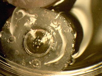


A304005_1_En_26_Fig2_HTML.jpg


Fig. 26.2
Both core and skirt are shown in this picture. The skirt is white due to collagen incorporated in the pores. The clear ring between the center and the skirt shows interdigitation between the two components


References


1.

Coassin M, Zhang C, Green WR, Aquavella JV, Akpek EK. Histopathologic and immunologic aspects of alphacor artificial corneal failure. Am J Ophthalmol. 2007;144(5):699–704.PubMedCrossRef


2.

Myung D, Koh W, Bakri A, Zhang F, Marshall A, Ko J, et al. Design and fabrication of an artificial cornea based on a photolithographically patterned hydrogel construct. Biomed Microdevices. 2007;9(6):911–22.PubMedCrossRef


3.

Chirila TV. An overview of the development of artificial corneas with porous skirts and the use of PHEMA for such an application. Biomaterials. 2001;22(24):3311–7.PubMedCrossRef


4.

Hicks CR, Werner L, Vijayasekaran S, Mamalis N, Apple DJ. Histology of AlphaCor skirts: evaluation of biointegration. Cornea. 2005;24(8):933–40.PubMedCrossRef

Mar 20, 2017 | Posted by in OPHTHALMOLOGY | Comments Off on Integrating Keratoprosthesis

Full access? Get Clinical Tree

Get Clinical Tree app for offline access