The microdebrider has been one of the most significant innovations in the field of rhinology. The use of this instrument for endoscopic sinus surgery offers the advantages of mucosal preservation, improved precision, expeditious tissue removal, and better visualization. Recent advances in microdebrider technology now permit 360 degrees, blade rotation, continuous tracking of the instrument using surgical navigation, and the ability to control bleeding with bipolar energy. A variety of specialty blades are also available, each attempting to address a specific operative limitation encountered during endoscopic surgery. To use these devices effectively and safely, the limitations of microdebriders must be appreciated.
Techniques in endoscopic sinus surgery (ESS) have continued to evolve, often propelled forward by technological advances in instrumentation. The microdebrider is one of the premier innovations in instrumentation for ESS. The use of this instrument has continued to grow more popular in recent years and has reduced reliance on traditional nonpowered sinus instruments, such as curettes and forceps. Microdebriders are preferred for ESS by many surgeons because the instruments spare adjacent mucosa during surgery and offer improved precision, more expeditious tissue removal, and better visualization.
The original design for what we now know as the microdebrider was patented by Urban in 1969 as a “vacuum rotary dissector.” It was initially used by the House group in the 1970s for morselizing acoustic neuromas, but later became more widely used in orthopedic surgery for arthroscopy. These devices were introduced for nasal surgery in 1994 by Setliff and Parsons, and successive models have appeared with improved design and versatility. The microdebrider is a cylindrical, electrically powered shaver supplied with continuous suction. The basic design consists of a hollow shaft with a rotating or oscillating inner cannula. Applied suction draws soft tissue into a port on the side of the tip when it is open and, as the blade rotates or oscillates back, the trapped tissue is sheared off between the inner and outer cannulas. The slower the speed of the inner blade, the larger the tissue bites are, as more tissue is able to be suctioned into the port before being cut off. Thus the faster the blade speed, the less aggressive the instrument.
The morselized pieces are small enough to be sucked down the instrument, aided by self-irrigating hand pieces, which provide a steady stream of saline through a separate set of tubing. Although individual pieces are small, the histology of the tissue is well preserved and tissue removal by microdebrider has been shown to be no different than piecemeal resection using conventional instrumentation with regards to pathologic examination.
Most microdebrider hand pieces have maintained the basic cylindrical design. These hand pieces are usually held as one would hold a scalpel, although some, such as the Diego microdebrider (Gyrus ACMI-ENT Division, Bartlett, Tennessee) have a pistol-grip design, which some consider more ergonomic. Ultimately, the choice of hand piece becomes a matter of surgeon comfort and preference, as the various microdebriders available are materially similar with respect to function.
With the proliferation of image-guidance systems for ESS, there was growing interest in being able to also track the end of a microdebrider during complex cases to minimize the number of movements required to establish and maintain surgical orientation. Optical-based navigation systems responded with “universal calibration” paradigms, which permit the navigation of virtually any rigid instrument by affixing an array of reflective spheres to the desired instrument. Some microdebrider manufacturers have also recently modified hand-piece designs to include a post that quickly couples with the hardware necessary for image-guided surgery calibration ( Fig. 1 ).

Microdebrider blades
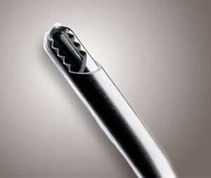
Various blade configurations have been developed for the microdebrider. The edges on both the inner and outer cannulas can be either straight-edged or serrated. The serrated edges allow for better gripping of soft tissue, while straight edges are less traumatic and more sparing of adjacent tissue ( Fig. 2 ).
Depending on the relative angles of the openings to the inner and outer cannulas, the cutting action can either be a guillotine or scissors action. Most microdebrider blades have a scissors-type action, with an angle between the openings of the inner and outer cannulas allowing for a traveling plane of resection, so that the shearing force is only applied to a small area of tissue at a time, maximizing efficiency. With a guillotine mechanism, the apertures of the two cannulas run parallel to one another and the entire bite of tissue is sheared off at once.
Blades can be set to rotate or oscillate. Oscillation typically runs at a slower speed (up to 5000 rpm) and is beneficial in soft tissue resection. At slower speeds, the port remains open longer, allowing more soft tissue to be drawn into the aperture before the cut is made. Forward and reverse settings are faster (up to 15,000 rpm) and allow a drill-like action more suitable for takedown of thicker bone, such as during takedown of the maxillary crest during septoplasty or septal spur resection, or during dacryocystorhinostomy. However, due to their relatively low revolution speed, these instruments are less efficient at removing thick bone compared with high-speed drills.
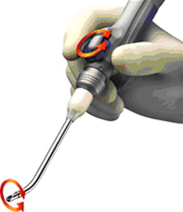
Newer microdebrider designs permit the shaver blade to be rotated on their primary axis to orient the aperture toward the tissue to be removed. Microdebrider blades are also available with a variety of prebent angles, with either a concave or convex bend ( Fig. 3 ). This allows improved access to some hard-to-reach areas, such as the paranasal sinuses, though these bent cannulas do tend to be more prone to clogging than straight cannulas. Some, such as the Straightshot M4 (Medtronic-Xomed, Jacksonville, Florida) ( Fig. 4 ), are prebent but also allow 360° tip rotation, accomplished by a wheel on the handpiece. Products of other manufacturers offer similar capabilities.

Stay updated, free articles. Join our Telegram channel

Full access? Get Clinical Tree


