Neuroendoscopic surgery encompasses minimally invasive approaches to the skull base using endoscopic techniques. There are unique technologic challenges with endoscopic endonasal skull base surgery, such as a limited working space, difficulty in visualization and identification of neurovascular structures and removal of tissue, hemostasis, and dural reconstruction. Technologic advances that have enabled this surgery include specialized operating suites, neurophysiologic monitoring, imaging and visualization technologies, dissection instrumentation, hemostatic materials, and reconstructive materials. Advances in each of these areas and the needs and challenges of the future of neuroendoscopic surgery are discussed.
Technology often drives progress, and that is true with endoscopic surgery of the cranial base. The introduction of endoscopes and endoscopic instruments for the treatment of sinus disease paved the way for more advanced surgeries that extended the limits of surgery to the orbit, ventral skull base, and upper cervical spine. There are unique technologic challenges with the endoscopic endonasal skull base surgery, such as a limited working space, difficulty in visualization and identification of neurovascular structures and removal of tissue, hemostasis, and dural reconstruction. Some of the technologic challenges of endonasal skull base surgery were met through simple modifications of existing sinus surgery instruments. Others are still limiting factors for continued progress. Technologic advances that have enabled this surgery include specialized operating suites, neurophysiologic monitoring, imaging and visualization technologies, powered instrumentation, hemostatic materials, and reconstructive materials.
Operating suite
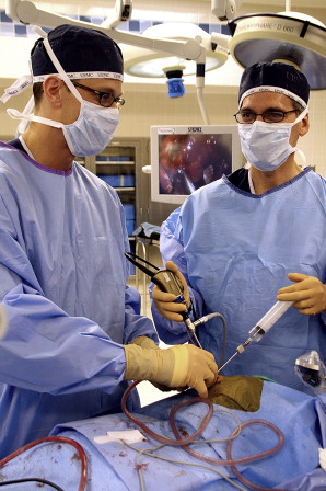
A specialized operating suite offers multiple enhancements for the benefit of the patient, surgeons, anesthesiologist, and other medical personnel. Access to the patient is facilitated by suspending equipment from booms mounted onto the ceiling. Multiple monitors provide both surgeons with comfortable viewing angles ( Fig. 1 ). Wall-mounted screens allow the operative assistants and anesthesiologists to monitor the progress of the surgery and respond appropriately to a surgical crisis. A telestration monitor facilitates communication between the surgeons and observing physicians who are learning. Anatomic structures can be highlighted, and directions provided. Other data are also imported into the screens to allow the surgeon to track multiple physiologic processes simultaneously, such as vital signs, neurophysiologic monitoring, and radiologic imaging.
Neurophysiologic monitoring
Neurophysiologic monitoring provides complete monitoring of the intraoperative environment. Modalities include somatosensory evoked potentials (SSEPs), brainstem evoked responses (BSERs), cranial nerve electromyography, and microvascular Doppler. SSEP consists of simultaneous stimulation of upper (median) and lower (tibial) extremity peripheral nerves, with monitoring of cortical brain responses. SSEP is a sensitive method of assessing the adequacy of hemispheric blood flow during carotid manipulation or injury. It also provides information concerning brainstem function and integrity. Inadequate perfusion (hypotension), especially during blood loss, can be detected by SSEP in the absence of other physiologic changes. SSEPs are monitored in all skull base surgeries because of the potential for carotid injury and major venous bleeding from the cavernous sinus. In rare cases, SSEP monitoring can alert the surgeon to developing complications, such as intracranial hemorrhage, and allow early intervention before a clinical examination or imaging.
BSER provides similar monitoring of brainstem function and is used during surgery in the region of the brainstem (transclival approach), especially when there is tumor compression of the brainstem. Electromyography of the cranial nerves provides information regarding the integrity of the cranial nerves and can detect manipulation of the nerve before injury. Stimulation of tumor tissues with an endoscopic probe during dissection can help localize the nerve and avoid injury. A microvascular Doppler is used to identify vessels such as the internal carotid artery (ICA) when it is displaced from its normal location or obscured by a tumor or scar tissue.
Imaging
Image guidance or intraoperative navigation is an essential technology for endonasal skull base surgery. The skull base is a “black box” and the location of important neurovascular structures is not always readily apparent. Preoperative computed tomography (CT) angiography provides good definition of bone structures and the ICA. Magnetic resonance imaging (MRI) provides better soft tissue detail, especially when there is intracranial extension. If both types of information are needed, image fusion is performed. Image guidance is used to identify normal anatomic structures, detect tumor margins, and assess the completeness of resection.
One drawback of image guidance technology is the absence of real-time imaging. Static images reflect the preoperative status of the patient. With large intracranial tumors, shift of soft tissues after debulking of the tumor renders the image guidance information regarding the tumor-brain interface inaccurate. The images can be updated with an intraoperative CT or MRI scanner with minimal delay ( Fig. 2 ). This allows the surgeon to assess the completeness of resection, detect movement of structures, and complete the tumor resection if warranted. In rare cases, an intraoperative scan can detect complications such as intracranial hemorrhage (eg, subdural hematoma) and allow surgical intervention before the patient leaves the operating room.


