Immunology of Infectious Systemic Diseases that Affect the Eye
Mitchell H. Friedlaender
Infectious diseases are intimately associated with the functions of the immune system. Much of our knowledge about basic mechanisms in immunology has been acquired by the study of host interaction with infectious agents. Immune mechanisms probably evolved to protect the individual from the destructive effects of parasitic organisms and their products. If these protective mechanisms are successful, they eliminate the infectious agents. Under certain conditions, however, immune mechanisms can behave as pathogenic forms of hypersensitivity. The eye might be directly infected by a variety of agents that produce systemic disease, or it could be a focus of hypersensitivity reactions to these agents.
Bacterial Systemic Infections
Tuberculosis
Mycobacterium tuberculosis is a facultative, intracellular, aerobic, acid-fast bacillus that is pathogenic only for humans. It produces mainly lung disease, but many extrapulmonary sites, including the eye, can also be affected. Cellular rather than humoral immune mechanisms are decisive in the host’s recovery from tuberculosis, and in its diagnosis, the tuberculin skin test (a manifestation of cellular immunity) is more useful than the measurement of serum antibody.
Immunopathology
Mycobacterium tuberculosis can survive and proliferate within phagocytic cells. It may escape the bactericidal effect of the macrophages by preventing the fusion of enzyme-containing lysosomes with phagosomes that contain the organisms. Lipids and waxes of high molecular weight constitute up to 60% of the bacterium’s cell wall and may be responsible for the responses of the host’s tissues to the tubercle bacillus. They may also account for the impermeability of the organism to tissue stains. Tuberculoproteins are responsible for the induction of hypersensitivity and for the reaction of the skin to tuberculin. A substance known as “cord factor” causes the serpentine growth of the tubercle bacillus and can also inhibit leukocyte migration and stimulate granuloma formation.
The host deals with M. tuberculosis principally by the mechanism of cellular immunity. Cell-mediated immunity to tuberculin can be passively transferred to uninfected experimental animals along with living lymphoid cells but not with immune serum. Immunity can also be transferred with the leukocyte extract transfer factor. Although patients with agammaglobulinemia produce no demonstrable antibodies, they can develop delayed hypersensitivity responses and normal resistance to the tubercle bacillus. Patients with defects in delayed hypersensitivity are more susceptible to the disease than persons normal in this respect.
Tuberculosis can affect nearly all of the ocular tissues. Granulomatous uveitis, the lesion most frequently encountered, can be associated with mutton-fat keratic precipitates, Koeppe nodules, and vitreous opacities. Diffuse posterior uveitis can also occur and, although rare with modern treatment, the disease can affect the lids, sclera, cornea, and retina. With the recent increase in tuberculosis in patients with acquired immunodeficiency syndrome (AIDS), the ocular manifestations of tuberculosis are increasing.1,2,3 Approximately 2% of patients with AIDS or human immunodeficiency virus (HIV) have ocular tuberculosis, and some of these have sight-threatening manifestations, such as choroidal granulomas, subretinal abscesses, and panophthalmitis.3a
Leprosy
Leprosy is a chronic infectious disease caused by Mycobacterium leprae, an obligate, intracellular, acid-fast bacillus (Fig. 2-32.1). This organism can invade nerves and skin and give rise to widespread clinical manifestations. Ocular lesions occur in 90% of lepromatous leprosy cases, and nearly one third of these produce significant visual loss.
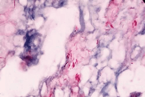 Figure 2-32.1. Mycobacterium leprae in skin biopsy. (Courtesy Mr. M. Okumoto) |
Immunopathology
The following three clinical types of leprosy are recognized: (a) lepromatous (nodular) leprosy is characterized by marked deficiency in cellular immunity and extensive infiltration of the tissues with M. leprae; (b) tuberculoid leprosy is distinguished by the preservation of immune responses and the absence of demonstrable bacilli within the tissues; and (c) intermediate or borderline leprosy has features of both the lepromatous and tuberculoid types. Patients with lepromatous leprosy are anergic to the lepromin skin test antigen, to which normal subjects have a positive delayed hypersensitivity reaction. They also fail to develop contact allergy to dinitrochlorobenzene (DNCB) and other contact sensitizers; they have depressed delayed hypersensitivity reactions to common bacterial antigens and they demonstrate prolonged skin allograft survival.4,5
Patients with lepromatous leprosy have significantly lower than normal levels of circulating T and B lymphocytes, and the T cells in the paracortical regions of their lymph nodes are severely depleted.6 Their peripheral lymphocytes show diminished blast transformation in the presence of the mitogen phytohemagglutinin. Animals thymectomized at birth, and therefore lacking cellular immunity, readily develop widespread infections with M. leprae.7 The infection is apparently related to the survival of the bacillus within macrophages. Recent electron microscopic studies have shown that the organism can escape from the phagolysosome and remain free in the macrophage cytoplasm.
In contrast to being deficient in cellular immunity, patients with lepromatous leprosy produce normal or greater than normal numbers of antibodies and may even produce autoantibodies. They have above-normal levels of both gamma globulin and the cryoproteins. Thyroglobulin antibodies and rheumatoid factor are found in 50% of patients and false–positive Venereal Disease Research Laboratory (VDRL) test results are seen in about 10%. Lupus erythematosus (LE) cells occasionally appear, and antibodies to both basement membrane and intercellular substances have been found in the skin of infected patients. The similarity between leprosy and the collagen vascular diseases, particularly LE, is indicated by their sharing such clinical manifestations as butterfly facial rashes, arthritis, depressed cellular immunity, and autoantibody production.
In lepromatous leprosy, the lids may show granulomatous inflammation,8 and nodules may appear on the face and eyelids. A hyperemic conjunctivitis may develop and may be followed by symblepharon and lid deformities. Corneal nodules, interstitial keratitis, iridocyclitis, and chorioretinitis have also been reported.9
Diagnosis
The lepromin skin test, which was introduced by Mitsuda, is performed with extracts of skin nodules from patients with leprosy. A positive intradermal test result leads to a tuberculin type of skin reaction 24 to 48 hours later, and a nodular reaction (the Mitsuda reaction) appears in about 7 days and peaks in 3 or 4 weeks. The test is useful in differentiating the types of leprosy in patients already known to have the disease. A positive lepromin skin test result indicates tuberculoid or nearly tuberculoid disease, and a negative skin test result indicates progression to lepromatous leprosy. Positive skin test findings are common in the healthy population because of cross-reactivity with other mycobacterial antigens.
Syphilis
Syphilis is caused by the spirochete Treponema pallidum, a motile, highly infectious agent that functions predominantly as an extracellular pathogen. It occurs naturally only in humans and is most easily transmitted by sexual contact. Both humoral and cellular immunity are important in the defense of the host against syphilis. Three stages of the diseases are clinically identifiable, and the eye may be involved in all three. Although severe, destructive syphilitic lesions have been far less common since the discovery of penicillin, the disease must be considered in the differential diagnosis of many diverse clinical problems.
Immunopathology
Immunity to T. pallidum depends on a complex interaction between humoral and cellular immunity. Several lines of evidence indicate that antibody is important in this infection. Partial immunity develops in the rabbit after the passive transfer of serum from immune animals. Immobilizing antibodies against treponemes can be found in the serum of patients who have had syphilis, especially advanced syphilis. A number of nonspecific or reaginic antibodies are found in the sera of patients who have the disease, and these form the basis of many serologic diagnostic tests. Immune complex deposition can play a role in the nephropathy associated with secondary syphilis in adults, and IgG may be found along the glomerular basement membrane in patients with congenital syphilis.10
Delayed hypersensitivity directed against treponemal antigens is absent in primary and early secondary syphilis; it develops late in the secondary disease and is found regularly in latent and tertiary disease. Because syphilis progresses through the primary and secondary stages despite the presence of antibodies that immobilize treponemes, it follows that antibodies developing during the course of an infection, at best, are only partially protective.
When viewed microscopically, the typical lesion of syphilis is a granulomatous inflammation. Granulomas, which contain reticuloendothelial cells (including epithelioid and giant cells), are thought to represent an immune response to such poorly soluble substances as foreign bodies, insoluble antigens, and microorganisms that cannot be easily eliminated. Granulomatous reactions clearly differ from typical delayed hypersensitivity reactions: their onset is much slower, and they may require weeks or even months to develop. Many granulomatous lesions are accompanied by vasculitis, suggesting that an associated immune complex disease may exist.
Diagnosis
During primary and secondary syphilis, the spirochete can be identified microscopically by dark-field examination. Serologic tests do not become positive until 14 to 21 days after the initial infection. Because spirochetes are difficult to identify in the late stages of the disease, serologic tests are extremely important. Two categories of tests—for reaginic antibodies and for treponemal antibodies—are available. Reaginic antibodies bear no relation to IgE; rather, they are antibodies directed against cardiolipin, a tissue-derived substance thought to be a component of mitochondrial membranes. Beef heart is used as a source of cardiolipin in many reaginic antibody diagnostic tests, examples of which are the VDRL, Hinton, and Kahn tests. False–positive results on these tests occur in such diseases as LE, leprosy, infectious mononucleosis, and hepatitis, however, probably because of the many tissues and microorganisms that have mitochondrial membranes.
Two tests for treponemal antibody are the T. pallidum immobilization test and the fluorescent T. pallidum antibody test (FTA). In the former, specific antibodies against the spirochete can be detected, whereas the latter uses spirochetes from infected rabbit testes as a substrate for the serum being tested. If positive, the bound antibody can be detected with fluoresceinated antihuman gamma globulin. Other nonpathogenic treponemes may be absorbed from the serum before testing, in which case the test is referred to as the “FTA antibody absorption” (FTA-ABS) test).
The FTA-ABS test result is positive in 80% to 100% of primary syphilis cases and the VDRL test finding is positive in 50% to 70%.11 In secondary syphilis, the findings for both tests are nearly always positive. The VDRL test result is less often positive after treatment, whereas the FTA-ABS test result remains positive for many years; although false–positive VDRL results are common, rarely is a false–positive FTA-ABS result encountered, except in systemic lupus. In latent syphilis, the VDRL test finding is negative in up to one third of patients.
Treponemes are sometimes detected in the aqueous humor of patients with uveitis, but they may be nonpathogenic organisms such as Treponema microdentium.12 Aqueous humor aspiration, followed by the FTA technique, may be useful when searching for treponemes.
A chancre may occur on the lids or conjunctiva in primary syphilis. A pinkish, macular rash may appear on the lids in secondary syphilis, and ulcerative blepharitis can cause scarring and loss of cilia. Iridocyclitis can occur in secondary syphilis, and chorioretinitis can be associated with late secondary and tertiary disease. Tertiary syphilis can produce distinctive lesions of the eye and central nervous system.
Interstitial keratitis, the hallmark of congenital syphilis, can be a hypersensitivity phenomenon.13 Other features of congenital syphilis are iridocyclitis, chorioretinitis, and malformation of the optic discs. Patients who are HIV positive may have an atypically dense vitritis14 or other forms of uveitis.15,16,17
Tularemia
Francisella tularensis, the organism that causes tularemia, is a pleomorphic gram-negative coccobacillus that requires special culture media for its isolation. As small a dose as 50 organisms of the more virulent type A form can produce infection in humans. Type B produces a milder disease. Infection is transmitted by intermediate rodent hosts, especially the rabbit. A substantial number of cases have also been reported in association with exposure to ticks. Other intermediate hosts are squirrels, cats, foxes, and raccoons. Tularemia is an occupational hazard for shepherds, mink ranchers, hunters, and butchers, all of whom may handle infected tissues. Purulent conjunctivitis and typical Parinaud’s oculoglandular syndrome can follow inoculation of the eye with F. tularensis.
Immunopathology
Although the host’s defenses against tularemia are not fully understood, cell-mediated immunity appears to be important. Immunity is associated with (a) a delayed hypersensitivity skin reaction to tularemia skin-test antigen, and (b) the greater-than-normal ability of rabbit macrophages to kill F. tularensis. In vitro tests for cell-mediated immunity are present in sensitized individuals. Inactivated vaccines have no protective effect, but a live, attenuated strain of the organism can protect both humans and animals. No evidence suggests that antibodies are protective in tularemia.
Diagnosis
Diagnosis is based on isolation of the organisms and a rise in agglutinating antibody titers, which usually occurs after the first 2 weeks of illness but can take much longer. In most cases, there is a fourfold or greater rise in agglutinating antibody titers. A skin test, in which a lyophilized, ether-extracted bacterial antigen is used, may be helpful but is not always available.
Brucellosis
Brucellosis is an infection acquired by humans through contact with infected tissue of cattle, pigs, sheep, and goats, or by ingestion of raw milk or milk byproducts. One of the most prevalent zoonoses in the Unites States, it is caused by several species of the genus Brucella, a small gram-negative bacillus. The causal species are B. abortus, B. melitensis, B. suis, and rarely B. canis. Veterinarians and abattoir workers are often intermediate hosts. The organism is transmitted from infected tissues to humans through cuts in the skin, conjunctival contact, ingestion of uncooked meat, or inhalation of organisms. The ocular lesions associated with Brucella infection are nummular keratitis, scleritis, optic neuritis, and uveitis.
Immunopathology
Brucella has a predilection for the reticuloendothelial system. Once ingested, the organisms are rapidly taken up by neutrophils and transported to the histiocytes of the liver, spleen, and lymph nodes. There, they enter a period of prolonged intracellular residence. They can remain intracellular for several weeks (rarely for months) while the phagocytes containing the organisms form noncaseating granulomas. Clinical symptoms occur when organisms are released from infected reticuloendothelial cells.
Immunity to Brucella seems to be cell mediated. Delayed hypersensitivity develops during experimental infection. The skin test lacks specificity, however, and has been discontinued for routine use in the United States.
Although antibodies in Brucella infection are not protective, agglutinating and complement-fixing antibodies can be measured. An IgA antibody can act as a blocking factor and produce spuriously low agglutinin levels, but interference by this factor can be avoided by using a Coombs’ test. Antibody measurement is important in the diagnosis of brucellosis, because the organism is fastidious and culturing is difficult.
Lyme Disease
Lyme disease, which is caused by the spirochete Borrelia burgdorferi, is transmitted by the deer tick.18,19 Systemic findings include a purpuric skin rash, “target” lesions, and sudden onset of arthritis involving the knees and ankle joints. Serologic and immunofluorescent tests are available for identifying the infection. Ocular findings include chronic intermediate uveitis,20 posterior uveitis,21 choriocapillaritis, neuroretinitis, retinal vasculitis, multifocal corneal infiltrates, and conjunctivitis.22,23 Treatment with antibodies, such as ceftriaxone, and steroids is generally indicated.24
Viral Systemic Infections
Varicella-Zoster
Varicella-zoster (V-Z) virus is the etiologic agent in varicella (chickenpox) and herpes zoster. Chickenpox is the more common clinical manifestation of the two. It is a common contagious disease of childhood characterized by fever, a papular, vesicular rash, and transmission by droplet infection. Herpes zoster, including herpes zoster ophthalmicus, is more common in middle and old age. It can occur in healthy individuals with no apparent precipitating cause and is then referred to as primary or spontaneous. Secondary, or symptomatic, herpes zoster develops in individuals whose natural immunity has been impaired by such factors as old age, malignancy, immunosuppressive therapy, chronic illness, or trauma. In patients infected with HIV, herpes zoster infections can be prolonged and chronic.25 The virus can also be a cause of acute retinal necrosis.26 Polymerase chain reaction (PCR) techniques have enabled detection of very small amounts of V-Z viral DNA from intraocular fluid samples of patients with acute retinal necrosis.26a
Immunopathology
The V-Z virus is neurotropic and produces a skin rash with a dermatome distribution. Most herpes zoster infections are caused by a reactivation of latent V-Z virus residing in the dorsal root of a ganglion. This usually occurs at a time when the host’s immunity is in some way compromised. The reactivated virus can travel along the nerve axons and produce the characteristic vesicular lesion of the skin in accordance with the distribution of the nerve. Infection can also follow re-exposure of the host to the virus by contact with a patient who has either chickenpox or zoster. In neither event can the affected immune-deficient individual respond adequately to ward off the viral infection.
It has been shown that varicella can be prevented by passive immunization with antibody to V-Z virus. The role of antibody in the prevention of zoster, however, and in the eventual recovery from either zoster or varicella is somewhat less clear. Hope-Simpson27 has suggested that a critical antibody level exists above which an individual is protected from latent virus in neuronal tissues, and that this level can be maintained by endogenous or exogenous stimulation by viral antigens.
Complement-fixing antibody usually appears early in the course of V-Z infection, and some observers have speculated that patients lacking such antibody early in the course of zoster are more likely to have disseminated zoster infection. Although patients with disseminated zoster do not synthesize detectable levels of complement-fixing antibodies until 2 or 3 weeks after the disease begins, some of them have been shown to have V-Z antibody when tested by a new, more sensitive method. By this method (immunofluorescence), it is possible to detect antibodies to membrane antigens of cells infected with the virus and to show a prompt rise in IgA, IgG, and IgM in both localized and generalized zoster.28 Dissemination can occur even in the presence of high levels of serum antibody. This means that a brisk serum antibody response to V-Z virus does not necessarily alter the course of the disease.
It has long been suspected that cellular immunity is important in the recovery from infection with V-Z virus.29 Patients with cell-mediated immunity deficiencies are more susceptible to zoster than patients with humoral immune deficiencies. Depression of cellular immunity from malignancy or treatment with immunosuppressive drugs also predisposes to the development of zoster. In vitro studies have shown that leukocytes from donors immune to varicella are more efficient in inactivating V-Z virus than leukocytes from donors susceptible to varicella.30 The leukocytes from the susceptible patients fail to reduce the V-Z virus titer. Thus, individuals who recover from V-Z virus infection have specific cell-mediated immunity against the virus. They seem also to have high local levels of interferon.
Vaccinia
Edward Jenner (1798) was the first to immunize a subject against smallpox by an inoculation with vaccinia. Vaccinia virus is only mildly pathogenic for humans and affords protection against infection with smallpox virus by cross-reacting with it. An ocular vaccinial lesion can occur after vaccination if the eye is accidentally inoculated, and generalized vaccinia can develop after the vaccination of immunodeficient subjects and patients with widespread dermatoses, especially atopic dermatitis.
Immunopathology
Approximately 8 days after an inoculation of the skin with vaccinia virus, a delayed hypersensitivity reaction develops at the inoculation site.31 The vaccinia virus contains antigens that cross-react with smallpox virus, protecting the vaccinated individual from smallpox. Vaccination of normal subjects produces no systemic disease unless the patient has been compromised.
The immune response to vaccinia virus is the result of an interaction between antibody, cell-mediated responses, and interferon. A healthy, immunocompetent subject responds to intradermal vaccination with a “primary take”: the host’s immune defenses eradicate the virus at the site of inoculation. Subsequent revaccination in the same subject produces a milder skin reaction that peaks in 4 or 5 days. This reaction is probably caused mainly by cell-mediated immune responses to the virus. Subjects with severe defects in cellular immunity almost always have a fatal response to smallpox vaccination. Subjects with defective antibody responses can develop severe necrotic skin reactions (vaccinia gangrenosum), but this can be treated successfully with vaccinia immune globulin or the drug thiosemicarbazone. These preparations, however, cannot prevent a fatal response to smallpox vaccination in patients with T-cell defects. It would seem, therefore, that both humoral and cellular immunity are important in the control of vaccinia virus infection. It may be that antibody participates in the reduction of the antigen load (i.e., the mass of virus particles), and that failure to do so may result in temporary immune paralysis of the T-cell system. If the T-cell system is inoperative, the virus will apparently not be eliminated despite the presence of antibody.
Immunization of rabbits with vaccinia virus vaccine by the intranasal route results in the appearance of IgA antibody activity in tears.32 If the vaccination is intradermal, however, the antiviral activity of the tears is associated with IgG. IgG is also the predominating serum immunoglobulin found in immunized animals. Antibody titers in both tears and serum can be raised by the interferon inducer poly I:C. Vaccination by either the intradermal or the intranasal route results in reduced shedding of the virus and a reduction in the rabbit’s clinical disease.33 High levels of serum-neutralizing antibody can be correlated with mild illness, but tear antibody is apparently not related to either illness or virus shedding. The lack of protection by neutralizing antibody in the tears suggests that the cellular immune mechanism plays a prominent role in vaccinia virus infection.
Rubella
The teratogenic potential of the rubella virus was first recognized by the Australian ophthalmologist Sir Norman Gregg.36 Infection during early pregnancy leads to congenital malformation of the eyes, heart, and ears of the fetus. The ocular defects in congenital rubella syndrome include cataract, microphthalmia, nystagmus, retinopathy, and transient corneal clouding. These defects develop in 30% to 60% of infants exposed fetally to rubella, with cataract and retinopathy occurring most often.
Immunopathology
The fetus is most susceptible to the effects of the rubella virus from day 20 to day 40 of gestation. The mechanism responsible for the malformations is not completely understood. It is believed that death of the infected cells, a change in rate of cell growth, or perhaps both of these mechanisms are important. The reason for the virus persistence in the fetus is not understood. Endogenous IgM antibodies and maternal IgG antibodies are present at birth, and the antibody responses of the fetus are apparently intact. Fetal cellular immunity is depressed, however, and this may be a factor in the inability of the fetus to clear virus from the tissues.
In postnatal rubella infection, antibodies to the virus increase rapidly, reaching maximal titers in 7 to 10 days. IgM antibody can be detected for approximately a month, but IgG antibody persists for many years.37 Antibodies to rubella virus can be assayed by such techniques as hemagglutination inhibition, neutralization of virus infectivity, complement fixation, indirect immunofluorescence, and immunoprecipitation. All of the assay methods (except complement fixation) yield similar results, with the titers peaking within 2 weeks and then gradually decreasing. Antibodies usually remain detectable for life, and individuals with antibodies in their sera are believed to be immune from reinfection.
Stay updated, free articles. Join our Telegram channel

Full access? Get Clinical Tree


