Immunology and Allergy


Immunology and Allergy


Mohamad Raafat Chaaban

Robert M. Naclerio


Understanding immunology is fundamental to understanding the pathophysiology, diagnosis, and treatment of many diseases. Allergic rhinitis (AR), with its symptoms of itch, sneeze, rhinorrhea, and congestion, is a classic example of an immunologically mediated disease. We focus on understanding the immunology of AR, which provides broader insights into the immunology of other disease processes that are important to otolaryngologists.

The prevalence of allergic disease in the United States is 20%, and appears to be increasing (1). A nationwide survey published in 2006 showed that 54.6 % of people in the United States tested positive for at least one allergen (2), highlighting the fact that many more people have positive allergy tests than have AR.

AR affects both children and adults, compromising their quality of life (3). The symptoms are either seasonal or perennial in nature. AR (4) is uncommon before the age of 5 (5,6) with the peak incidence occurring between 17 and 22 years (7).

The profound economic impact of allergy is also increasing. Between 2000 and 2005, the cost of treating AR almost doubled from $6.1 billion to $11.2 billion, with more than half of this money spent on prescription medications (8). Costs related to lost productivity and missed work, and health care costs incurred by exacerbation of coexisting medical conditions caused by allergy are more difficult to quantify (9).


THE UNIFIED AIRWAY

In the past several years, there have been some interesting developments in the field of immunoglobulin E (IgE)-mediated diseases. Several studies have shown pathophysiologic parallels between AR and asthma after challenge with antigen. These include hyperresponsiveness to irritants, cell types recruited to sites of inflammation, cytokines produced (10,11), and adhesion molecules expressed after challenge (12).

Increased recognition of the link between the upper and lower airway has led to various terms for respiratory disease involving both, including: “the unified airway,” “the united airway,” “chronic allergic respiratory syndrome,” and “allergic inflammatory airway syndrome” (13). Although the linkage has not been fully characterized, there is speculation that a bidirectional linkage exists. Rhinitis can affect asthma by way of the nasobronchial reflex, secreted cytokines, upregulation of circulating cells, or microaspiration (14,15).

In addition to pathologic studies, several studies have shown that AR is a risk factor for the development of asthma, and increasingly so with greater severity of rhinitis (16,17,18). In a retrospective study of health claims for asthma patients, those patients with concurrent rhinitis doubled their annual medical resource utilization and costs (19).

The clinical practice guidelines for asthma entitled Expert Panel Report 3: Guidelines for the Diagnosis and Management of Asthma, developed by the National Asthma Education and Prevention Program of the National Heart, Lung and Blood Institute released in August 2007, stated: “The Expert Panel recommends that clinicians evaluate patients who have asthma regarding the presence of rhinitis/sinusitis diagnosis or symptoms. it is important for clinicians to appreciate the connection between upper and lower airway conditions and the part the connection plays in asthma management” (20). Treatment of rhinitis may enhance asthma control (21,22,23,24), and early treatment of allergies may prevent the development of asthma (22,23,24).

Interestingly, patients treated with low-dose fluticasone propionate nasal spray had a significantly lower risk of experiencing an asthma-related hospitalization and incurred lower asthma-related and overall health care costs
compared with those who used montelukast (25), further demonstrating the connection between the upper and lower airways.


THE IMMUNE RESPONSE

The immune system identifies and destroys elements foreign to the body while recognizing and protecting selfcomponents. The inability to properly recognize “self” results in the broad class of diseases known as autoimmunity disorders. Many autoimmune disorders affect the ear, nose, and throat. For example, a cell-mediated immune response is thought to be the etiologic precipitant for the development of relapsing polychondritis, a rare multisystem autoimmune disease, which targets cartilaginous tissues and often involves the nose. Wegener granulomatosis, a vasculitic disease that can be accompanied by ulceration of the nasal mucosa, is also thought to have an autoimmune etiology. Evidence points to auto-reactive T cells aiding in the production of antineutrophil cytoplasmic antibodies, which may be pathogenic in cell-mediated induced injury.


INNATE AND ADAPTIVE IMMUNITY


Innate Immune System

The innate immune system is the first line of defense against foreign substances. It comprises both physical barriers and an array of receptors and antimicrobial compounds. The sinonasal barrier comprises ciliated epithelial cells, subepithelial glands, goblet cells, and the mucus blanket. This epithelium provides a barrier via tight junctions that prevent passage of pathogens across the mucosal surfaces and mucociliary transport to eliminate pathogens trapped in secreted mucus. Nasal glandular products such as lactoferrin, defensins, and lysozyme are the initial defense against infection and function irrespective of prior exposure to the pathogens. Impairment of the innate immune system provides one theory as to how inflammation leads to the development of chronic rhinosinusitis (CRS) (26).

The cells associated with innate immunity include neutrophils, monocytes, mast cells, eosinophils, basophils, and dendritic cells (DCs). These cells are activated in the presence of microbes and work to rid the body of infection. They use pattern recognition receptors (PRRs) found on their surfaces, intracellular compartments and secreted in the bloodstream to opsonize bacteria, activate coagulation and complement cascades, induce phagocytosis and apoptosis, and implement proinflammatory signaling pathways (27).

There are several classes of PRRs: Toll-like receptors (TLRs), RIG-I-like receptors, Nod-like receptors, and C-type lectin receptors (27). PRRs recognize highly conserved DNA sequences that are necessary for the survival of many microorganisms. These highly conserved sequences, known as pathogen-associated molecular patterns (PAMPs), include peptidoglycan and lipoteichoic acid from gram-positive bacteria, lipopolysaccharide (LPS) from gram-negative bacteria, and RNA from viruses. The responses of the innate immune system against the PAMPs signal the adaptive immune system to develop memory and subsequent longer-lasting immune responses. TLRs, first described in 1994, are the most studied of the various PRR classes. They are transmembrane glycoproteins with an extracellular N-terminal leucine-rich domain and an intracellular C-terminal domain, which is itself known as Toll/interleukin (IL-1) receptor (TIR) because of its homology to IL-1 (28).

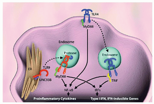
Recognition of the various PAMPs by TLRs on the surface of monocytes, macrophages, DCs, and mast cells initiates inflammatory responses, which induce cytokine signaling that can be Th1 or Th2. This signaling can be categorized as either myeloid differentiation primary response (MyD88)-dependent (Fig. 25.1), which is used by all TLRs except for TLR3, or TIR containing adaptor-inducing interferon (IFN)-γ-dependent pathways (29). TLRs respond to specific conserved sequences or to molecules present on invading microbes; for example, TLR4 responds to lipid A, a component of LPS, whereas TLR3 recognizes double-stranded RNA. Inflammatory cells express different classes of TLRs, depending on their lineage and maturity (30). The spatial distribution of TLRs ultimately determines the microbes they encounter and whether these elements are recognized as being self or nonself (31,32). Some TLRs are expressed on the cell surface, whereas others are expressed intracellularly within endosomes (Table 25.1).


Figure 25.1 Cell biology of TLR signaling. TLR4 is located at the plasma membrane and translocates to the endosomal compartment on stimulation. Although MyD88-dependent signaling occurs without endosomal translocation of TLR4, TIR-domain-containing adapter-inducing interferon-β (TRIF)-dependent signaling requires dynamin-dependent translocation. In a resting cell, TLR9 is localized at the endoplasmic reticulum (ER), but is translocated to the endosome on stimulation, where a protease or proteases cleave TLR9. IRF, interferon regulatory factor; TRIF, Toll/IL-1 receptor domain containing adaptor-inducing interferon-β. (Adapted from Kumagai Y, Shizuo A. Identification and functions of pattern-recognition receptors. J Allergy Clin Immunol 2010;125:985-992.)


TABLE 25.1 SUMMARY OF TLRS


TLR Location


PAMPs Recognized


TLR1


Cell surface


Triacyl lipopeptides (bacteria and mycobacteria)


TLR2


Cell surface


Hemagglutunin protein (viruses), peptidoglycan, and LTA (Grampositive bacteria), lipoarabinomannan (mycobactria)


TLR3


Endosome


ssRNA, dsRNA viruses


TLR4


Cell surface


LPS (Gram-negative bacteria), glycoinositolphospholipids (Trypanosama), mannan (Candida), fusion protein (respiratory synctial virus)


TLR5


Cell surface


Flagellin, an important protein in bacteria motility, adhesion, and invasion


TLR6


Cell surface


LTA (Gram-positive bacteria), zymosan (Saccharomyces)


TLR7


Endosome


ssRNA viruses


TLR8


Endosome


ssRNA viruses


TLR9


Endosome


dsDNA viruses, unmethylated CpG motifs


TLR11


Cell surface


Profilins from toxoplasmosis gondii, uropathogenic Escherichia coli


dsDNA, double stranded deoxyribonucleic acid; LPS, lipopolysaccharide; LTA, lipoteichoic acid; PAMP, pathogen-associated molecular patterns; ssRNA, single stranded ribonucleic acid; TLR, toll-like receptor. Adapted from Ooi EH, Psaltis AJ, Witterick IJ, et al. Innate immunity. Otolaryngol Clin N Am 2010;43: 473-487.


Natural killer (NK) cells originate in the bone marrow and participate in the innate immune system. NK cells simultaneously express both T-cell markers (CD3, T-cell receptor [TCR]-αβ) and NK cell markers (CD56, CD16, CD95, and CD178) (33). A large fraction of NK cells are referred to as invariant NK cells, and they are characterized by the expression of a single unique TCR-α rearrangement. Upon activation of NK cells, they are capable of rapid and substantial production of cytokines, including IL-4, which is an important cytokine in allergic pathogenesis (34).

NK cells were first known for their capability to lyse tumors without any prior priming or immunization. They have been shown to have diverse functions ranging from secreting cytokines such as INF-γ, TNF-α, and granulocyte monocyte-colony stimulating factor (GM-CSF) and chemokines (lymphotactin, RANTES (Regulated upon Activation, Normal T-cell Expressed, and Secreted), and macrophage inflammatory protein [MIP]-1α/MIP-1β) after activation (35) to serving as intermediaries between the innate and adaptive immune systems.

NK cell precursors express IL-2 receptor once they commit to the NK lineage. As they mature, further receptor diversity is procured, and the mature NK cell exhibits specific major histocompatibility complex (MHC) receptors, including 2B4, CD38, CD7, and killer-cell immunoglobulin (Ig)-like receptor. Upon completion of maturation, NK cells migrate to the peripheral blood and congregate in the spleen, lymphoid organs, lungs, and liver. There are two different types of NK cells—those that participate in killing and those that focus on cytokine production (36).

Another feature of innate immunity is the complement system. The complement system is the primary humeral mediator of antigen-antibody reactions. Initially, complement was thought to play a major role only in innate immunity against invading pathogens. It was discovered subsequently that the complement system plays an important role in the adaptive immune system by being involved with both T and B cells (37). It has also been shown to play a role in tumor growth (38), tissue regeneration, and in diseases like hemolytic uremic syndrome and age-related macular degeneration (39).

The complement consists of at least 30 chemically and immunologically distinct proteins, which can interact with each other, with antibodies, and with cell membranes. The proteins exist either as soluble in the blood or as membraneassociated. There are three routes by which the complement system is activated: the classical pathways, the lectin pathway, and the alternative pathway. All of the pathways converge at C3 (which is the most abundant complement protein found in the blood), resulting in the formation of activation products and the membrane attack complex (C5b-9) (40).

The biologic activity of complement is manifested in three ways. First, certain complement proteins bind to or opsonize foreign particles. Specific cellular receptors for these complement proteins then mediate the binding and uptake of the opsonized particles by polymorphonuclear leukocytes and monocytes. Second, the small fragments of proteolytic cleavage from complement proteins diffuse readily, and they can bind to neutrophils and macrophages, causing chemotaxis and cell activation. Similar receptors on lymphocytes and antigen-presenting cells bind antigen-antibody complexes and enhance specific immune responses such as phagocytosis and clearance of apoptotic cells. Third, complement causes lysis by the
insertion of a hydrophobic “plug” into lipid-membrane bilayers, allowing osmotic disruption of the target cell. Deficiencies in complement lead to severe infectious or autoimmune diseases.

Complement may play a role, in the nasal mucosa, in an immediate-type hypersensitivity reaction by stimulating mast cells. Studies of asthma have implicated increases of anaphylatoxins C3a and C5a at sites of inflammation after segmental allergen provocation of the bronchus in asthma patients (41), which can contribute to cellular recruitment of inflammatory cells.

Another important player in the innate immune response is the numerous antimicrobial peptides that are expressed in the sinonasal and lower airway epithelium (26). These include lactoferrin (42), lysozyme (43), cathelicidin (44), defensins (45), SP-A and -D (surfactant protein) (46,47), acid mammalian chitinase (47), collectins (48), serum amyloid A (49), and secretory leukocyte proteinase inhibitor (50). The most abundant of the antimicrobial peptides in nasal secretions are the lysozyme, lactoferrin, and secretory leukocyte proteinase inhibitor (51). In addition to being referred to as endogenous antibiotics by being secreted in response to microbes, these peptides also play a role in activating or suppressing effector cells of the innate and adaptive immune system. Human cathelicidin, LL-37, is chemotactic for neutrophils, monocytes, and T cells (52). It has also been shown that SP-D can cause a shift from a Th1 to a Th2 cytokine response, inhibit lymphocyte proliferation (53), and regulate DC responses (54).

The defensins, composed of α-defensins and β-defensins, are expressed on cells such as neutrophils, monocytes, epithelial cells, and paneth cells (55). Paneth cells, which are located at the base of intestinal glands, secrete α-defensins when exposed to bacteria and thus contribute to the gastrointestinal barrier. Degranulation of cells that express defensins occur in a dose-dependent manner as part of the host defense in response to infection at epithelial surfaces (55). It is well known that phagocytes and leukocytes are recruited to sites of infection via chemotactic factors (56). Defensins induce chemotaxis of the immature DCs to the sites of infections (56).

The mRNA of HD5, an α-defensin, is expressed in variable amounts in nasal epithelial cells (57). β-Defensins in mice induce maturation of DCs via TLR4 (58). Because DCs are involved in both antigen (Ag)-specific immune responses and Th1/Th2 polarization, defensins may contribute to interactions between the innate and the adaptive immune system via their influence on DCs. Cathelicidin is an antimicrobial protein expressed by epithelial cells, monocytes, NK cells, mast cells, and γδT cells. LL-37, a cathelicidin peptide, participates in host antimicrobial immunity by recruiting phagocytes, immature DCs, and T cells to sites of inflammation (52). Both LL-37 and human β-defensin-2 cause mast cell degranulation, with consequent histamine release (59). Eosinophil-derived neurotoxin is implicated in antiviral roles and has been shown to play a role in DC development, and like defensin, it may also play a role in bridging of the innate system with the adaptive immune system (60).

Other roles of the antimicrobial peptides have been postulated. Biofilms, which are implicated as a possible cause of CRS (61,62), are organized communities of microbial cells that are attached to living surfaces and are composed of exopolysaccharide matrix encased in a self-produced exopolysaccharide matrix and attached to an inert or living surface (63). In vitro, synthetic Cathelicidin LL-37 shows antibiofilm properties at subbactericidal concentrations when cultured with Pseudomonas aeruginosa (64). In animal models, synthetic LL-37 successfully eradicated biofilm of Pseudomonas sinusitis (65).


Adaptive Immune System

Innate immunity controls and eliminates infection with the help of the complement system, NK cell activation, and TLR activation. If the innate immune system is unable to mount an inflammatory response sufficient to remove pathogens, the adaptive immune response is activated. The adaptive immune system, in contrast to the innate immune system, is highly specific in nature and benefits from gene rearrangement. Developing B and T lymphocytes obtain randomly assembled Ig receptors and TCR from specific gene segments. The variable, diversity, and joining (VDJ) segments that are flanked by recombination signal sequences are recombined with either addition or subtraction of nucleotides, creating immense variability in the receptor (66). Whereas the remodeling process that regulates chromatin accessibility of target gene segments remains unclear, it is evident that these processes tremendously expand the diversity of antigen recognition.

The adaptive immune system utilizes many of the same cells as the innate immune system. One major difference between the two responses is the mode of recognition of antigens. Whereas the innate immunity recognizes microorganisms through components present on their cell walls via broad receptor recognition, the adaptive immune system recognizes specific antigens that are processed and presented by antigen-presenting cells in the context of MHC receptors. In cell-mediated adaptive immunity, priming occurs during the first exposure to antigen by MHC II on antigen presenting cells (APCs) with the help of costimulatory molecules. TCR receptors on the T cell associate with the CD3 receptor and, in turn, activate a series of phosphorylation steps within the cell, resulting in the formation of either CD4+ or CD8+ T cells. Activation of T-helper (Th) cells allows the secretion of many cytokines and mediators which, in turn, activate effector cells such as macrophages. Also, in humeral adaptive immunity, the primed Th cell comes into contact with B cells expressing the same antigen. Interaction between these two cell types and linking of CD40 and CD40L on B and activated Th cells, respectively,
causes B-cell activation and differentiation into antibodysecreting plasma cells (67).

A major difference between the adaptive and the innate immune system is the capability of memory in the adaptive system. Most activated T cells function as effector cells and later undergo apoptosis. Some of them, however, differentiate and persist in the host for many years to provide rapid protection on subsequent antigen exposure. The exact mechanism by which certain subsets of T cells become memory cells, and under what signaling conditions, is under investigation.


THE LINK BETWEEN THE INNATE AND ADAPTIVE IMMUNE SYSTEMS

There has been increasing evidence regarding the cross talk between the innate and adaptive immune responses. TLRs, being part of the innate immunity, play an important regulatory role in the function of antigen-presenting cells (68). It has been found that the signals that spur the induction of the adaptive immune system are many, but ample evidence now suggests that activation of TLRs on APCs results in upregulation of costimulatory molecules that induce type-I INF responses and facilitate APC-T-cell interactions that are necessary for the adaptive immune system (69). In one study, Jang et al. (70) found that IL-6 and IL-10 produced by DCs in response to Mycobacterium tuberculosis occurred largely as a result of the contribution of TLR2 responses.

DCs express TLR, which are able to recognize pathogenderived molecules or endogenous signals released by damaged or dying cells (71). Immunologic tolerance results from exogenous antigen presentation of DCs in the absence of direct PRR stimulation (72). PRRs on DCs also play an essential role in driving the cell production of Th1 and Th17-polarizing cytokines such as IL-12, IL-6, transforming growth factor (TGF-β), and IL-23 (68,69,70,71). There is also evidence that DCs can be primed for Th-2 polarization through OX40/OX40 ligand interactions, which are thought to be facilitated by the basophil production of IL-4 (71,72).

TLR activation through several pathways contributes to polarization to Th1 immune responses—leading to the hypothesis that this polarization may prevent Th2 immune responses and allergy. Studies in allergen-challenged TLR4-defective mice showed reductions in airway inflammation with eosinophils, allergen-specific IgE levels, and Th2 cytokine production, compared with wild-type mice (73). TLR activation may also lead to Th2 responses. TLR-2-stimulated DCs in vivo produce abundant IL-10, which favors a Th2 response (74). All of these developments have led to hypotheses that TLRs play a role via DCs in linking the innate immune system to adaptive immune deviation in allergy.

Trompette et al. (75) reported on a more intimate link discovered between TLR4 signaling and allergic sensitization to specific antigens. The major allergens of house dust mites are homologs of MD-2, the secreted LPS-binding member of the TLR4 signaling complex. Functional homology suggests that Der p2/LPS may drive airway Th2 inflammation in vivo in a TLR4-dependent manner, retaining this ability in the absence of MD-2. These data further suggest that Der p2 tends to generate effector lymphocyte responses because of its ability to activate the innate immune system through TLR4 signaling (76,77,78,79). A further understanding of the influence of TLRs on Th1/Th2 is likely to help us better understand the mechanisms of allergy.

Immunohistologic studies on sinonasal biopsy specimens have shown that TLR4 is localized in the submucosal seromucinous glands and the TLR2 in the stroma deep to the epithelial surface (80). Reduced mRNA expression of TLR2 was reported in turbinate tissue biopsies of patients with allergic or non-AR as compared to controls (81). It has also been shown that the TLR2 mRNA expression of TLR3 and TLR4 mRNA was reduced in patients with nasal polyps and with fungal colonization, which may represent a link between fungus and TLR expression in nasal polyps (82).

Another link between innate and adaptive immunity is through the NK cells. They have been found in several studies either to activate DC maturation (83), or they are activated by DCs to increase in numbers, secrete IFN-γ, and then to modulate or, in essence, create negative feedback of the afferent limb of the immune response by killing immature DCs (84). This concept is important in that it suggests a complex interplay of cells in the innate regulation of the immune response by NK cells prior to their activation in the adaptive immune response.


CELLS OF THE IMMUNE SYSTEM

The cells of the immune system are derived from pluripotent hematopoietic cells. They develop along two distinct lineages: the myeloid and the lymphoid. The myeloid stem cell lineage is further divided into granulocytes, megakaryocytes, platelets, and erythrocytes. Granulocytes play an important role in allergic responses as early responders in inflammation; they phagocytose bacterial pathogens and secrete important cytokines.

Pluripotent stem cells also differentiate along the lymphoid stem cell line, of which T cells, B cells, and NK cells are major players in the immune response to allergy (85).


Neutrophils

Blood neutrophils are composed of two interchangeable subpools: the “circulating pool” and the “marginal pool.” For the “circulating pool” of neutrophils to reach the site of acute inflammation, they must pass through a series of coordinated steps that include adherence to endothelium, extravascular migration, chemotaxis, membrane recognition, attachment to foreign particles, phagocytosis, fusion of lysosomes, degranulation, and a burst of oxidative metabolism.


Neutrophils are recruited and activated by Th17 cells in either a direct or an indirect way. The direct way is through IL-8 production, and the indirect way is by inducing the production of CSF and CXCL8 (86,87). For their adhesion to the vascular endothelium, they need chemokines and selectins.

Once they reach the site of inflammation, the neutrophils recognize particles by the opsonins attached to them. These opsonins include Igs to which the neutrophils exhibit Fc receptors and the C3b fragment of complement. After phagocytosis, the processes described for mononuclear phagocytes also apply to neutrophils.

In regard to their role in allergy, neutrophils are recruited during the late response to antigen challenge, but they are rarely seen to increase during seasonal allergen exposure, making their role in allergic disease uncertain.


Eosinophils

Eosinophils are produced in the bone marrow from CD34+ hematopoietic precursors. They circulate in the blood and localize to inflamed tissues. Many cytokines are important in the recruitment of eosinophils to sites of inflammation, but the most important are IL-1, IL-5, and TNF (88). Eosinophils possess adhesion molecules (ligands), which assist in their movement toward chemokines such as RANTES, MIP-1α, eotaxin, and monocyte chemoattractant protein-4 (MCP-4), secreted by inflamed tissues (85).

Eosinophil function results from elaboration of a variety of cytokines, proteins, peroxidases, and enzymes. One of these, a major basic protein, is cytotoxic and helminthotoxic. Also elaborated are eosinophil peroxidase, eosinophil-derived neurotoxin, Charcot-Leyden crystal protein, and eosinophil cationic protein (85).

Survival of eosinophils in tissues is based upon their need for several growth factors, such as IL-5, IL-3, and GM-CSF. In the absence of growth factors, they undergo programmed cell death (apoptosis). IL-33 enhances the survival of eosinophils, causes the production of superoxide anion, and activates eosinophils as effectively as does IL-5 (89). Blocking of the receptors of IL-33, namely ST2, reduced the survival of eosinophils and the production of IL-8 (89).

Konya et al. (90) reported that endothelial cell-derived prostaglandin (PG) I-2 attenuated the migration of eosinophils in vitro. In addition, upon treatment of the endothelial cells with a cyclooxygenase inhibitor, eosinophil migration and adhesion were blocked. These results suggest that selective PGI-2 receptor agonists might have beneficial effects in allergic inflammation.


Basophils

Basophils are granulocytes that possess high-affinity IgE receptors. They contain histamine and other mediators, including cytokines. They can contribute to anaphylaxis by releasing histamine. They express IL-4, and contribute to allergic reactions at tissue sites such as the nose, lungs, and skin (91,92).


Monocytes

Monocytes originate in the bone marrow from pluripotent stem cells and then are released into the blood. They produce PGs, IFNs, proteases, and cytokines. They play a role in immunomodulation with their expression of histamine H4 receptor, which is involved in the perpetuation of allergic inflammation. Histamine H4 mediates mast cell migration toward CXCL12, which is a constitutive chemokine (ligand of CXCR4 and CXCR7) that is expressed in the skin and in the airway epithelium and plays a significant role in allergic airway diseases (93,94).

Upon migration from the blood into tissues, monocytes mature into macrophages or APCs, which are either dendritic or Langerhans cells interspersed in the epithelial layer of the nasal mucosa and skin. It has been found that this maturation is associated with upregulation of H-1 (histamine) receptors (95). In the proliferation of immature macrophages, mitogens such as CSF, which is produced by fibroblasts, lymphocytes, and monocytes, play an important role. During inflammation, both migration and proliferation increase dramatically (96).

Macrophages are the most abundant immune cell in the airways in the absence of inflammation (96). They play an important role in the initiation and regulation of the immune response by producing Th1-type cytokines (IL-12, IL-18, TNF-α, and IFN-α/β). Macrophages that produce IL-12 increase the bronchial responsiveness associated with eosinophil migration. Macrophages producing IL-1 stimulate T-cell function, and present immune molecules to lymphocytes (97), displaying the same major histocompatibility determinants used by T cells (98). IL-1 increases production of PGs and leukotrienes, which may alter vascular permeability and bronchial tone. IL-1 also induces the production of acute-phase proteins, including complement components, fibrinogen, and clotting factors, and increases the activity of adhesion proteins (e.g., intracellular adhesion molecule [ICAM]-1). Substances chemotactic for macrophages include C5a anaphylatoxin, bacterial products such as N-formylmethionyl peptides, and products from stimulated B and T lymphocytes (99).

Also important are substances that inhibit migration away from sites of inflammation: lymphokines (macrophage inhibitory factor and macrophage activation factor) and proteolytic enzymes produced during activation of complement (factor Bb).

The capacity for macrophages to recognize opsonized particles resides in their receptors, which bind the Fc portion of Igs and the C3 components of complement, which facilitate phagocytosis and subsequent killing of ingested microbes and tumors as follows. Particles bound to specific or nonspecific membrane receptors are surrounded
by the macrophage cell membrane, forming phagocytic vesicles. Endocytic vacuoles become secondary lysosomes after fusion with primary lysosomes. Within the lysosomal compartment, the contents are digested at acid pH by more than 40 hydrolytic enzymes. After ingestion of particles, macrophages as well as neutrophils undergo a respiratory burst, observed as a dramatic increase in the consumption of oxygen and activation of a membrane-associated oxidase. This oxidase reduces molecular oxygen to superoxide anion, which, in turn, dismutates to hydrogen peroxide. Superoxide and hydrogen peroxide interact to give rise to hydroxyl radicals and singlet oxygen. These reactive metabolites of oxygen exert antimicrobial and antitumor effects.

Another group of effector molecules synthesized by macrophages includes nitric oxide and reactive nitrogen intermediates. The macrophage itself is protected from these oxygen metabolites by its glutathione peroxidase and catalase. Numerous soluble agents, including antigen-antibody complexes, C5a, ionophores, and tumor promoters, can trigger the respiratory burst without phagocytosis (97).


Lymphocytes


T Lymphocytes

The cells primarily responsible for immune recognition are lymphocytes, which have surface-specific receptors for antigenic determinants of foreign molecules. Certain lymphocytes confer the ability to recognize host cells that are infected with intracellular microorganisms through recognition of epitopes presented on the cell’s surface. It is therefore imperative that lymphocytes also retain the ability to recognize self-antigens in addition to foreign elements in order to be effective in targeted destruction.

The T cells make up 60% to 70% of peripheral lymphocytes. They are concentrated in zones where they are likely to meet antigen, namely, the paracortical areas of lymph nodes and of the spleen. They originate in the thymus and participate in cell-mediated immunity. T cells confer high degrees of specificity within the clone, and variability across all T cells through the recombination of the antigenspecific portion of their CD3-TCR. In addition to CD3, T cells have a variety of other surface markers that are important in allergy. CD4 and CD8 are markers on certain subsets of T cells and enhance the signal that occurs when a T cell comes into contact with an appropriate antigen-MHC complex borne by APCs. A second signal is provided via CD28 that interacts with the costimulatory molecules B7-1 and B7-2 that are present on APCs (97).

When the T cells leave the bone marrow, they leave as the pre-T cells CD4-CD8-DN (double negative) cells. It is not until gene segment rearrangements occur during their maturation that they will have a gene coding a full-length TCR protein. From a pre-T cell, sequential rearrangements of two TCR genes leads to surface expression of an αβ or γδ TCR (100). Double positive T cells, that is, T cells that are both CD4 positive and CD8 positive, only survive if they are able to recognize self-antigens presented in that particular individual’s MHC molecules. They thus undergo positive selection. The T cells then drop either the CD4 to become CD8+, or cytotoxic T cells, or retain only the CD4+ molecules, to become CD4+ T helper cells. Deeper in the thymus, negative selection occurs, which selects for T cells that bind too strongly to self-antigens and causes these cells to apoptose. These two selection processes allow for tolerance of self by the immune system as well as positive selection of T cells that are able to recognize peptides displayed in that individual’s unique MHC I and MHC II molecules.

γδ T cells, which only comprise 2% of T cells, are thought to play a role in mucosal immune defense because they accumulate in epithelial sites of inflammation and in some mouse models, γδ T cells do play a role in allergic inflammation (101), however, evidence for the role of γδ T cells in allergic inflammation is conflicting.


CD4+ T Cells

CD4+ T cells interact with cells in the context of MHC-II protein, which is present on APCs. These cells develop into Th1 and Th2 cells. Upon leaving the thymus, CD4+ cells enter the lymphoid organs, where their naïve TCR receptor comes into contact with antigen-MHC complexes on APCs. The type of APC, the costimulatory molecules produced, in addition to the local cytokine environment, all play a role in determining whether the Th cell will become a Th1 or Th2 cell (102). Th1 and Th2 cells are only able to be distinguished from each other by the substances they produce. Th1 cells secrete IFN-γ, IL-2, TNF-α, and TNF-β, which are important for eliminating intracellular pathogens. In contrast, Th2 cells secrete IL-4, IL-5, IL-6, and IL-13, which are important for the elimination of extracellular organisms.

It is also important to mention that peptides are not the only target of T-cell response, as T cells recognize a variety of lipid antigens by means of a family of nonpolymorphic genes (CD1) (103) that evolved to present lipids and glycolipids to the mammalian immune system (104,105). CD1a is expressed on DCs and macrophages. The CD1 protein is implicated in pollen capture and processing (106,107). It is speculated that this recognition by CD1+ DCs and CD1-restricted T cells is involved in the rapid handling of the foreign inhaled grain and initiates the allergic response (107).


CD8+ T Cells

CD8+ cells interact in the context of MHC-I, which is present on all cells. The role of CD8+ cells in allergy is not clear, although some studies hypothesized that CD8+ cells may prevent the development of airway hyperresponsiveness (108,109). CD8+ cells secrete cytokines of the Th1 type and function primarily as cytotoxic cells promoting the death of recognized infected cells.

Dichotomous findings with respect to CD8+ cells led to attempts to further characterize the subpopulations of
CD8+ cells. One study showed the different cytokine profiles of CD8 cell subpopulations stimulated by dendritic cell 1 (DC1) and dendritic cell 2 (DC2). Specifically, IL-10 producing-CD8 cells activated by DC2s were presumed to play a regulatory and possibly an immunotolerant role in the immune response (110). CD8 cells are thought to prevent the induction of immune responses early in the sensitization process, but had a limited role once airway sensitization had occurred (111). Other studies point to the homing capabilities of CD8 cells. Those cells that migrate to lymphoid organs are named central memory cells, whereas those that home to nonlymphoid tissue are called effector cells in inflammatory processes. Elucidation of how and when activated T cells commit to memory cell lineage is still unknown. One interesting development is that memory CD8+ T cells are more efficient in TCR signaling and better augment downstream signaling than are naïve or effector cells (112). This supports the finding that symptoms of AR may continue to increase in patients later in the allergy season despite decreasing allergen counts.


Treg Cells

T cells previously called T suppressor cells, are now called T regulatory (Treg) cells. Treg cells are CD4+CD25+ T cells that express the IL-2 receptor marker (CD25). Previously known for taking part in preventing autoimmunity and transplant rejection, they are now recognized as immunomodulators in the Th1/Th2 paradigm. The Treg subset called Tr1 cells (inducible Treg cells) can produce large amounts of IL-10 and TGF-β (113), which can down regulate allergenspecific Th1 and Th2 responses (114).


Figure 25.2 Potential mechanisms of conventional allergen immunotherapy. High-dose allergen exposure during immunotherapy results in both immune deviation of Th2 responses in favor of a Th0/Th1 response and the generation of IL-10- and TGF-β-producing CD4+CD25+ T cells, and possibly Tregs. IFN-γ-induced activation of bystander macrophages and/or other cells represents an alternative source of these inhibitory cytokines. During subsequent natural environmental exposure to allergens, the activation and/or maintenance of the usual atopic Th2 T cell response is inhibited. Additionally, these cytokines induce preferential switching of B cell responses in favor of IgG and IgG4 antibodies (and possibly IgA antibodies under the influence of TGF-β). IgG may also inhibit IgE-facilitated allergen binding to antigen-presenting cells with subsequent downregulation of IgE-dependent Th2 T-lymphocyte responses. Blue arrows represent immune response pathway to natural exposure (low-doses Ag and IgE); green arrows represent immune response pathway to immunotherapy (high dose Ag); red blocked lines represent inhibition (high-dose Ag); dotted lines represent possible means of action not yet proven. (Adapted from Robinson DS, Larché M, Durham SR. Tregs and allergic disease. J Clin Invest 2004;114:1389-1397.)

Treg cells play a role in areas of tolerance. Children who outgrew their milk allergy (called tolerant children) demonstrate significantly higher peripheral Treg cells compared to children who continued to experience allergy to cow’s milk (115).

In a cohort study of over 150 children at age 6 from atopic families, CD4+CD25+ T cells were highest during pollen allergy season, which suggests that these cells represent a mixture of regulatory and activated cells (116). Furthermore, in another study, the number of Treg cells to dust mite allergen were found to be similar in both nonatopic and atopic individuals, cautioning against a simplistic view that atopic diseases are caused only by a deficiency of Treg cells (117). Treg cells also play a role in immunotherapy by secreting IL-10 and TGF-β and inducing B cell Ig class switch toward IgA and IgG4 (Fig. 25.2).


B Lymphocytes

B lymphocytes, like T lymphocytes, leave the bone marrow and localize to peripheral lymphoid tissues. They aggregate in follicles within lymphoid organs, where they coordinate the humeral response. When stimulated by antigen via their IgM surface receptors or B cell receptors (BCRs), BCRs associated ligands aggregate to form signaling complexes, whose strength is modulated and propagated via second
messengers such as diacylglycerol, inositol triphosphate, and intracellular calcium release. B cells differentiate into plasma cells. Plasma cells are responsible for secreting different classes of Igs that mediate humeral immunity. The synthesis of Igs together with heavy chain class switching can occur in the nasal mucosa (118) and be induced in response to allergen exposure (119). IL-4, IL-13, and CD40 are the elements necessary to promote synthesis of IgE and Ig heavy chain class switching in the nasal mucosa. Wise et al. (120) and Durham et al. (121), showed activation of these mediators following allergen provocation. Secondary lymphoid organization can also be seen within nasal polyps (122). Thus class switching can occur outside lymph nodes (123,124), and in the sinonasal tissue.


Dendritic Cells

DCs express receptors such as members of the TLR family that enable them to recognize pathogen-derived molecules or endogenous signals released by dying or damaged cells (125). DCs also express receptors of cytokines like TNFs or IFNs, allowing these cells to recognize a wide spectrum of pathogens like viruses or parasites.

DCs are thought to have two distinct origins. The majority are from a myeloid progenitor, which bears CD11c, CD33, and CD13 cell surface markers and is called myeloid DCs. A smaller subset of DCs are called “plasmacytoid DCs” (pDCs) because of their similarity to antibodysecreting plasma cells. These cells bear a high number of CD123 cell surface markers, which respond to IL-3 and, in turn, produce type I IFN. Characteristics of the pDCs link them to both lymphoid and myeloid precursors (125). The mechanism by which DCs help establish T-cell memory and tolerance, has yet to be determined, but at least in a mouse asthma model, if pDCs are depleted and animals are exposed to an allergen, then IgE sensitization, airway eosinophilia, and Th2-cell cytokine production occurs, which is not seen in mice with pDCs (126).

In AR, cytokines such as IL-4, IL-5, and IL-13 produced by activated cells play a role in inflammation. In cultures of pDCs and CD4+T cells of grass pollen allergic patients, IL-2, IL-5, IL-10, and IL-13 production is increased and upon exposure to grass allergen, CD4+ cells grow and pDCs will produce IL-4, IL-5, and IL-13 with repeated stimulation (127). This further supports the theory that pDCs have effects on stimulating allergen-specific (memory) Th2 responses in the presence of mucosal inflammation.

In their immature forms, DCs in the periphery of the body primarily function to sample antigen. The maturation of DCs occurs after TLR-ligand stimulation (128), upregulation of MHC classes I and II, and upregulation of costimulatory molecules such as CD40, CD80, and CD86 (129). These mature DCs then go on to interact with naïve CD4+ and CD8+ cells.

Another role of DCs is polarizing of T cells into Th2-type effector cells. In animal models of allergic airway inflammation, dendritic-cell-like APCs resembling epidermal Langerhans cells preferentially uptake antigen and present it to CD4+ cells locally in the lung (130), supporting the work of others showing that DCs act to induce Th2 responses (131,132) and that they are necessary to induce a chronic inflammatory eosinophilic response (133,134).

Langerhans cells, another subset of DCs, are the subpopulation that plays a role in T-cell responses in the nasal submucosa in seasonal allergies (135). Other studies comparing functional DC subtypes in normal human upper airways as opposed to the characterizations made in mouse models found that the majority expressed a macrophage-like phenotype (CD11b+CD14+CD64+CD68+RFD7+) (136).


IMMUNOGLOBULINS

Igs are glycoproteins composed of 82% to 96% polypeptide and 4% to 18% carbohydrate components. They account for approximately 20% of the total plasma proteins. Igs function to tag and aggregate toxins and bacteria. They facilitate the activation of the complement system as well as the activation of macrophages, neutrophils, and lymphocytes to clear foreign materials. Binding of antibody to the Fc receptor of mast cells sensitizes these cells for immediate type hypersensitivity.

Ig diversity is attained via multiple processes. The human heavy chain (VH), D, and junctional (JH) heavy chains are found on chromosome 14. There are 46 functional VH segments per haploid genome, with considerable polymorphism within certain segments. V segments vary in two specific regions called complimentarily-determining regions that form part of the antigen-binding site of antibodies. There are seven families of D segments that code for diversity in the Ig repertoire. Functional JH segments, six in number, are located downstream of the D region. Combinatorial rearrangement of this large number of genes codes for diversity by utilization of recombinant activation genes (RAG1 and RAG2) to facilitate binding to recombinant signal sequences. DNA strand breakage and rejoining at splice sites account for much of the diversity observed in Ig function; however, the recombination process incorporates additional methods that greatly increase the variety of expressed antibodies, which includes the addition or removal of nucleotides to segments and somatic point mutation of rearranged genes. All completely assembled Ig molecules contain an equal number of heavy (H) and light (L) polypeptide chains (Fig. 25.3). In a healthy individual, there are nine isotypes of Igs and four different classes, they are: IgG, IgA, IgM, and IgE.

In normal adults, IgG, which has the most prominent role in memory immune responses, constitutes approximately 75% of the total serum Igs. The relative concentrations of the four subclasses are as follows: IgG1, 60% to 70%; IgG2, 14% to 20%; IgG3, 4% to 8%; and IgG4, 2% to 6%. IgG can fix complement, with the subclasses’ ability to fix complement as follows: IgG3 > IgG1 > IgG2. IgG4,
although unable to fix complement by the classic pathway, can utilize the alternative complement pathway. IgG4 also plays a role in immunotherapy, as shown in Figure 25.2.


Figure 25.3 Antibody structure. Antibodies are Y-shaped, flexible molecules consisting of two heavy and two light chains linked together by disulfide bonds. The light and heavy chains are composed of constant (CL, CH1, CH2, CH3) and variable (VL, HL) regions. (Adapted from Moser M, Leo O. Key concepts in immunology. Vaccine 2010;31;28(Suppl 3):C2-C13.)

IgA, constituting approximately 15% of the total serum Igs, predominates in body secretions. There are two subclasses, IgA1 and IgA2. The helper T cells in the lymphoid tissues of the gastrointestinal and respiratory tracts switch the B cells from IgM to IgA secretion. Secretory IgA, because of its abundance in saliva, tears, and bronchial and nasal secretions, as well as mucous secretions of the small intestine, provides the primary defense mechanism against local mucosal infections. Its main function may be to prevent access of foreign substances to the general immunologic system. Besides its traditional role in extracellular antibody function, IgA can neutralize viruses intracellularly, can provide an internal mucosal barrier by intercepting antigens and ferrying them through the epithelium, and, after binding to the surfaces of some leukocytes, can activate the alternate pathway of complement activation. Its role in food tolerance to antigens in the digestive system is currently under study. In a study by Frossard et al. (137), a significant increase in the antigen-specific IgA-producing cells was seen in a group of mice tolerized to food allergy.

IgM constitutes 10% of serum Igs and normally exists as a pentamer with a molecular weight of 900,000. IgM antibody predominates in the early immune response and along with IgD is the major Ig expressed on the surface of B cells and is far and away the most efficient Ig in complement fixiation. The enormous size of this pentamer Ig can cause dangerous obstruction in high concentration in areas such as small venules. After an early rise, the IgM response declines and is replaced by IgG of the same antigen specificity. The fetus makes IgM before birth, but maternal IgM is too large to cross the placenta. Thus, IgM antibody to a specific organism in newborn serum indicates intrauterine infection.

IgD is a monomer that is normally present in serum in trace amounts (0.2% of total Igs) and whose function remains unknown. IgD (with IgM) predominates on the surfaces of human B lymphocytes, and thus its most important role may be as a receptor.

Normally, IgE comprises only 0.004% of the total serum Igs, but it binds with very high affinity to mast cells and basophils via the Fc region, thus playing an important role in allergic responses. Like IgD and IgG, IgE normally exists in monomeric form. IgE is produced by B cells after the initiation of two important signals: IL-4 or IL-13 targeting of the gene that causes switch recombination, and interaction of activated-T-cell CD40 ligand (CD40L) with CD40 on the surface of B cells to initiate deletional switch recombination (138). Although these mechanisms are not fully understood, the formation of IgE in the atopic or allergic patient plays a major role in the disease process. IgE is also produced locally in nasal mucosa in patients with AR (139).

IgE antibodies fixed to mast cells undergo cell surface cross-linking upon combination with allergen, which then triggers the release of preformed inflammatory mediators such as histamine, tryptase, and protease from mast cells and basophils. IgE probably promotes the survival of mast cells by binding to its high-affinity receptor, FcεR I, on the mast cell, which leads to increased expression of antiapoptotic molecules (140). The continued survival of proinflammatory cells in the context of allergic inflammation may be a factor in the chronic symptomatology of the disease even after the removal of allergens. A subunit of the FcεRI receptor, the β chain, probably promotes transport of FcεRI to the surface of mast cells and basophils and hence amplifies the downstream events of the allergic response several-fold in those cells (141). The role of FcεRIβ as a candidate gene for atopy modulation is unclear.

IgE also binds to macrophages, neutrophils, eosinophils, and DCs via low affinity receptors. In macrophages, FcεRI cross-linking results in production of IL-10. Expression of cell surface FcεRIα is present on neutrophils from asthmatic patients (142), indicating that neutrophils may play a role in the allergic response via an IgE-dependent activation mechanism. Human eosinophils were found to secrete IL-10 upon engagement of the receptor (143). Targeting IgE has proven clinically useful, as demonstrated by omalizumab, a monoclonal antibody against IgE, which decrease serum and cell-surface IgE levels and has been useful in the care of severe allergic asthma since approved by the U.S Food and Drug Administration (FDA) in 2003.

Also important are strategies for modulating IgE synthesis by blocking of transcription factors for IgE production. In a mouse model of asthma, mice lacking T-cell-specific
T-box transcription factor (T-bet), a transcription factor that skews toward Th1 and away from Th2 differentiation, had airway changes resembling an asthma phenotype (144). Other studies that under way are those of blocking synthesis of cytokines such as IL-4 and IL-13 that promote production of IgE. Anti-CD23 antibodies in their role of inhibiting IgE production are also being studied.


CYTOKINES, INTERFERONS, AND CHEMOKINES

Cytokines are soluble mediators that have growth, differentiation, and activation functions for immune responses. Cytokines can be grouped into those that are predominantly APCs or T-lymphocyte derived; that predominantly mediate cytotoxic (antiviral and anticancer), humeral, cell-mediated (Th1 and Th17), or allergic immunity (Th2); or that are immunosuppressive (Treg cells) (145). Table 25.2 lists the cytokines produced by lymphocytes, and Table 25.3 is a complete list of cytokines.


TABLE 25.2 Th LYMPHOCYTE FAMILIES


Family


Cytokine Repertoire


Cytokines Involved in Differentiation


Transcription Factors Involved in Differentiation


Th1


IFN-γ, TNF-α, TNF-β, GM-CSF, IL-2, IL-3


IL-12: activates STAT4, leading to expression of T-bet; induces IL-18R expression


IL-18: upregulates IL-12R, further induces IFN-γ expression


IL-27: activates STAT4, leading to increased expression of T-bet and IFN-γ


IFN-γ: increases expression of T-bet by increasing expression of STAT1; negative regulator of Th17 and Th2


T-bet: master regulator of Th1 cells; potentiates production of IFN-γ and IL-12Rβ2; suppresses Th2 and Th17 differentiation


STAT4: produced in response to IL-12 and potentiates production of IFN-γ


STAT1: increases expression of T-bet; negative regulator of Th17


Th2


IL-2, IL-3, IL-4, IL-5, IL-9, IL-13, IL-24, IL-25, IL-31, TNF-α, GM-CSF


IL-4: activates STAT6, leading to expression of GATA-3; negative regulator of Th17, IL-19, IL-25, IL-33


TSLP: promote differentiation and survival of Th2-like cells


GATA-3: master regulator of Th2 cells; potentiates IL-4 expression; suppresses expression of Th1 differentiation and cytokines expression (IFN-γ)


MAF: contributes to IL-4 production once a Th2 program is established; inhibition of Th17 differentiation


STAT6: promotes Th2 cell differentiation; negative regulator of T-bet expression and Th1 differentiation


NFAT: increases transcription of IL-4


Th9


IL-4, IL-9


TGF-β: induces the high IL-9 phenotype of Th2-like lymphocytes


Th17


IL-17 (IL-17A), IL-17F, IL-21, IL-22


IL-6: differentiation factor for the generation of Th17 cells TGF-β, IL-21


IL-23: support the differentiation and function of Th17 cells in the additional presence of IL-6


RORγt (retinoic acid-related orphan nuclear receptor) is the master regulator of Th17 cell differentiation


STAT3: activated by IL-6 and essential for Th17 differentiation


nTreg/iTreg


IL-10


TGF-β: differentiation factor for the generation of nTreg


cells IL-10: important for differentiation of peripheral iTreg cells, role in nTreg development uncertain


IL-2: promotes survival, proliferation, and survival of nTreg cells through their constitutive expression of CD25


FOXP3: master regulator of thymus-derived nTreg cells


Th3


TGF-β, IL-10


Adapted from Commins SP, Borish L, Steinke JW. Immunologic messenger molecules: cytokines, interferons, and chemokines. J Allergy Clin Immunol 2010;125(2 Suppl 2):S53-S72.


Chemokines are a group of small (8 to 12 kD) proteins with the ability to affect cell migration or chemotaxis. These cells include the neutrophils, monocytes, lymphocytes, eosinophils, fibroblasts, and keratinocytes. To date, 52 chemokines and 20 chemokine receptors have been described, which are listed in Table 25.4, and their involvement in diseases in Table 25.5.


TABLE 25.3 CHARACTERISTICS OF CYTOKINES


Only gold members can continue reading. Log In or Register to continue

Stay updated, free articles. Join our Telegram channel

May 24, 2016 | Posted by in OTOLARYNGOLOGY | Comments Off on Immunology and Allergy

Full access? Get Clinical Tree

Get Clinical Tree app for offline access

Cytokine


Structure


Size Molecular Weight


Receptors


Cell Sources


Cell Targets


Major Functions