Genetic Hearing Loss


Genetic Hearing Loss


John Greinwald

Catherine Hart


Hearing impairment is the most common sensory deficit. It occurs in 2 to 3 per 1,000 live births and affects approximately 28 million Americans (1). It can be congenital or acquired, developing any time during infancy, childhood, or adulthood. Of children younger than 18 years of age, 5% have a hearing impairment, and that incidence increases to 40% to 50% by age 75. Among school-aged children, 2% have a hearing loss that interferes with educational achievement.

Most cases of hearing loss are attributed to nongenetic causes (e.g., middle-ear fluid). Advances in technology and mass screening programs, however, have resulted in an ongoing increase in the identification of specific heritable forms of deafness, most of which are expressed as sensorineural losses. Heritable hearing loss can be transmitted through various patterns of inheritance. Most authors attribute 75% to 80% of genetic deafness to autosomal recessive patterns and 18% to 20% to autosomal dominant patterns. The remainder are classified as X-linked or mitochondrial. Additionally, deafness can present as an isolated finding (nonsyndromic) or can be associated with other pathologic findings (syndromic) such as craniofacial anomalies.

This chapter presents an overview of heritable hearing loss within the framework of these basic genetic principles. We discuss a number of genes that are linked to a wide spectrum of hearing disorders and briefly describe the relevance of genetic advances to case management.


BASIC GENETIC PRINCIPLES

Human genes are molecular codes for inherited factors. Genes are arranged linearly on 23 pairs of chromosomes. These chromosomes consist of 22 pairs of autosomes and 1 pair of sex chromosomes, comprising a total of 46 chromosomes. Males have an X and a Y pair of sex chromosomes, whereas females have two X chromosomes. The location of a gene on a chromosome is termed a locus. Each chromosome pair carries a distinctive set of gene loci, and a given gene can have several alternative codes, which are referred to as alleles. The actual genetic code (genotype) for a specific trait consists of either two identical alleles (homozygous) or two disparate alleles (heterozygous). The physical manifestation of a trait, referred to as the phenotype, is determined by which alleles are present and how they interact. An allele is considered dominant if its presence results in a specific phenotype. It is considered autosomal recessive if both alleles are required for the expression of its phenotype. An X-linked recessive gene is present in only one allele (hemizygous) in males because the Y chromosome does not carry an allele complementary to the X chromosome. A specific DNA base pair change at a specific allele is considered a truncating or nontruncating mutation.

A nontruncating (or missense) mutation alters the codon resulting in an altered amino acid product. A truncating (or nonsense) mutation results in a nonfunctional protein due to premature termination of transcription. Truncating mutations can also occur due to mutations at the exon splice sites, which result in nonfunctional protein production.

Dominant traits are transmitted in a vertical fashion from one generation to another. A 50% chance exists that an affected heterozygous individual will transmit the abnormal gene to offspring. Penetrance is the ability of a gene to manifest any of the phenotypic characteristics related to that gene. In some dominant disorders, not all persons carrying the affected gene display the disease phenotype. This occurrence is called incomplete penetrance. Dominant disorders also can have variable expressivity, whereby family members present with different manifestations of the affected gene. It is thus presumed that environmental influences or interaction with other genes can modify phenotypic expression.


Figure 102.1 Basic gene structure.

In the absence of consanguinity, an autosomal recessive trait is usually seen in an offspring in small nuclear families. The offspring of heterozygous (i.e., carrier) parents have a 25% risk of being affected. Occasionally, being heterozygous for two different genes (double heterozygous) can cause a condition. An X-linked recessive trait can lack phenotypic expression if it is carried by a heterozygous female, but male offspring of this female would have a 50% chance of inheriting the gene. Expression of the gene would then occur because of the absence of a second X chromosome. A female offspring of an X-linked female carrier has a 50% risk of being a carrier of such a trait. Because males inherit their father’s Y chromosome and not his X-linked recessive gene, they are unaffected and do not become carriers. In contrast, females inherit their father’s X-linked recessive trait and, as such, do become carriers of the affected gene.

Heritable disorders that are caused by abnormalities at the chromosomal level and involve extra or absent chromosomal material are characterized by developmental delays and varied congenital anomalies, except when they involve the sex chromosomes. The nature and severity of the resultant disorders depend on the quantity and origin of the chromosomal material involved. In persons with trisomy, three copies of an entire chromosome are present. Trisomy 21 (Down syndrome) is the least severe autosomal trisomy. Trisomy 13 (Patau syndrome) and trisomy 18 (Edwards syndrome) are less common but more severe. Other autosomal types of trisomy usually are lethal. In the condition known as monosomy, which is also lethal, only one chromosome rather than a pair is present. Embryos with sex chromosome abnormalities are quite viable, with the exception of a conceptus with only a Y chromosome. Persons with one extra X or Y chromosome or with only one X chromosome have mild clinical features such as seen in Klinefelter syndrome (47,XYY or 47,XXY) and Turner syndrome (45,X).

It is known that disparate genotypes can produce a similar phenotype. This phenomenon, which is referred to as genetic heterogeneity, often makes it difficult to identify causative genes. In nonsyndromic hearing loss, gene identification is difficult in that there is a high degree of heterogeneity and an absence of features that clearly define clinical subtypes. In syndromic hearing loss, however, distinct subtypes as well as interfamilial differences assist in the identification of these genes. The identification of autosomal recessive nonsyndromic hearing loss genes is facilitated through the study of both inbred and isolated populations, which reduce genetic heterogeneity. Studies of large families (kindreds) can facilitate the identification of autosomal dominant hearing loss genes.

A detailed description of the process involved in gene identification is beyond the scope of this chapter. In brief, however, this process entails the following three steps: A gene is first localized to a specific chromosomal region. Physical mapping techniques are then used to isolate DNA from that region. Through the efforts of the Human Genome Project, nearly all of the 30,000 human genes have been mapped, thereby allowing for the bioinformatic identification of candidate genes. Step three involves sequencing analysis of candidate genes to identify the specific mutation associated with a particular disease.

Each gene generally consists of DNA that codes for (a) the actual protein(s) (exons); (b) interspersed areas between exons, which are called introns; and (c) untranslated regulatory regions before and after the exons, which are respectively referred to as upstream and downstream (Fig. 102.1). The upstream untranslated region has specific areas of DNA that are bound by regulatory proteins. These areas regulate the function of the gene, and this region is called the promoter. Occasionally, one common promoter may exist for several genes, which is called a locus control region.


AUTOSOMAL RECESSIVE DISORDERS

Most genetic hearing disorders are transmitted by an autosomal recessive mode of inheritance, and of the hearing disorders presenting in childhood, approximately 80% are inherited recessively. In most patients, hearing loss is nonsyndromic, but 10% to 20% of patients display syndromic disorders. The identification of recessive syndromes involving hearing loss, thus, necessitates a diligent search for other syndromic components. Because of the asymptomatic status of heterozygous carriers of a recessive gene and the 25% inheritance risk, it is often difficult to distinguish between nongenetic disorders and those that are recessively inherited. In both clinical situations, a single affected child generally presents in a family with no known history of hearing loss.


Nonsyndromic Recessive Disorders

To date, linkage studies have identified at least 85 loci for autosomal recessive nonsyndromic hearing loss (ARNSHL) (2). These loci are given the prefix DFNB; DFN signifies deafness, whereas B signifies a recessive mode of inheritance. Thirty seven causative ARNSHL genes have been identified (Table 102.1). DFNB1 accounts for about one half of all ARNSHL and, as such, is among the most active areas of clinical research. As implied by its designation,

DFNB1 was the earliest autosomal recessive gene locus to be successfully mapped and characterized. The gene at the DFNB1 locus is gap junction beta 2 (GJB2), which produces a protein called connexin 26 (
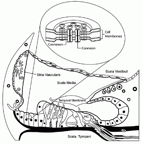
1). Connexins comprise a family of gap junction proteins that function as intercellular channels. These channels allow ions and low molecular weight molecules to travel from cell to cell (Fig. 102.2). The supporting cells of the organ of Corti express several gap junction proteins, including connexin 26. Although the function of connexin 26 in the inner ear has not been conclusively determined, it is thought to be involved in potassium recycling. In the normal ear, a sound stimulus causes depolarization of hair cells, which is mediated by the influx of potassium ions. These ions are thought to transverse through connexin molecules back to the stria vascularis. From the stria vascularis, ions are actively pumped back into the endolymph to reset its electrical gradient (Fig. 102.2).


TABLE 102.1 SELECTED GENETIC DEFECTS ASSOCIATED WITH HEARING IMPAIRMENT


Gene (Protein)


Locus


Mode of Inheritance or Syndrome


Phenotype


CDH23(cadherin 23)


DFNB12


ARNSHL


SNHL


Usher 1C


AR syndromic


Congenital hearing loss, retinitis pigmentosa, and variable vestibular areflexia


COCH (cochlin)


DFNA9


ADNSHL


Onset of hearing loss occurs between ages 20-30; profound at high frequencies and variable progression to anacusis by age 40-50. Variable vestibular dysfunction


COL2A1 (collagen 2A1)


Stickler type 1


AD syndromic


Robin sequence, myopia, joint degeneration, variable degree of hearing loss


COL11A1(collagen 11A1)


Stickler type 2


COLL11A2(collagen 11A2)


Stickler type 3


Stickler type 1 phenotype without ocular problems


COL4A3(collagen 4A3)


Alport


X-linked


Progressive high-frequency SNHL, nephritis


COL4A4 (collagen 4A4)


AR syndromic


COL4A5 (collagen 4A5)


AD syndromic


GJB2 (connexin 26)


DFNA3A


ADNSHL


Severe to profound SNHL


DFNB1A


ARNSHL


Mild to profound SNHL


GJB6 (connexin 30)


DFNB1B


ARNSHL


Severe to profound SNHL


DFNA3B


ADNSHL


High frequency to profound SNHL


DDP (deafness or dystonia peptide)


DDP (previously called DFN1)


X-linked syndromic


Early onset deafness with mental retardation


DIAPH1 (diaphanous)


DFNA1


ADNSHL


Fully penetrant progressive, low-frequency SNHL


EDNRB (endothelin receptor)


Waardenburg type 4


AR syndromic


WS with Hirschsprung disease


EYA1 (eyes absent 1)


Branchio-oto-renal


AD syndromic


Conductive, sensorineural, or mixed hearing loss with branchial remnants and renal anomalies


KVLQT1 (potassium-gated voltage channels)


Jervell and Lange-Nielsen


AR syndromic


Severe to profound SNHL, syncope, prolonged QT


KCNE1 (potassium channel voltage-gated, risk-related subfamily member 1)


MITF (microphthalmia-associated transcription factor)


Waardenburg type 2


AD syndromic


Same as type 1 without dystopia canthorum


MYO7A (myosin 7A)


DFNB2


ARNSHL


Progressive SNHL with variable vestibular and ocular findings


DFNA11


ADNSHL


Moderate SNHL


Usher 1B


AR syndromic


Profound congenital deafness, vestibular areflexia, and progressive retinitis pigmentosa


NDP (norrin)


Norrie


X-linked syndromic


Ocular symptoms, progressive SNHL, and mental retardation


OTOF (otoferlin)


DFNB9


ARNSHL


Severe to profound SNHL


PAX3 (paired box gene 3)


Waardenburg type 1


AD syndromic


White forelock, heterochromia iridis, SNHL, and dystopia canthorum


SLC26A4 (pendrin)


DFNB4


ARNSHL


Enlarged vestibular aqueduct, euthyroid goiter


Pendred


AR syndromic


Mondini dysplasia and SNHL


POU3F4 (POU domain 3, transcription factor 4)


DFN 3


X-linked


Stapes fixation


POU4F3 (POU domain 4, transcription factor 3)


DFNA15


ADNSHL


Progressive hearing loss


TCOF1 (treacle)


Treacher Collins


AD syndromic


Lower eyelid coloboma, micrognathia, microglossia, zygomatic arch hypoplasia, macrostomia, hearing loss


TECTA (α-tectorin)


DFNA8/12


ADNSHL


Prelingual severe to profound SNHL


DFNB21


ARNSHL


USH1C (USH1C)


Usher 1C


AR syndromic


Profound congenital deafness, vestibular areflexia, and progressive retinitis pigmentosa


USH2A (USH2A)


Usher2A


AR syndromic


Congenital sloping hearing loss, normal vestibular function, and progressive retinitis pigmentosa


MTRNR1


12rRNA


mitochondrial


Mild to severe hearing loss, usually associated with aminoglycoside


MTTS1


tRNAser (UCN)


mitochondrial


Mild to severe hearing loss, can be progressive


AD syndromic, autosomal dominant syndromic; ADNSHL, autosomal dominant nonsyndromic hearing loss; AR syndromic; autosomal recessive syndromic; ARNSHL, autosomal recessive nonsyndromic hearing loss; SNHL, sensorineural hearing loss.


Adapted from Van Camp G, Smith RJH. Hereditary Hearing Loss Homepage. Hereditaryhearingloss.org. For a comprehensive and current list of genes related to hearing impairment, please refer to this website.


Figure 102.2 Location of gap junctions within the supporting cells of the organ of Corti. Six connexins comprise a connexon. Connexons form gap junctions, which function as intracellular channels.

DFNB1 is estimated to cause up to 50% of all ARNSHL in many populations. Of patients presenting with hearing impairment of 70 dB or greater, nearly 40% have DFNB1; of those with a mild to moderate hearing loss, only 10% to 15% have this gene (3, 4). GJB2 mutations have been found in patients from many different ethnic backgrounds, and specific mutations vary with patient ethnicity. The deletion of a guanine at nucleotide position 35 of the gene (35delG) is one of the most common mutations that results in autosomal recessive hearing impairment, particularly in patients of European descent. The carrier frequency for the 35delG deletion in Europe is 1 in 50. A deletion of a thymine at position 167 (167delT) is often present in the Ashkenazi Jewish population, with a carrier frequency of 1 in 25. A 235delC mutation occurs more frequently in Asians. Among the white population in the United States, the frequency of heterozygous carriers of mutations in GJB2 is approximately 1 in 40. This carrier frequency is almost as common as that in cystic fibrosis, which has a carrier frequency of approximately 1 in 24. The high frequency of mutations and the small size of this gene make screening and molecular genetic diagnosis feasible.

Additionally, a unique genetic mutation has been found in the GJB6 gene. This mutation appears to present in carriers of GJB2 mutations (double heterozygote state). A large deletion (170 to 340 kilobases [kb]) upstream and including the 5′ portion of the GJB6 gene has been found to cause severe hearing loss (5). Interestingly, GJB2 and GJB6 are located in close proximity on chromosome 13, and this deletion may interfere with a common promoter element for the two genes (locus control region). Approximately 10% of patients with hearing loss who have one abnormal GJB2 allele also have the GJB6 deletion (6).

GJB2 mutations can create altered protein expression, subcellular localization, or abnormal functionality of the connexin complex. GJB2 mutations cause a wide range of
hearing loss phenotypes, ranging from mild to profound hearing impairment. These mutations can be nontruncating or truncating. The type of mutation can produce varying levels of hearing loss. For example, a biallelic truncating mutation produces a severe to profound hearing loss in nearly all patients (e.g., 35delG/35delG). In contrast, the presence of one nontruncating mutation (e.g., V37I) commonly causes a milder form of hearing impairment (2).

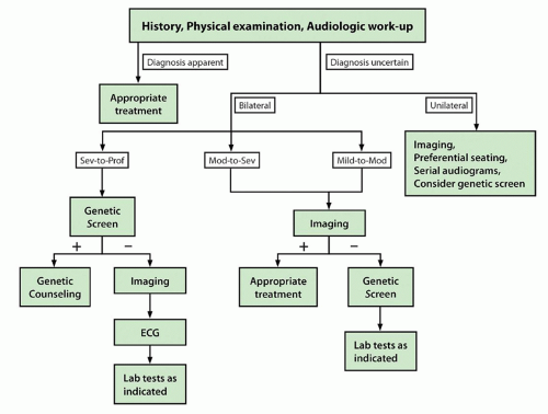
The clinical significance of DFNB1 mutations lies in diagnostic evaluation and case management. Genetic testing is now routinely part of the algorithm in the evaluation of hearing loss (7). Genetic testing should be the first line of evaluation of children with bilateral ARNSHL of 60 dB or greater (Fig. 102.3). For those with unilateral or mild bilateral hearing impairment, genetic testing should be considered after temporal bone imaging is performed. If the imaging study is normal, genetic testing is then appropriate. If the imaging study demonstrates enlarged vestibular aqueduct or Mondini deformity, SLC26A4 (Pendred) testing should be performed. New genetic platforms will allow rapid screening for multiple genes at one time (e.g., “Ear-gene,” Cincinnati Children’s Hospital Medical Center, Cincinnati, OH). Recent data have shown that gene chip platforms are highly accurate and will likely become instrumental in diagnostic evaluation of sensorineural hearing loss (SNHL) (8).

The prognosis for children with hearing loss related to GJB2 is variable and has been shown to correlate with the genotype. This is particularly important in light of new hearing screening programs that are increasingly detecting hearing loss in very young children. Genetic information can assist in predicting the actual hearing phenotype, which is valuable considering the emphasis on early (age 12 months) cochlear implantation. For example, if a nontruncating mutation is present, a child would be more likely to have mild hearing loss. Conversely, the presence of biallelic truncating mutations in a child in whom an auditory brainstem response (ABR) test at 1 month of age showed severe hearing loss would lead the practitioners to recommend early aggressive treatment. A high likelihood would exist of recommending cochlear implantation to ensure the child’s ability to communicate verbally. Interestingly, research indicates that patients with DFNB1-related hearing loss who meet criteria for cochlear implantation have better outcomes than those with congenital hearing loss from other causes (9). Such findings emphasize that genotype can have clinical significance, and further study aimed at elucidating this particular association is ongoing.


Figure 102.3 Algorithm for the diagnosis of genetic hearing disorders.

Most other ARNSHL genes have been identified from isolated consanguineous families, and the prevalence of mutations in these genes in the general population has yet to be determined. A technology now being developed (resequencing microarray), however, will enable the rapid reading of a large amount of DNA.

Another ARNSHL gene worthy of note is otoferlin (OTOF). This gene was originally found to be the cause of hearing loss in several isolated families (DFNB9) (Table 102.1). Although the function of OTOF is not certain, it is hypothesized that this gene is a calcium sensor involved in hair cell exocytosis (10, 11). Research has shown that in some patients, OTOF is also responsible for auditory neuropathy (AN) (12). AN is a unique form
of SNHL characterized by absent ABR waveforms, present otoacoustic emissions, and variable behavioral thresholds. The prevalence of AN in patients with SNHL ranges from 0.5% to 15%. The etiology of AN is quite heterogenous with approximately 40% of cases having a genetic basis. It can be autosomal recessive, as in OTOF; autosomal dominant; X-linked; or syndromic. Some patients with AN also have other peripheral neuropathies or central nervous system disease. The treatment of AN can be challenging, involving the judicious use of FM devices and hearing aids as well as cochlear implantation. A better understanding of the molecular basis of this disease will allow for more accurate diagnosis and a more individualized treatment approach.


Syndromic Recessive Disorders


Jervell and Lange-Nielsen Syndrome

Jervell and Lange-Nielsen (JLN) syndrome is a congenital disorder associated prolonged QT interval leading to episodes of arrhythmias and syncope that occur as early as the second or third year of life. These episodes are caused by a cardiac conduction defect and can lead to sudden death. Beta-adrenergic blockers (e.g., propranolol) and implantable cardioverter defibrillators have proved effective in managing the cardiac component of the syndrome.

The degree of hearing loss can vary, but it is almost always severe to profound. Clinicians should consider this diagnosis in children with hearing loss and unexplained syncope or in cases of a family history of syncope or sudden death. Patients suspected of having JLN syndrome should have an electrocardiogram; results will show large T waves and prolongation of the QT interval.

One form of JLN has been attributed to homozygosity for mutations of a potassium channel gene called KVLQT1 (13) (Table 102.1). Other mutations in this gene cause the dominantly inherited long QT syndrome (Romano-Ward syndrome), which does not include hearing loss in the phenotype. Not all families with JLN syndrome map to this gene, clearly indicating genetic heterogeneity. Mutations also have been found in another potassium channel gene, KCNE1 (14). In some families, heterozygous carriers of JLN syndrome show a prolonged QT interval but are otherwise asymptomatic.


Pendred Syndrome and Enlarged Vestibular Aqueduct Syndrome

First described in 1896, Pendred syndrome is a disorder in which hearing impairment is associated with abnormal iodine metabolism; the typical result is a euthyroid goiter. The gene that causes Pendred syndrome has been identified and is named SLC26A4 (or PDS

Only gold members can continue reading. Log In or Register to continue

Stay updated, free articles. Join our Telegram channel

May 24, 2016 | Posted by in OTOLARYNGOLOGY | Comments Off on Genetic Hearing Loss

Full access? Get Clinical Tree

Get Clinical Tree app for offline access