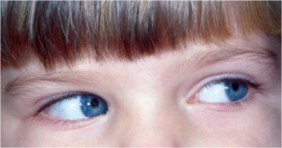
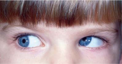
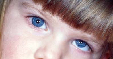
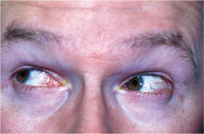
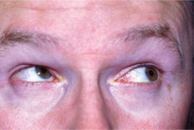
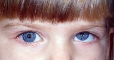
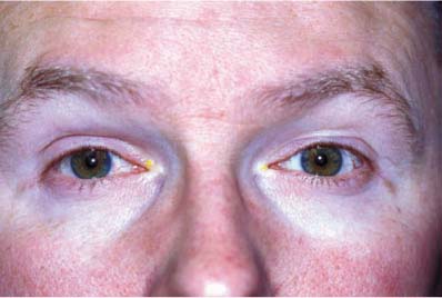
Chapter 31 The fourth (trochlear) nerve (IV) supplies the superior oblique muscle. Palsies of this cranial nerve may be congenital or acquired, but congenital palsies can also become symptomatic later in life. Involvement of the trochlear nerve can occur in isolation or combined with other cranial nerve palsies. The initial examination should confirm the diagnosis of fourth nerve palsy, determine if it is congenital, and assess for any accompanying orbital or neurologic abnormalities. The patient should be seen by an ophthalmologist or neuro-ophthalmologist within a week to obtain baseline measurements and a complete examination. Fourth nerve palsy is the most common form of acquired vertical diplopia in adulthood.1,2 Binocular vertical diplopia is usually worse in gazing away from the affected side, in unilateral cases. There may be horizontal and torsional components to the diplopia, particularly in downgaze, is common in bilateral cases. Hypertropia occurs on the side of the fourth nerve palsy (Figs. 31–1, 31–2, 31–6, and 31–7). The hypertropia is greater in lateral gaze to the side opposite the palsy and on head tilt to the same side. For example, a left fourth nerve palsy causes a left hypertropia greater in right gaze and left head tilt (Figs. 31–3, 31–5, and 31–8). Often the patient unconsciously tilts the head away from the eye with the fourth nerve palsy. This reduces the hypertropia and sometimes eliminates the double vision (Fig. 31–4). Rarely, the patient tilts the head toward the eye with the palsy, so as to maximally separate the two images and thus help in ignoring one of the images. The inaction of the superior oblique causes the eye to excyclotort. Sometimes this is manifest as torsional diplopia. The two images are not parallel. If the patient looks at a horizontal line, the images form a V tilted to the right or left (< or >). The narrow portion of the V points in the direction of the affected eye, because the hypertropic excyclotropic eye sees an image that is lower and incyclotorted in space. A bilateral fourth nerve palsy often causes only a small vertical deviation in primary gaze. The hypertropia “reverses” in lateral gaze (right hypertropia in left gaze and left hypertropia in right gaze). A vertical deviation is present with head tilt and reverses direction with head tilt in the opposite direction (bilateral positive head tilt). The patient may assume a chin-down position, to avoid downgaze, where the diplopia is worse. In most cases, excyclotorsion of greater than 10 degrees occurs. The torsion can be measured using a double Maddox rod. The most common cause of bilateral palsy is head trauma. FIGURE 31–1 Left IV N palsy with left hypertropia in primary gaze. FIGURE 31–2 Left IV N palsy, left gaze, eyes look orthophoric. FIGURE 31–3 Left IV N palsy, right gaze with overshoot of left eye from inferior oblique overaction. FIGURE 31–4 Left IV N palsy, right head tilt, eyes look orthophoric. FIGURE 31–5 Left IV N palsy, left head tilt with increase in left hypertropia. Ask the patient to look down. If the fourth nerve is functional, the top of the globe will rotate in toward the nose. Look at a blood vessel on the conjunctiva to see the incyclotorsional movement. If it is absent, then fourth nerve palsy is present. The following indications should raise the question of alternate diagnoses: FIGURE 31–6 Right IV N palsy, eyes look orthophoric in primary gaze. FIGURE 31–7 Right IV N palsy, right gaze, eyes look orthophoric. FIGURE 31–8 Right IV N palsy, left gaze with overshoot of right eye from inferior oblique overaction. The most common etiology of fourth nerve palsy in children7
FOURTH NERVE PALSIES
URGENCY OF EVALUATION
DIAGNOSIS
SYMPTOMS
Demographics
Diplopia
SIGNS
Hypertropia
Head Tilt
Excyclotorsion
Bilateral Fourth Nerve Palsy

Fourth Nerve Function in the Presence of a Third Nerve Palsy
Red Flags
 Diplopia or ptosis, which is worse in the evenings (suggests myasthenia gravis; see Chapter 34)
 Lid retraction/stare, lid lag on downgaze, proptosis, resistance to forced ductions (suggests Graves’ ophthalmopathy; see Chapter 38)
 Redness of the eye, chemosis, pain (suggests orbital inflammation; see Chapter 40)
 Headache, fever, weight loss, difficulty swallowing, jaw tiredness with prolonged chewing (suggests temporal arteritis in persons over 50; see Chapters 18 and 47)
 History of facial trauma, anesthesia of lower eyelid (suggests orbital floor fracture with entrapment of the inferior rectus)
 Association with other cranial nerve palsies (suggests infectious process, infiltrative process, myasthenia gravis or cavernous sinus lesion; see Chapter 33)
 Gaze evoked amaurosis (suggests an intraorbital lesion)
 Clicking sound on vertical eye movements (suggests trochlear inflammation)
 Association with brainstem or cerebellar symptoms or signs (suggests skew deviation; see Chapter 35)
DIFFERENTIAL DIAGNOSIS1,3–6
Congenital Fourth Nerve Palsy
![]()
Stay updated, free articles. Join our Telegram channel

Full access? Get Clinical Tree


