Fluorescence Angiography of Choroidal and Retinal Tumors
James J. Augsburger
Fluorescence angiography has been used extensively in the evaluation of tumors of the choroid and retina during the past four decades. Several excellent reviews on the subject of fluorescein and indocyanine green (ICG) angiography of intraocular tumors have been published,1,2,3,4,5,6,7 and interested readers are referred to these sources for additional information on this subject. The role of fluorescence angiography in a patient with an intraocular tumor is largely documentary in nature, but it can be of differential diagnostic value in certain cases. The essential clinical importance of fluorescence angiography in the evaluation of most choroidal and retinal tumors appears to be its ability to reveal patterns inconsistent with the presumptive diagnosis rather than to elucidate pathognomonic features.8 The role and importance of fluorescence angiography in various tumors of the choroid and retina are summarized in the remainder of this chapter.
CHOROIDAL TUMORS
Both fluorescein and ICG angiography are used currently to evaluate many choroidal tumors. Because ICG angiography tends to image choroidal blood vessels substantially better than does fluorescein angiography, it appears to be the preferred method for evaluating most choroidal tumors. However, because fluorescein angiography has been used more extensively, both the fluorescein and ICG angiographic features of the following selected choroidal tumors are described and illustrated.
CHOROIDAL NEVUS
The choroidal nevus is a benign uveal melanocytic tumor of limited growth potential. The typical lesion (Figs. 1, 2, 3, and 4) appears as an ill-defined, gray-brown choroidal mass that is usually less than 5 mm in maximal basal diameter and less than 1 mm in thickness. The basal margins of typical lesions often blend into the normal surrounding choroid in a feathered or striate pattern. Drusen and retinal pigment epithelial (RPE) pigment clumps commonly develop on the surface of these tumors.
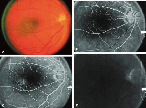 Fig. 1. Typical melanotic choroidal nevus. A. Ill-defined gray-brown macular choroidal lesion with bland surface features. B-D. Fluorescein angiogram of lesion. B. Laminar venous phase frame showing intact retinal vasculature overlying nevus but virtually no hypofluorescence corresponding to choroidal lesion. C. Full venous phase frame showing granular hyperfluorescence corresponding to minor retinal pigment epithelial alterations and drusen on surface of tumor. D. Late-phase frame showing continued hypofluorescence of choroidal lesion with mild staining of surface drusen. |
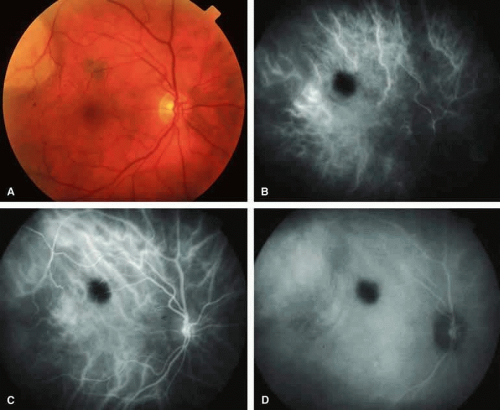 Fig. 2. Typical melanotic choroidal nevus. A. Small gray choroidal nevus one disc diameter above fovea. The pale lesion at upper left margin of photograph is a choroidal melanoma that was the main focus of the following angiogram. B-D. Indocyanine green (ICG) angiogram of lesion. B. Early-phase frame showing intense hypofluorescence of nevus. C. Intermediate-phase frame showing sustained hypofluorescence of nevus. D. Late-phase frame showing sustained hypofluorescence of nevus. |
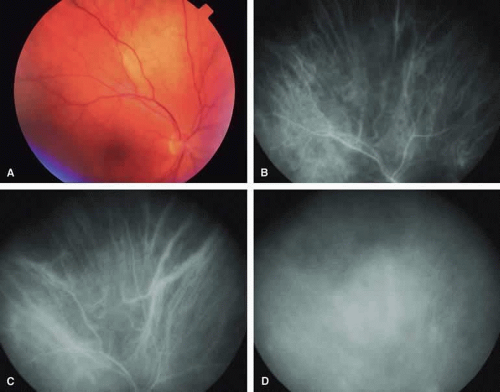 Fig. 3. Amelanotic choroidal nevus. A. Ill-defined pale yellow choroidal nevus superior to optic disc. B-D. Indocyanine green (ICG) angiogram of lesion. B. Early-phase frame showing mild hypofluorescence of nevus and visibility of large-caliber choroidal arteries passing through lesion. C. Intermediate-phase frame showing features similar to those of the previous frame. D. Later phase frame showing ill-defined mild hyperfluorescence of lesion. |
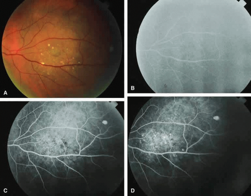 Fig. 4. Melanotic choroidal nevus with drusen and retinal pigment epithelial (RPE) alterations. A. Gray-brown choroidal nevus with numerous white drusen and dark gray pigment clumps on its surface. B-D. Fluorescein angiogram of lesion. B. Venous phase frame showing retinal arteries and veins but no convincing sign of the underlying choroidal nevus. C. Later venous phase frame showing mild granular hyperfluorescence and hypofluorescence centrally corresponding with drusen and RPE pigment clumps overlying choroidal nevus. D. Late venous phase frame showing increased multifocal hyperfluorescence corresponding to drusen and persistent foci of hypofluorescence corresponding to RPE alternations. |
Fluorescein and ICG angiographic features of choroidal nevi have been described by numerous authors.1,2,3,4,5,6,7,9,10,11,12 Neither fluorescein nor ICG angiography appears to be particularly helpful for evaluation of a typical choroidal nevus. Ophthalmoscopy is usually sufficient to allow one to decide with reasonable certainty whether a small melanotic choroidal tumor is a typical choroidal nevus or an equivocal lesion that may be either a large nevus or small choroidal melanoma. No well-designed clinical study has ever demonstrated significant independent differential diagnostic value of fluorescence angiography for clarifying this differential diagnosis.8
Typical Melanotic Choroidal Nevus
Fluorescein angiography of a typical choroidal nevus with bland surface features (see Fig. 1) shows the entire lesion to be hypofluorescent relative to the adjacent uninvolved choroid throughout the study. No large-caliber choroidal blood vessels are usually identifiable within the lesion. The retinal vasculature overlying the lesion appears well defined and normal on fluorescein angiography.
ICG angiography of a typical melanotic choroidal nevus (see Fig. 2) shows better definition of the basal area of the lesion than does fluorescein angiography. The entire lesion appears completely and uniformly dark throughout the ICG angiogram. Only the larger retinal blood vessels overlying the nevus are usually demonstrated on ICG angiography.
Amelanotic Choroidal Nevus
Approximately 10% to 15% of choroidal nevi are largely or completely amelanotic clinically. Fluorescein and ICG angiography of an amelanotic choroidal nevus (see Fig. 3) tend to show less prominent hypofluorescence of the lesion than they do with darkly melanotic nevi. Because of the lack of intracellular melanin pigment within the nevus cells, some large-caliber choroidal blood vessels running through the nevus may be visible in the region of the mass (see Fig. 3B and C). These choroidal blood vessels are better defined by ICG angiography than by fluorescein angiography. Amelanotic choroidal nevi often appear mildly hyperfluorescent in late-phase frames (see Fig. 3D).
Choroidal Nevus with Drusen and Clumps of RPE Hyperplasia
If a choroidal nevus has drusen and RPE alterations on its surface (see Fig. 4A), fluorescein angiography (Fig. 4B, C, and D) tends to show patchy or stippled window defect hyperfluorescence corresponding to foci of RPE depigmentation, fluorescence blockage by clumps of RPE hyperplasia on the surface of the lesion, and late staining of at least some of the drusen. These features are not usually as evident on ICG angiography as they are on fluorescein angiography.
Choroidal Nevus Versus Melanoma
Small melanocytic choroidal tumors larger than 5 mm in diameter and thicker than 1 mm but without clearly invasive clinical features (e.g., nodular eruption through Bruch’s membrane, retinal invasion) may be either benign nevi or small choroidal melanomas. Several features of these lesions have been identified as prognostic of the likelihood of subsequent lesion enlargement,13 a surrogate indicator of the malignant potential of the tumor. Features suggestive of low growth potential and probable benign histology include thickness less than or equal to 1.5 mm, uniform gray-brown coloration of the lesion, and drusen and RPE clumping on the surface of the lesion. In contrast, features suggestive of higher growth potential and probable malignant histology include thickness greater than 1.5 mm, nonuniform coloration of the lesion, prominent clumps of lipofuscin pigment on the surface of the lesion, and serous subretinal fluid overlying and surrounding the lesion. None of these features is a reliable indicator of the underlying histologic nature of the tumor; however, the greater the number of unfavorable features, the greater the likelihood of lesion enlargement if it is followed without treatment after initial detection.
Choroidal Nevus Versus Melanoma with Bland Surface Features
Several authors have evaluated fluorescein and ICG angiography as differential diagnostic tools for differentiating between large benign choroidal nevi and small malignant choroidal melanomas.9,10 An example of such a lesion evaluated by fluorescein angiography is presented as Figure 5. Some common assertions based on these studies include the following. If the lesion is a nevus, fluorescein angiography is likely to show hypofluorescence of the lesion throughout the study, absence of large-caliber intralesional blood vessels, and lack of late smudgy hyperfluorescence resulting from leakage from these vessels. In contrast, if the lesion is a melanoma, fluorescein angiography is likely to show at least some intralesional tumor blood vessels, some patchy or smudgy fluorescein leakage from those blood vessels, and at least patchy or smudgy late fluorescence of the tumor. ICG angiography is more likely to reveal intralesional blood vessels within bland appearing small melanocytic choroidal lesions (especially darkly melanotic lesions) than is fluorescein angiography, and lesions with prominent intralesional blood vessels are generally regarded as more likely to be melanomas. Unfortunately, there have been no confirmatory angiographic-histopathologic correlation studies of small melanocytic choroidal tumors in the nevus versus melanoma category to confirm or refute these assertions. At best, this author regards the fluorescence angiographic features of bland appearing small melanocytic choroidal tumors as suggestive but not confirmatory of the pathologic nature of the tumor.
 Fig. 5. Choroidal nevus versus melanoma. A. Partially melanotic and partially amelanotic choroidal tumor with prominent clumps of retinal pigment epithelial (RPE) pigment and drusen on surface. B-D. Fluorescein angiogram of lesion. B. Laminar venous phase frame showing poorly defined hypofluorescence of choroidal lesion and area of window defect hyperfluorescence corresponding to amelanotic portion of lesion. C. Full venous phase frame showing persistent mild hypofluorescence corresponding to melanotic portion of lesion, increased hypofluorescence corresponding to RPE thinning and drusen overlying lesion, and foci of intense hypofluorescence corresponding to RPE clumps on lesion. D. Late-phase frame showing persistence of features shown in the prior frames plus more numerous hyperfluorescent foci corresponding with additional drusen. |
Choroidal Nevus Versus Melanoma with Prominent Lipofuscin Pigment Clumps
If one obtains a fluorescein angiogram on a small melanotic choroidal lesion (nevus versus melanoma) that has prominent clumps of lipofuscin pigment on its surface (Fig. 6), the pigment clumps appear intensely hypofluorescent throughout the study. This appearance is attributable to the complete blocking of choroidal fluorescence by the lipofuscin. ICG angiography does not show lipofuscin pigment clumps on the surface of the tumor as well as fluorescein angiography does.
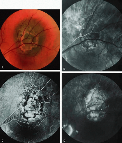 Fig. 6. Melanotic choroidal nevus versus melanoma with prominent clumps of overlying orange pigment. A. Darkly melanotic choroidal lesion with prominent clumps of overlying orange pigment (lipofuscin). B-D. Fluorescein angiogram of lesion. B. Arterial phase frame showing irregular hypofluorescence and hyperfluorescence corresponding to lesion. C. Laminar venous phase frame showing generalized window defect hyperfluorescence of mass plus intense choroidal fluorescence blockage by orange pigment. D. Late-phase frame showing multiple pinpoint foci of hyperfluorescence at retinal pigment epithelial (RPE) level, leakage of fluorescein into the overlying and surrounding subretinal fluid, and partial obscuration of the blockage of choroidal hypofluorescence corresponding to the orange pigment on the surface of the lesion. |
Choroidal Nevus Versus Melanoma with Overlying Serous Subretinal Fluid
A blister of serous subretinal fluid sometimes develops over and around a presumed choroidal nevus (Fig. 7A), especially if the lesion is located in the macula.11 If one performs a fluorescein angiogram on a small melanocytic choroidal lesion (nevus versus melanoma) that has shallow overlying serous subretinal fluid (see Fig. 7B, C, and D), one or more hyperfluorescent leak sites may show up slowly at the RPE level as the study progresses. In some cases, fluorescein will clearly leak from those foci into the overlying serous subretinal fluid. ICG angiography does not show hyperfluorescent leak sites at the RPE level as well as fluorescein angiography does.
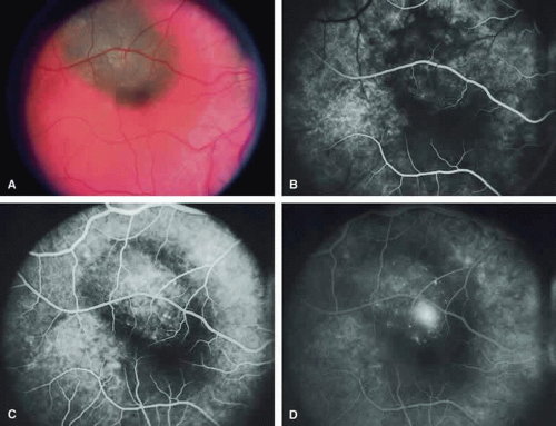 Fig. 7. Melanotic choroidal nevus versus melanoma with overlying serous subretinal fluid. A. Bland gray-brown choroidal lesion involving upper portion of macula with shallow overlying and surrounding blister of serous subretinal fluid. B-D. Fluorescein angiogram of lesion. B. Arterial phase frame showing patchy choroidal hypofluorescence corresponding to choroidal lesion. C. Full venous phase frame showing faint zone of choroidal hypofluorescence corresponding to marginal zone of lesion, mild hyperfluorescence corresponding to the central portion of the lesion, and discrete pinpoint foci of hyperfluorescence at the retinal pigment epithelial (RPE) level on the lesion. D. Later phase frame showing smudge hyperfluorescent focus of fluorescein leakage into subretinal fluid. |
Choroidal Nevus Versus Melanoma with Choroidal Neovascular Membrane
A choroidal neovascular membrane occasionally develops from the surface of a small melanocytic choroidal tumor (nevus versus melanoma).12 This vascular structure can usually be anticipated because of the presence of ophthalmoscopically evident hemorrhagic or exudative subretinal fluid overlying a portion of the tumor (Fig. 8A). Fluorescein angiography in such cases (see Fig. 8B to D) generally reveals the neovascular membrane as a relatively well-defined vascular network that fluoresces brightly in the early frames of the study and leaks progressively by the late frames. If the subretinal fluid is grossly hemorrhagic, ICG angiographymay show the choroidal neovascular network better than does fluorescein angiography.
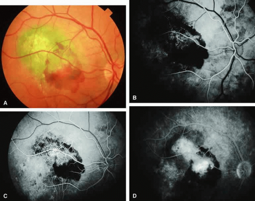 Fig. 8. Melanotic choroidal nevus versus melanoma with overlying choroidal neovascular membrane. A. Same lesion shown in Figure 7 following spontaneous accumulation of subretinal blood and hard exudates associated with development of choroidal neovascular membrane overlying lesion. B-D. Fluorescein angiogram of lesion. B. Arterial phase frame showing abnormal vascular network deep to retina, partially obscured by blockage corresponding to subretinal blood. C. Laminar venous phase frame showing greater extent of abnormal vascular network overlying lesion and zone of window defect hyperfluorescence extending inferiorly through the macula. D. Late-phase frame showing smudgy hyperfluorescence from abnormal subretinal blood vessels, persistent blockage of choroidal fluorescence by the subretinal blood, and persistence of later hyperfluorescence due to retinal pigment epithelial (RPE) thinning extending inferiorly through the macula. |
Fluorescence Angiographic Detection of Lesion Enlargement
Small melanocytic choroidal lesions in the nevus versus melanoma category are frequently monitored periodically following initial documentation for enlargement or other indicators of possible malignant behavior. Fluorescein angiography sometimes defines the basal area of these lesions better than does fundus photography, but ICG angiography is clearly the preferred technique for this purpose. If one elects to follow a small melanocytic choroidal tumor without treatment, comparative fluorescence angiography can be used to identify slight lesion enlargement that is not clearly evident by ophthalmoscopy or comparison of fundus photographs. Because benign choroidal nevi can and do enlarge, however, detection of subtle lesion enlargement by fluorescence angiography probably has limited differential diagnostic value in these lesions.
CHOROIDAL MALIGNANT MELANOMA
Choroidal malignant melanoma is the most common primary malignant intraocular neoplasm of adults. It has substantial local growth potential in addition to its well-known propensity to metastasize and, thereby, prove fatal to the host. Fluorescence angiography can provide evidence in support of the diagnosis of choroidal melanoma but is rarely if ever sufficient by itself to establish the diagnosis. Several ophthalmoscopically detectable features of clinically diagnosed melanocytic choroidal tumors are strongly indicative of their malignant potential. These include tumor diameter substantially greater than 7 mm, tumor thickness substantially greater than 3 mm, prominent intralesional large-caliber blood vessels, nodular apical eruption of the tumor through Bruch’s membrane, invasion of the overlying retina by the tumor, and a prominent nonrhegmatogenous retinal detachment with shifting subretinal fluid.
Several distinct fluorescein and ICG angiographic patterns have been associated with choroidal melanomas.1,2,3,4,5,6,7,14,15,16,17,18,19,20,21 These distinct patterns depend on the degree of pigmentation of the tumor cells, the thickness of the tumor, the presence or absence of eruption of tumor through the overlying Bruch’s membrane, and the presence or absence of retinal invasion by the tumor.
Melanotic Choroidal Melanoma Without Invasive Features
The typical choroidal melanoma that has not broken through Bruch’s membrane generally appears as a gray-brown to dark brown, dome-shaped, solid choroidal mass (Fig. 9A). The overlying retina commonly appears normal except that it is draped over the choroidal tumor. Fluorescein angiography (see Fig. 9B, C, and D) typically shows the tumor to be relatively hypofluorescent during the early frames of the study. If prominent clumps of orange lipofuscin pigment are present on the surface of the tumor, they will appear intensely hypofluorescent throughout the study because of blockage of the underlying choroidal and tumor vascular fluorescence. During the arterial phase frames of the study, several ill-defined large-caliber deep intralesional blood vessels may be identifiable against the generally hypofluorescent background. As the study continues, these large intralesional blood vessels leak progressively so that the surface of the lesion tends to appear hyperfluorescent by the late frames. Fluorescein may also accumulate in the overlying retina and retinal pigment epithelium as tiny discrete pinpoint foci of intense hyperfluorescence. If serous retinal detachment is present overlying and around the tumor, the fluorescein will leak through the retinal pigment epithelium and accumulate in the subretinal fluid by the late frames. The retinal vascular pattern of the overlying retina commonly appears unremarkable in these cases.
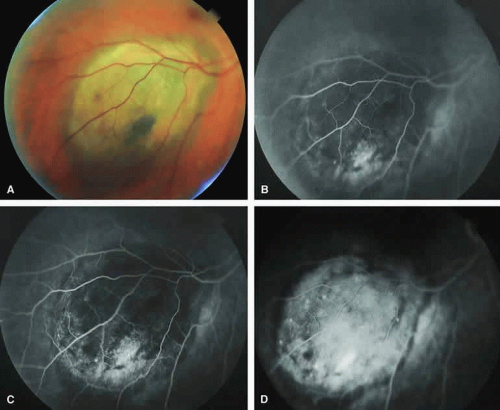 Fig. 9. Typical melanotic choroidal melanoma without invasive features. A. Nodular gray-brown nodular choroidal tumor with linear clumps of orange lipofuscin pigment and central whitish discoloration of overlying retinal pigment epithelium. B-D. Fluorescein angiogram of lesion. B. Venous phase frame showing mild hypofluorescence of most of tumor but with ill-defined hyperfluorescent focus near inferior margin. C. Later venous phase frame showing persistent generalized hypofluorescence of mass, increased smudgy and punctate hyperfluorescence near the inferior and temporal margins of lesion, and choroidal fluorescence blockage by lipofuscin pigment overlying mass. D. Late-phase frame showing diffuse hyperfluorescence of subretinal fluid and outer retina overlying mass, persistent hypofluorescence corresponding to the margin of the lesion, persistent choroidal fluorescence blockage corresponding to the lipofuscin pigment clumps, and several pinpoint dots of intense hyperfluorescence overlying the mass. |
If ICG angiography is performed on a similar tumor (Fig. 10), the tumor mass generally appears more intensely hypofluorescent throughout the study and its intrinsic blood vessels appear more clearly visible than on fluorescein angiography. ICG slowly accumulates within the extracellular space of the tumor so that the tumor usually appears mildly hyperfluorescent, at least in part, in the late frames.
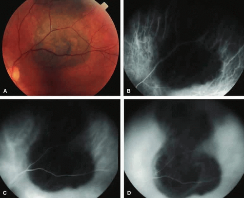 Fig. 10. Typical melanotic choroidal melanoma without invasive features. A. Ill-defined bilobed melanotic choroidal mass with prominent orange lipofuscin pigment clumps on its surface. B-D. Indocyanine green (ICG) angiogram of lesion. B. Early-phase frame showing generalized hypofluorescence of mass but with large-caliber choroidal blood vessels passing through it. C. Later phase frame showing persistent hypofluorescence and better definition of choroidal lesion. Some choroidal blood vessels are still visible through the mass. The lipofuscin pigment clumps produce limited choroidal fluorescence blockage. D. Late-phase frame showing persistent hypofluorescence blockage of most of mass but some smudgy hyperfluorescence of marginal aspects of lesion. |
Amelanotic Choroidal Melanoma Without Invasive Features
If the choroidal melanoma being evaluated is relatively amelanotic (Figs. 11A and 12A), its large-caliber intralesional blood vessels will tend to show up more distinctly on both fluorescein angiography (see Fig. 11B, C, and D) and ICG angiography (see Fig. 12B, C, and D) than they would in a darkly melanotic choroidal melanoma. Despite its amelanotic color, the cellular component of the mass tends to be at least mildly hypofluorescent relative to the adjacent uninvolved choroid during the early frames of the study. As with melanotic melanomas, the mass usually appears at least mildly hyperfluorescent in the late-phase frames (see Figs. 11D and 12D).
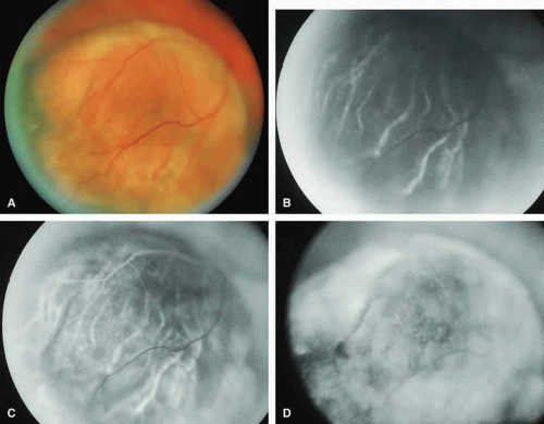 Fig. 11. Amelanotic choroidal melanoma without invasive features. A. Amelanotic, golden-yellow choroidal tumor with prominent visibility of intralesional blood vessels. B-D. Fluorescein angiogram of lesion. B. Choroidal filling phase frame showing generalized hypofluorescence of lesion but with well-defined intralesional large-caliber blood vessels not connected to retinal vasculature. C. Retinal arterial phase frame showing even more prominent intralesional large blood vessels against persistently hypofluorescent tumor. D. Late-phase frame showing diffuse hyperfluorescence of surface of lesion resulting from exuberant leakage of fluorescein into subretinal space and lesion. |
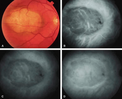 Fig. 12. Amelanotic choroidal melanoma without invasive features. A. Pale yellow macular choroidal tumor with tiny clumps of disrupted retinal pigment epithelial (RPE) pigment on its surface. B-D. Indocyanine green (ICG) angiogram of lesion. B. Early-phase frame showing prominent fluorescent large-caliber intralesional blood vessels against the background of a generally hypofluorescent choroidal mass. C. Later phase frame showing persistent hypofluorescence of the mass except for the prominent intralesional blood vessels. D. Late-phase frame showing increase in fluorescence of mass compared with surrounding normal choroid and marginal hyperfluorescence resulting from ICG accumulation in the overlying subretinal fluid. |
Choroidal Melanoma With Nodular Eruption Through Bruch’s Membrane
If a choroidal melanoma has erupted through Bruch’s membrane (Figs. 13A and 14A), it forms an apical nodule that is generally hypomelanotic and contains many large-caliber blood vessels. Fluorescein angiography of these tumors (see Fig. 13B, C, and D) typically shows hypofluorescence of the base of the lesion during the early frames, relatively rapid filling of the prominent blood vessels in the apical nodule during the venous and recirculation frames, and intense late staining of the apical nodule resulting from progressive fluorescein leakage by the late-phase frames. Similarly, ICG angiography of these tumors (see Fig. 14B, C, and D) also shows relative early hypofluorescence of the tumor base, early filling of prominent intralesional blood vessels within the apical nodule, intense staining of the apical nodule by the recirculation phase frames, and persistent late hyperfluorescence of the mass.
Stay updated, free articles. Join our Telegram channel

Full access? Get Clinical Tree


