Fig. 7.1
A partially pigmented cutaneous melanoma in the lateral aspect of the lower eyelid of the right eye, near the lateral canthus; the lesion changed in shape before the photograph was taken (Photograph courtesy of Dr. Peter A. Martin)

Fig. 7.2
High magnification of superficial cutaneous melanoma of the lower eyelid margin without involvement of the palpebral conjunctiva (Photograph courtesy of Dr. Peter A. Martin)
It is likely that most eyelid melanomas evolve through the lentigo maligna precursor lesion [10, 13, 14, 16]. Lentigo maligna is a slowly-developing non-palpable pigmented macule, usually on exposed cutaneous surface of elderly patients. It slowly enlarges in size, although some areas may undergo regression. The lesions change shape and size and may change color from tan to brown to black. When there is progression to lentigo maligna melanoma, the invasive areas are usually marked by small nodular formations and are usually dark brown or black, although invasion may occur without any obvious clinical changes. Theoretically, eyelid melanomas may evolve through dysplastic nevi that affect the eyelid or as melanomas of the superficial spreading type. Nodular melanomas are exceptionally rare among these already rare tumors, and small heavily pigmented nodules at the eyelid margin may well represent pigmented basal cell carcinomas.
Eyelid melanoma can often involve the eyelid margins (Fig. 7.3). In such cases the mucocutaneous junction may be breeched and the palpebral conjunctiva may be involved. It is often difficult to know whether the melanoma originates in the skin or in the conjunctiva. Such cases have a worse prognosis, and some relate this to the conjunctival involvement that may grow unseen for many years [8].
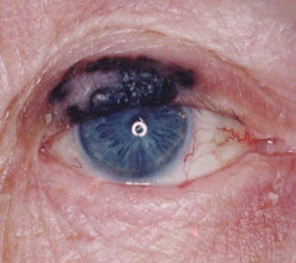
Fig. 7.3
Large cutaneous melanoma of right upper eyelid with involvement of the eyelid margin (Photograph courtesy of Dr. Peter A. Martin)
7.5 Diagnosis and Differential Diagnosis
The clinical examination of pigmented periocular lesions requires the ophthalmologist to provide an exceptional level of illumination (beyond that typically available in ophthalmic examining lanes that are used for refraction, slit-lamp examination, and funduscopy). One should be prepared to turn the room lights up to maximum level and to cast additional illumination on the affected area by a dedicated light near the examination chair. Magnification may help the ophthalmologist to detect subtle changes in a lesion’s color and texture, and the lens used for indirect ophthalmoscopy may be especially useful in this setting. Ophthalmologists should also not overlook the utility of the slit lamp in examining the eyelids for malignancies of all types, not only pigmented eyelid lesions. The slit lamp provides exceptional magnification and illumination, critical to achieving a focused clinical differential diagnosis.
Most nevi of the eyelid margin are nodular, a reflection of the space-occupying characteristics of the intradermal collection of nevus cells. Lesions that feature a roughened or exaggerated skin texture are more likely to be of epithelial origin, even if pigmented. Thus, melanocytes may generate pigmentation within seborrheic keratoses, but such lesions should never be mistaken for melanomas because (a) the precursor lesions of most melanomas in the periocular skin are flat (pigmented seborrheic keratoses are elevated) and (b) seborrheic keratoses feature an irregular surface texture, while invasive melanoma typically produces a smooth nodular surface in the context of an otherwise flat precursor lesion in which skin markings are unaffected.
As mentioned above, basal cell carcinomas may be pigmented by virtue of the generation of excess melanin pigment in an otherwise “mundane” basal cell carcinoma of the nodular type. Such lesions are rare and may be mistaken for malignant melanomas of the nodular type. The treatment of pigmented nodular lesions of the eyelid involves total resection, so the initial treatment of these lesions is identical regardless of the eventual histological diagnosis. Spitz nevi may be confused clinically and histologically for melanomas of the nodular type on and around the eyelid (Chap. 3).
7.6 Histopathologic Features
Lentigo maligna is remarkable for epidermal atrophy in the context of effacement of the rete and solar elastosis. Upon this background, atypical melanocytes populate the basal layers of the epidermis and may be identified along adnexal structures such as the pilar units of the eyelash. Ophthalmologists should realize that in the nomenclature of contemporary dermatopathology, these lesions may be called “melanoma in situ,” meaning that atypical melanocytes are within the epidermis, confined above the epidermal basement membrane. The term “melanoma in situ” is roughly equivalent to what would be called “primary acquired melanosis (PAM) with atypia” in the conjunctiva; however the term “PAM” is not used in cutaneous pathology and the term “melanoma in situ” is typically not used in describing the pathology of the conjunctiva.
Any breach of the epidermal basement membrane by atypical melanocytes renders the lesion a malignant melanoma. Should the invasive component arise in the context of an intradermal melanocytic lesion featuring melanocytes in a pagetoid distribution, one might then state that the melanoma is of a superficial spreading type. The type of melanoma (lentigo maligna melanoma or superficial spreading melanoma) does not influence the clinical behavior of the lesion.
Clark’s microstaging of melanoma [17] does not apply to the eyelid skin because in this location, the dermis is not stratified into papillary and reticular zones and there is no subcutaneous fat in the eyelid (if one encounters adipose tissue in the examination of an eyelid biopsy, then the pathologist should conclude that the surgeon violated the orbital septum). The major prognostic parameter for melanomas of the eyelid skin is the depth of invasion measured by a calibrated ocular micrometer from the top of the granular layer of the epidermis to the point of deepest invasion into the dermis [18]. Other prognostic factors of importance in cutaneous melanoma at other body sites include the presence of ulceration (a poor prognostic sign), which is seldom seen in primary eyelid melanomas. Cell type, so significant among the histological characteristics of uveal melanoma, does not appear to play an independent role in this histological prognosis of eyelid melanomas.
7.7 Treatment
7.7.1 Excision
There is a general consensus that complete surgical excision with free surgical margins of normal skin is the treatment of choice for cutaneous malignant melanoma in general and eyelid melanoma in particular. However, the ideal width of the surgical margins that are necessary in order to prevent recurrences is a matter of controversy. Harris et al [19]. recommended simple excision for in situ melanoma, 1 cm margins for tumors of 1 mm thickness or less, 2 cm margins for tumors of 1–4 mm depth, and 2 cm or greater margins for more than 4 mm depth. However, because of difficulties in eyelid reconstruction, most studies exclude eyelid melanomas from these recommendations. In order to attain adequate functional and cosmetic lid reconstruction, early diagnosis and treatment are very essential in managing eyelid melanomas.
Modified “slow” Mohs’ surgery (mapped serial excision) using paraffin sections has been recommended by several experts as the treatment of choice in cases of lentigo maligna and lentigo maligna melanoma [10, 13, 20, 21]. They found that this technique offers a high early cure rate in conjunction with tissue conservation. They also found that the recommendation of 1 cm margins for melanoma of less than 1 mm thick is insufficient for complete excision. Cook and Bartley [1] recommended modified Mohs’ technique using frozen tissue as treatment of choice, but the use of frozen tissue sections for melanoma is controversial because of freeze artifacts that make accurate interpretation difficult. A recent survey of 44 cases did not find that margins of excision have a statistically significant effect on local, regional, or distant recurrence [22].
Resection of periorbital and eyelid melanomas is challenging because of the important anatomic structures in this region [23]. The challenge lies in the need to provide the best functional and aesthetic results and to still resect the primary lesion with the intent of effecting the cure and protecting the eye. The surgeon should not compromise the adequate margins of resection in order to facilitate periorbital reconstruction. The type of reconstruction performed depends on the size of the surgical defect and its location (e.g., primary closure, full-thickness skin grafts, upper lid myocutaneous flaps, cheek advancement flaps, cervicofacial flaps, inferiorly-based nasolabial flaps, transconjunctival flaps, frontalis muscle flaps, and medial transposition Z-plasty) (Chap. 10) [23]. The needs of most patients can be met by one procedure, but in difficult cases two or more procedures are required.
< div class='tao-gold-member'>
Only gold members can continue reading. Log In or Register to continue
Stay updated, free articles. Join our Telegram channel

Full access? Get Clinical Tree


