 Eyelid Sebaceous Gland Tumors
Eyelid Sebaceous Hyperplasia and Adenom
General Considerations
Sebaceous glands of the eyelid area include the Meibomian glands of the tarsus, Zeis glands of the cilia, and sebaceous glands of the caruncle. Each can give rise to hyperplasia, adenoma, or adenocarcinoma (sebaceous carcinoma) (1,2,3,4,5,6,7,8,9,10,11,12,13,14). There is an intriguing relationship between sebaceous gland tumors and the Muir-Torre syndrome (MTS) (5,6,7,8,9,10,11,12,13).
Muir-Torre Syndrome
MTS is an autosomal-dominant disorder characterized by sebaceous gland adenoma or carcinoma, keratoacanthomas, and gastrointestinal malignancies and other systemic tumors. The number of sebaceous adenomas in MTS can range from 1 to 100 (8,9,10).
A patient with one or more cutaneous sebaceous adenom as has a greatly increased chance of developing internal malignancies. The internal cancer can become clinically apparent long after the detection of the sebaceous tumor or it can precede it. The associated systemic malignancies can also be multiple (5,6,7,8). Immunohistochemistry has suggested that lack of expression of the MSH2 mismatch repair gene is an indicator of MTS and may be useful in screening such patients (10,11).
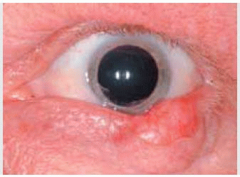
Clinical Features
Eyelid involvement with sebaceous gland hyperplasia is common in middle-aged or older individuals and is apparently unrelated to MTS (2). It occurs as one or more focal tan-yellow papules or as a diffuse thickening of the eyelids (3). Sebaceous adenoma, which can be associated with MTS, is clinically similar, but is slightly larger, appearing as a yellow nodule with a smooth surface.
Pathology
Sebaceous hyperplasia is composed of well-demarcated lobules of mature sebaceous glands usually located around a dilated sebaceous duct. In contrast, sebaceous gland adenoma is composed of two types of cells: Mature sebaceous cells and poorly differentiated basal cells (2). The tumor lobules do not usually open into a distinct duct. In some cases, there may be histopathologic overlap between sebaceous hyperplasia and adenoma, making histopathologic classification difficult. The sebaceous tumors seen with MTS often demonstrate typical protrusions of sebaceous tumor cells through the basement membrane into the epidermis (11).
Management
Complete resection is generally the best management. In patients with multiple small lesions, electrodessication or cautery and trichloroacetic acid are effective (14). As a part of management, any patient with one or more cutaneous sebaceous adenomas should be evaluated for gastrointestinal malignancy or other tumors of the MTS.
Selected References
1. Font RL. Eyelids and lacrimal drainage system. In: Spencer WH, ed. Ophthalmic Pathology. An Atlas and Textbook. 4th ed. Philadelphia: WB Saunders; 1996:2278-2282.
2. Elder D, Elenitsas R, Ragsdale BD. Tumors of the epidermal appendages. In: Elder D, Elenitsas R, Jaworsky C, et al., eds. Lever’s Histopathology of the Skin. Philadelphia: Lippincott-Raven; 1997:765-768.
3. Jakobiec FA, To K. Sebaceous tumors of the ocular adnexa. In: Albert DM, Jakobiec FA. Principles and Practice of Ophthalmology. 2nd ed. Philadelphia: WB Saunders; 2000:3400-3401.
4. Bhattacharya AK, Nayak SR, Kirtane MV, et al. Sebaceous adenoma in the region of the medial canthus causing proptosis. J Postgrad Med 1995;41: 87-88.
5. Muir G, Yates-Bell AJ, Barlow KA. Multiple primary carcinomata of the colon duodenum and larynx associated with keratoacanthoma of the face. Br J Surg 1967;54:191-195.
6. Torre D. Multiple sebaceous tumors. Arch Dermatol 1968;98:549-551.
7. Rulon DB, Helwig EB. Cutaneous sebaceous neoplasms. Cancer 1974; 22:82.
8. Tillawi I, Katz R, Pellettiere V. Solitary tumors of meibomian gland origin and Torre’s syndrome. Am J Ophthalmol 1987;104:179-182.
9. Jakobiec FA. Sebaceous adenoma of the eyelid and visceral malignancy. Am J Ophthalmol 1974;78:952-960.
10. Rishi K, Font RL. Sebaceous gland tumors of the eyelids and conjunctiva in the Muir-Torre syndrome: a clinicopathologic study of five cases and literature review. Ophthal Plast Reconstr Surg 2004;20:31-36.
11. Font RL, Rishi K. Sebaceous gland adenoma of the tarsal conjunctiva in a patient with Muir-Torre syndrome. Ophthalmology 2003;110:1833-1836.
12. Singh AD, Mudhar HS, Bhola R, et al. Sebaceous adenoma of the eyelid in Muir-Torre syndrome. Arch Ophthalmol 2005;123:562-565.
13. Demirci H, Nelson C, Shields CL, et al. Eyelid sebaceous carcinoma associated with Muir-Torre syndrome in two cases. Ophthal Plast Reconstr Surg 2007;23:77-79.
14. Griffith DG, Salasche SJ, Clemons DE. Sebaceous eyelid tumors. In: Griffith DG, Salasche SJ, Clemons DE, eds. Cutaneous Abnormalities of the Eyelid and Face. New York: McGraw-Hill; 1987:225-228.
Eyelid Sebaceous Carcinoma
General Considerations
Sebaceous carcinoma is an important neoplasm that occurs most frequently in the periorbital area, usually the eyelid. It has received a great deal of attention in the ophthalmic literature (1,2,3,4,5,6,7,8,9,10,11,12,13,14,15,16,17,18,19,20,21,22,23,24,25,26,27,28,29,30,31,32,33,34,35,36,37,38,39,40,41). It can exhibit aggressive local behavior and metastasize to regional lymph nodes and distant organs. Historically, this neoplasm has been notorious for masquerading as other benign and malignant lesions, resulting in delays in diagnosis and higher morbidity and mortality. Hence, it is important for the ophthalmologist to be cognizant of the clinical features of and current therapy for periorbital sebaceous carcinoma. Recently, greater awareness of this neoplasm has resulted in earlier diagnosis and provided the opportunity for less aggressive therapy (12,13,31,32). Although ophthalmologists have become more familiar with the clinical variations of periorbital sebaceous carcinoma, there remain delays in diagnosis and misdirected therapy (12).
Stay updated, free articles. Join our Telegram channel

Full access? Get Clinical Tree


