Evaluation of Ocular Motor Function
Richard London
Alignment of the eyes during early years of life is critical for the development of normal binocular vision and sensory fusion. Always determine alignment from the most natural condition to the less natural conditions. That is, start with the normal, undisrupted viewing conditions sometimes referred to as associated viewing conditions, where both eyes are allowed to take their natural alignment in an attempt to view an object of regard. Examples of tests under associated viewing conditions that detect the presence of an ocular deviation are observation, Hirschberg, Bruckner, and unilateral cover tests. When possible, the magnitude of the deviation can be confirmed by dissociated viewing conditions such as the neutralization of the angle with an alternate cover test. These tests prevent the eyes from seeing simultaneously and, thus, are less natural because this does not occur in the routine viewing of the world.
Assessment of ocular alignment begins with the first observation of the patient. Notice any assumed head postures. Head turns may be indicative of horizontal rectus palsy, a null point for nystagmus, or special conditions such as Duane retraction syndrome. Head tilts are suggestive of vertical deviations—usually superior oblique palsies (Fig. 16.1). It is also helpful to review old photos (“Fat scan” or family album tomography). The fact that a patient adopts an assumed head posture usually suggests fusion potential and should be viewed as a positive sign for future therapy.
The observation begins from the time the patient is greeted, and continues during the taking of history. The goal is to obtain an assessment of the patient’s head posture under natural and un–self-conscious conditions. An estimate of the magnitude of the assumed head posture in degrees should be attempted.
Following observation, more objective measurements of the angle of deviation are accomplished by light reflex tests and cover tests. Whereas the light reflex tests are the simplest, quickest, and easiest to perform on infants and young children, they are not as sensitive as cover tests. All of these tests require controlling the patient’s attention and accommodation.
All measurements of alignment should first be done with the patient allowed to manifest the assumed head posture, if any is present. This permits the clinician to judge the effectiveness of the attempted compensation. The head is then straightened to the forced primary position and the alignment remeasured to determine the actual magnitude of the deviation that the patient must overcome to maintain fusion.
Light Reflex Tests
Bruckner
The least obtrusive measurements are those where the patient simply looks at a light source held by the examiner. These measurements are determined by the Hirschberg and Bruckner
tests. The Bruckner test permits a qualitative judgment regarding the alignment of the eyes and anisometropia (1,2). Suspicion of the presence of these two major contributors to the development of amblyopia, therefore, can be increased or decreased by a quick and simple screening test. Evidence suggests that the Bruckner test is more reliable when the patient is older than 8 months of age (3). The direct ophthalmoscope is used as the light source for testing. A +1.00 D lens is placed in the ophthalmoscope to partially compensate for working distance and the large circular target is aimed at bridge of nose of the patient from approximately 50 cm away. Both pupils are visualized simultaneously and the brightness of the red reflexes compared. The eye with the brighter reflex is presumed either strabismic or more anisometropic (4). Pathology that affects the normal red reflex will also be identified by this test. This includes retinal detachment, gross retinal pathology; corneal, lenticular, and media opacities; anisocoria can possibly confound the result as well. It is reported that the sensitivity of the Bruckner test is 3 to 4 prism diopters (5). This test is particularly useful support as part of a battery of tests to help compensate for inherent limitations of each individual test with young children (6,7,8).
tests. The Bruckner test permits a qualitative judgment regarding the alignment of the eyes and anisometropia (1,2). Suspicion of the presence of these two major contributors to the development of amblyopia, therefore, can be increased or decreased by a quick and simple screening test. Evidence suggests that the Bruckner test is more reliable when the patient is older than 8 months of age (3). The direct ophthalmoscope is used as the light source for testing. A +1.00 D lens is placed in the ophthalmoscope to partially compensate for working distance and the large circular target is aimed at bridge of nose of the patient from approximately 50 cm away. Both pupils are visualized simultaneously and the brightness of the red reflexes compared. The eye with the brighter reflex is presumed either strabismic or more anisometropic (4). Pathology that affects the normal red reflex will also be identified by this test. This includes retinal detachment, gross retinal pathology; corneal, lenticular, and media opacities; anisocoria can possibly confound the result as well. It is reported that the sensitivity of the Bruckner test is 3 to 4 prism diopters (5). This test is particularly useful support as part of a battery of tests to help compensate for inherent limitations of each individual test with young children (6,7,8).
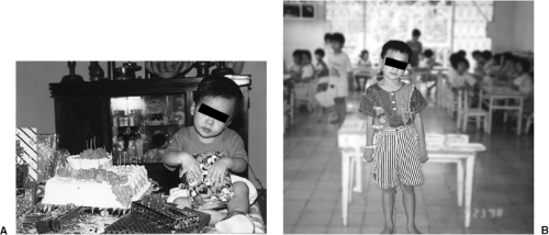 Figure 16.1. A: Infant manifests left head tilt resulting from a right superior oblique palsy. B: Same child manifesting same head tilt several years later. |
A commercially available instrument that makes use of the Bruckner reflex is the MTI (Medical Technologies, Inc) photoscreener. A Polaroid photograph is taken that enhances the retinal reflex and allows a documented, simultaneous comparison between the eyes. The MTI is reported to be effective for children as young as 6 months of age. It is potentially useful as a screener for use by nurses and in schools. It has been reported easier for pediatric residents to interpret compared with direct viewing of the Bruckner reflex (9).
Hirschberg Test
The Hirschberg test is also performed at a distance of about 50 cm from the patient, with a light aimed directly at the bridge of the patient’s nose. The light source should be sufficiently large to allow simultaneous viewing of both corneas, and is best done with a light of adjustable intensity to allow for patient comfort. The surrounding room should be sufficiently dark to reduce peripheral distracting elements. Various techniques to capture the child’s attention at the light source must be used to complete the measurement. Depending on the clinician’s talents, cooing, cat, bird, or dog sounds may be used. If using a transilluminator, it also helps to place the light source through a small toy to capture the child’s attention.
The light source will produce light reflexes, the first Purkinje image, behind the patient’s
pupil (although we often use the misnomer corneal light reflex). The difference in the location of these reflexes between the corneas results from the magnitude of the ocular deviation. The accepted conversion is 1 mm of displacement equals 22 prism diopters of deviation (10,11,12), although for flatter corneas, a more accurate conversion may be 27 prism diopters (13). This may be relevant in infants because they tend to have flatter corneas.
pupil (although we often use the misnomer corneal light reflex). The difference in the location of these reflexes between the corneas results from the magnitude of the ocular deviation. The accepted conversion is 1 mm of displacement equals 22 prism diopters of deviation (10,11,12), although for flatter corneas, a more accurate conversion may be 27 prism diopters (13). This may be relevant in infants because they tend to have flatter corneas.
It is important to estimate the location of the light reflexes from the center of the pupil. Because this point is not physically present, many clinicians attempt to estimate the location from the physical edge of the pupillary margin. This is subject to error because of the possibility of anisocoria and, therefore, unequal reference points. Some have suggested estimating from the limbus, but this is a relatively large distance. Additionally, the standard recording is based on distance from the center of the pupil. Use of any other reference point will necessitate an additional conversion.
With patients who have dark irides, it is often difficult to determine where the pupil ends and the iris begins. For these patients, using the direct ophthalmoscope as the light source facilitates the determination of the Hirschberg reflex. By placing a +1.00-D lens in the ophthalmoscope and viewing the corneas simultaneously, the pupils will show a red reflex used in the Bruckner assessment and a pinpoint of light in the background of the red reflex that is the reference for the Hirschberg test. This method is so quick and effective that I tend to use it regardless of iris color.
Determine the strabismic magnitude by estimating the position of the fixating eye’s reflex compared with that of the deviating eye. By convention, a reflex located nasally to the center of the pupil is called positive (+), whereas that temporal to the center is called negative (–). Remember, a light in a nasal location on the cornea projects temporally in the retina, whereas a temporal corneal reflex projects nasally. The common position of the corneal light reflex is +0.5 mm, representing the slightly temporal location of the fovea. A range of normality exists, however, and small deviations from the accepted norm should not be viewed with concern. The important factor for angular estimation is the difference between the reflexes. For example, if the right eye has a reflex of +0.5 mm and the left eye -1.0 mm, the difference between them is 1.5 mm. The accepted conversion into prism diopters is 22 prism diopters per millimeter. In the above example, the ocular deviation would be approximately 33 prism diopters left esotropia, with the right eye being the fixating eye and the left eye turned inward, yielding a temporal corneal reflex.
At times, the corneal light reflexes are so close to the expected norm that it is difficult to know which is the fixating and which is the turned eye. For instance, the right eye reflex is +0.5 mm and the left eye -0.5 mm. Which is the fixating eye? To be certain, look at the monocular corneal light reflexes. This monocular reflex is angle lambda, which is defined as the angle between the center of the entrance pupil and the visual axis. It is often misnamed angle kappa. Angle kappa, by definition, is measured at the nodal point of the eye. Because that point is not clinically accessible, the best we can do is assess angle lambda. In the example above, if we cover the left eye and the right eye moves so its angle lambda is now -0.5 mm, and we cover the right eye and the left eye maintains its -0.5 mm angle lambda, then we can state that this patient has a 22 prism diopter right exotropia. If it was the left eye that changed during angle lambda assessment, the patient has a 22 prism diopter left esotropia.
A potential misleading finding occurs in the rare case when a patient has unequal angle lambdas. In these cases, a unilateral cover test on either eye will result in no refixation movement and, therefore, no change in angle lambda in either eye.
Occasionally, a patient can have eccentric fixation sufficiently large to be seen on angle lambda testing. Most clinicians believe that a 0.5-mm deviation is the smallest that can be determined with good confidence. This translates into a minimal eccentric fixation of 11 prism diopters—a very large amount. When viewed binocularly as the Hirschberg test, however, eccentric fixation has no effect. In the very rare situation where the degree of eccentric fixation is equal to the angle of deviation and both are
sufficiently large to be seen on these tests, no movement will be observed when one eye is covered. Although this provides a good academic exercise, its true occurrence is fleetingly rare.
sufficiently large to be seen on these tests, no movement will be observed when one eye is covered. Although this provides a good academic exercise, its true occurrence is fleetingly rare.
Krimsky Test
To improve quantification of the Hirschberg test, a modification was made known as the Krimsky test. A prism is held before one eye until the light reflexes appear in the same location in each eye. The prism can be placed before either eye, and each method has its proponents. Remember, a prism placed before the fixating eye will result in a version movement of both eyes in the direction of the apex. Following insertion of the prism, the fixating eye sees a displaced image and refixates it. Because of Hering’s Law, the deviating eye moves in the same direction. In comitant deviations, the eye will move a degree equal to stimulus prism. Prism is inserted until the light reflex in the deviating eye matches that of the fixating eye before prism insertion. Those who favor this method argue that it is easier to see the light reflex with no prism in front of the eye. Two potential problems exist with this technique, however: first, the clinician must remember the initial location of the fixating eye’s reflex; and second, if the deviation is not comitant, the measured angle of deviation will be the secondary angle, often much larger than the primary angle, which is measured with the dominant eye fixating. For this reason, when incomitancy is suspected based on evaluation of ocular motilities (below), the Krimsky must be done with the measuring prism placed before the deviating eye. Thus, no version response is expected. This is the second method. In fact, this is the method I recommend being used exclusively. The criticism for this method is that the clinician must view the light reflex through a prism. I find this is easily done, and this method has the advantages of direct comparison of the light reflexes—no need to rely on memory—and it works with both comitant and incomitant deviations, with no need to change methods for different types of cases.
Prism position and orientation are always important during angle neutralization. The apex points to the direction of deviation of the eye. Be sure to hold the base-apex line parallel to the cornea. Do not stack prism in the same direction. Horizontal prism can be stacked with vertical prism, but the stacking of prism in the same direction results in a greater magnitude deviation than indicated by the number written on the prisms. For instance, stacking a 40- and 16-prism diopters results in an angle of 76 prism diopters rather than the expected 56 prism diopters.
Cover Testing
Unilateral Cover Testing
If the child is old enough to fixate accurately for cover testing, it is usually not necessary to perform the light reflex tests. The most natural of the cover tests is the unilateral cover test (UCT), also know as the cover-uncover test. The patient is allowed to view the fixation target without external barriers to fusion. One eye is then covered while the uncovered eye is observed to determine any movement toward refixation. The occluder, or examiner’s thumb for infants, should be held in place for 1 to 2 seconds to allow time for a deviated eye to pick up fixation. An inward refixation movement occurs with exotropia, whereas an outward movement indicates esotropia, downward with hypertropia and upward with hypotropia. No movement means that the uncovered eye is showing no manifest deviation. Following the covering of the eye, the occluder is then removed and the patient is allowed to refixate on the target. Remember, the UCT is essentially just a probe of the habitual alignment of the eyes. Following each probe, allow the patient several seconds to return to habitual deviation. Then, the other eye is probed with occlusion. The results of this testing will be findings of orthotropia, esotropia, exotropia, hypertropia, hypotropia, or some combination.
Care must be taken not to telegraph which eye will be covered. If the patient can anticipate which eye will be occluded, those with small angle alternating strabismus will switch fixations so quickly as to not be observed to have movement on the actual cover test. Bring the occluder from a central location, in front of the nose or from the center of the forehead, so that the patient
cannot anticipate which eye will be occluded. Vary the pattern of occlusion following the return to the habitual deviation so that the patient does not get into a rhythm.
cannot anticipate which eye will be occluded. Vary the pattern of occlusion following the return to the habitual deviation so that the patient does not get into a rhythm.
The UCT is performed at distance and near. With young children, a distance target with color that produces sound is often required to hold the patient’s attention. Videos can be useful for very young children, but are too large to allow accurate determination of small angle strabismus. A discrete target (e.g., an animated animal that makes noise) is preferred. If children are old enough, call their attention to specific markers on the target. For instance, ask the patient: “What color is the elephant’s eye?” “His nose?” This allows for most accurate fixation. Following distance testing, the UCT should be repeated at near.
The accuracy of this test depends on accommodation being locked onto the target of regard. Small pictures on fixation sticks make good targets. Again, ensure fixation and accommodation by asking questions of the child regarding the fixated picture.
The three most common errors in performing an appropriate UCT at near are (a) telegraphing the eye to be occluded; (b) using a target that does not lock accommodation; and (c) holding the fixation target too far from the child. A young child does not hold objects at 40 cm. Testing a child who is routinely visually examining objects at 25 cm at the adult standard of 40 cm will almost guarantee missing many accommodative esotropic conditions.
Remember, a patient can have a strabismus at one distance or position of gaze and not another, or, a constant deviation can occur at one location and an intermittent one at another. To determine the impact of a strabismus on visual development, the results of the UCT must be viewed along with the monocular visual acuities and stereopsis and, if the child is old enough, status of correspondence. (see Chapters 2, 7, 10 and 17).
Alternate Cover Testing
Unlike the UCT, the alternate cover test (ACT) demands that no opportunity is provided for the eyes to fuse or return to their habitual state. The goal is to determine the magnitude of the full deviation of the eyes in a fusion-free state. Thus, the ACT alone cannot determine whether a phoria or strabismus is present. It does, however, allow measurement of the challenge to fusional vergence and how it changes at different distances and different positions of gaze.
To encourage full dissociation, the occluder should be held over one eye for several seconds, then rapidly moved to the other eye, allowing no opportunity for fusion. The movement should shift occlusion between the eyes like a toggle switch. One is covered, then the other, with no pause between. The uncovered eye is observed to determine refixation, and the direction of this refixation is interpreted as with the UCT.
Measurement of the dissociated deviation is accomplished by neutralizing the movement with prism. A prism bar or loose prism is held before the deviating eye for patients who are strabismic, or either eye for those who are phoric. To neutralize a deviation, the base is held in the direction that the eye moves to pick up fixation. Ideally, the prism is held behind the cover paddle so that one eye clearly provides a fixation and accommodative control. As the ACT continues, prism is increased or decreased until no refixation movement is seen in the eye under the prism. Note, at times, a rebound or redress occurs in the uncovered eye even when the deviation is totally neutralized by prism (14). Equality of this redress should be considered the neutral point.
Again, remember when neutralizing the deviation with prism, to resist the temptation to stack prism in the same direction. To do so yields a significantly greater power because of the artificially increased angle of combined prism. You can stack horizontal and vertical prism and maintain accurate readings.
If the eye under the prism is neutralized, but movement is still observed in the other eye, some additional conditions must be considered. Of most importance is a true incomitant deviation resulting from a palsy of an extraocular muscle. Accommodative differences between the two eyes resulting from residual hyperopia (natural or induced) can mimic this condition in primary gaze.
If a deviation is incomitant, it will be equal in the cardinal positions of gaze or with either
eye fixating. With incomitant deviations, the deviation will change in different positions of gaze or in primary gaze when the fixating eye is changed, because of Hering’s Law. In the latter case, the standard method of recording is OD fixating = 20 Δ ET; OS fixating = 35 Δ ET. After determining the magnitude of the eyes’ deviation in primary gaze at distance, determine the angle in up, down, left, and right gazes (Fig. 16.2


eye fixating. With incomitant deviations, the deviation will change in different positions of gaze or in primary gaze when the fixating eye is changed, because of Hering’s Law. In the latter case, the standard method of recording is OD fixating = 20 Δ ET; OS fixating = 35 Δ ET. After determining the magnitude of the eyes’ deviation in primary gaze at distance, determine the angle in up, down, left, and right gazes (Fig. 16.2
Stay updated, free articles. Join our Telegram channel
Full access? Get Clinical Tree
