Evaluation of Accommodation
Ida Chung-Lock
Importance of Evaluating Accommodation
The evaluation of accommodation as part of a comprehensive eye examination is the standard of eye care for pediatric patients, because not only are accommodative deficiencies and inefficiencies associated with poor academic performance, but they can also signify a neuropathy or systemic abnormality. Accommodative problems have been linked to reading problems and learning disabilities in children (1). Studies note that 61% to 85% of children with learning problems (2,3) and 88% of those with diagnosed learning disabilities (4) have accommodative disorders of amplitude or facility. Accommodative deficiencies reported in healthy young individuals can be associated with convergence insufficiency, altitude changes, emotional stress, and refractive errors (2,5,6,7,8). Accommodative dysfunctions have been found to be associated with a wide variety of pathologic conditions. Ocular conditions include intraocular inflammation and premature sclerosis of the crystalline lens (8,9). Neurologic conditions affecting accommodation include those secondary to head trauma, neuralgic disease affecting the third cranial nerve, pharmacologic agents primarily parasympathetic, and encephalitis (9,10). Systemic conditions that have an impact on accommodation include myasthenia gravis, botulism, diphtheria, diabetes mellitus, Wilson’s disease, vascular hypertension, Guillian-Barré syndrome, chicken pox, tuberculosis, influenza, whooping cough, measles, dental caries or infections, endocrine disorders, anemia, arteriosclerosis, tonsillar infections, infectious mononucleosis, syphilis, and food poisoning (8,9,10,11,12,13). The varied causes of accommodative dysfunction make it essential for the clinician to evaluate accommodative function and interpret the findings in the context of the patient’s history and other clinical findings to reach an appropriate management plan.
Aspects of Accommodative Function to Evaluate
The exact prevalence of accommodative dysfunction is not well documented. The prevalence of accommodative dysfunction is reported to be present in 60% to 80% of patients with binocular dysfunction, but less than in 10% of an optometric clinic population (1,2).
Accommodative insufficiency, by far the most common of the accommodative disorders, is diagnosed primarily with the amplitude test. The other two aspects of accommodative function frequently tested clinically are accommodative response and accommodative facility. To separate the accommodative system from the binocular system and thereby remove possible contamination of the accommodative findings secondary to inefficiencies of binocularity, testing should be carried out under
monocular conditions. Clinical tests are available to evaluate the various aspects of accommodative function: push-up, pull-away, minus lens, dynamic retinoscopy, and visually evoked potentials to evaluate amplitude; monocular estimate method, Nott retinoscopy, fused cross-cylinder, book retinoscopy, bell retinoscopy, and photorefraction to evaluate accommodative response; plus and minus flipper lenses, and distance rock test to evaluate facility; and plus and minus added lenses to evaluate range of relative accommodation. Monocular accommodative facility and monocular amplitude tests give accommodative findings without interference from the binocular system, whereas the monocular estimate method, fused cross-cylinder, binocular accommodative facility test, and negative and positive relative accommodation tests results are derived under binocular conditions.
monocular conditions. Clinical tests are available to evaluate the various aspects of accommodative function: push-up, pull-away, minus lens, dynamic retinoscopy, and visually evoked potentials to evaluate amplitude; monocular estimate method, Nott retinoscopy, fused cross-cylinder, book retinoscopy, bell retinoscopy, and photorefraction to evaluate accommodative response; plus and minus flipper lenses, and distance rock test to evaluate facility; and plus and minus added lenses to evaluate range of relative accommodation. Monocular accommodative facility and monocular amplitude tests give accommodative findings without interference from the binocular system, whereas the monocular estimate method, fused cross-cylinder, binocular accommodative facility test, and negative and positive relative accommodation tests results are derived under binocular conditions.
Clinical Tests
Accommodative Amplitude
The amplitude of accommodation can be measured monocularly or binocularly. Measured monocularly, it is the maximal dioptric value produced by the accommodative system. Measured binocularly, it is the maximal dioptric value produced in the presence of convergence. Binocular measures are usually higher than monocular measures by 0.20 to 0.60 D because of convergence accommodation, although the measures can vary on repeated measurements and among different patients (6). Accommodative amplitude can be measured subjectively with the push-up method, the pull-away method, or the minus lens technique, and objectively using dynamic retinoscopy.
Push-up Method
Procedure
With one eye occluded, move the target, generally a 20/20 to 20/30 letter or detailed picture, toward the patient’s unoccluded eye at a rate of 5 cm/s (1). Encourage the patient to keep the target clear and to report when the target first starts to blur. The inverse of the measured distance in meters from the near point of accommodation (defined as the first sustained blur) to the patient’s spectacle plane is the maximal amplitude of accommodation expressed in diopters (15). This should be repeated with the other eye, and can be performed binocularly with presentation of the target along the midline. Patients should be wearing their distance manifest refraction and ambient lighting should provide sufficient illumination on the target as it approaches the patient’s eyes, but not so bright as to cause abnormal pupillary constriction. The push-up method has been shown to be reliable (16).
Pull-away Method
Procedure
The pull-away or push-away method involves moving a 20/20 to 20/30 letter target away from the patient’s spectacle plane until the patient can identify the stimulus (15) (Fig. 18.1). A variation of the push-up method, it is performed under similar conditions, except the target is initially presented directly in front of the unoccluded eye and is then moved away from the patient. The inverse of the measured distance at which the patient can correctly identify the target from the spectacle plane is the monocular amplitude of accommodation. The pull-away method is advantageous over the push-up method because the correct identification of the target gives a more reliable finding of the minimal amplitude of accommodation in patients with questionable subjective responses. The amplitude obtained with the pull-away method is comparable with the conventional push-up method (15,17,18,19).
Minus Lens Method
Procedure
The minus lens method is performed at 40 cm through the distance manifest refraction with a 20/30 target or, if the visual acuity is less than 20/20, choose a target that is two lines below the best acuity. The test is performed monocularly with the addition of lenses in -0.25-D steps until the patient reports first sustained blur. The absolute value of the lens addition +2.50 D for the working distance compensation is the total amplitude of accommodation for that eye.
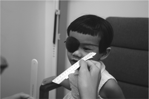 Figure 18.1. Procedure for pull-away method to measure monocular amplitude of accommodation. |
Dynamic Retinoscopy
Procedure
Dynamic retinoscopy gives an objective measure of accommodative amplitude (20). While the patient is viewing a near target, a 20/20 to 20/30 letter or picture equivalent, the clinician moves the retinoscope and the target together toward the patient. A change in reflex from a broad, bright and fast with motion reflex (indicating a normal lag of accommodation) to a narrow, dimmer and slower reflex, indicates focusing is lost and the amplitude of accommodation is exceeded (21,22).
Visually Evoked Potentials
Procedure
Visually evoked potentials (VEP) can be used to objectively measure accommodative amplitude. While viewing a phase-alternated checkerboard pattern, the amplitude is determined by having the patient view the stimulus through decreasing plus power lenses and then minus lenses until the measured VEP response produces a nearly constant value (23). Although VEP is a feasible method to determine amplitude of accommodation, the time and equipment involved limits its clinical utility, particularly when other methods are available.
Accommodative Response
Accommodative response is the actual amount of accommodation generated in response to a stimulus. Because of depth of focus, the accommodative response does not have to equal the stimulus demand exactly, and is usually less than the accommodative stimulus (24). The difference between the accommodative stimulus and accommodative response is the lead or lag of accommodation. Accommodative response is clinically most commonly measured objectively with the monocular estimate method (MEM) or without lenses using Nott retinoscopy. Using Nott retinoscopy, accommodative responses in children as young as 3 years of age was measured to be accurate to within 0.50 D (25).
Subjectively, it is most commonly measured using the fused cross-cylinder method (FCC).
Subjectively, it is most commonly measured using the fused cross-cylinder method (FCC).
Monocular Estimate Method
Procedure
The MEM is performed with age-appropriate reading material at the plane of the retinoscope light held at the patient’s habitual near working distance (as determined by asking the patient to hold reading material) or at the patient’s Harmon distance (the distance from the patient’s elbow to the third knuckle) (21) (Fig. 18.2). This distance is often approximately 20 to 25 cm (8–10 inches) for a young child. For infants or toddlers, a detailed toy or shiny object is a suitable target (21). The patient’s eyes should be in down-gaze when viewing the target, similar to a normal reading posture. Room illumination should be such that the reading target is adequately illuminated while allowing the clinician to easily view the retinoscopic reflex. The patient wears the appropriate lenses, such as a distance correction, a tentative near prescription, or no lenses. With both eyes open, lenses are quickly interposed monocularly, plus lenses for with motion and minus lenses for against, motion, to neutralize the reflex, leaving the lens in place no longer than half a second in order not to change the accommodative response or binocular fixation (26). The lens that neutralizes the reflex is the measured accommodative response. If astigmatism is present, the two major meridians can be neutralized separately. It is important to measure the response only when the patient is actively viewing the target presented. The use of magnetic fixation cards with premanufactured square cut-outs attached to the Welch Allyn retinoscope facilitates the MEM technique (Fig. 18.3).
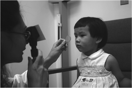 Figure 18.2. Procedure for monocular estimate method (MEM) retinoscopy to measure accommodative response. |
Nott Retinoscopy
Procedure
Nott retinoscopy is similar to MEM, except that to neutralize the reflex, the retinoscope
is moved closer or further from the patient while the target remains at a fixed distance (27,28). Thus, the target cannot be affixed to the retinoscope. The test is performed with the fixation distance at 40 cm and initial observation through the retinoscope at 50 cm. The retinoscope is then moved away from the patient if with motion is seen, and closer if against motion is seen until the reflex is neutral. The dioptric difference between the target and the position of the retinoscope at the point of neutrality is the measured accommodative response. When performing Nott retinoscopy, definite fixation, active accommodation, and alignment of the target and retinoscope beam within 10°, are important in order not to induce erroneous findings (29,30). Nott retinoscopy is said to be superior to MEM because it does not present the possible contamination of results from the neutralizing lens itself (27,31). Nott retinoscopy has been shown to be a repeatable and valid objective technique for assessing accommodative lag (32).
is moved closer or further from the patient while the target remains at a fixed distance (27,28). Thus, the target cannot be affixed to the retinoscope. The test is performed with the fixation distance at 40 cm and initial observation through the retinoscope at 50 cm. The retinoscope is then moved away from the patient if with motion is seen, and closer if against motion is seen until the reflex is neutral. The dioptric difference between the target and the position of the retinoscope at the point of neutrality is the measured accommodative response. When performing Nott retinoscopy, definite fixation, active accommodation, and alignment of the target and retinoscope beam within 10°, are important in order not to induce erroneous findings (29,30). Nott retinoscopy is said to be superior to MEM because it does not present the possible contamination of results from the neutralizing lens itself (27,31). Nott retinoscopy has been shown to be a repeatable and valid objective technique for assessing accommodative lag (32).
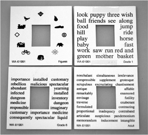 Figure 18.3. Set of magnetic fixation cards for patient fixation during monocular estimate method (MEM) retinoscopy. Available from Lombart Instrument Co., 5358 Robin Hood Road, Norfolk, VA 23513 . |
Bell Retinoscopy
Procedure
Bell retinoscopy is an objective test of accommodative response using a silver chrome bell attached to a thin metal rod as a target (33). With the retinoscope at 50 cm from the patient, the bell is held at the clinician’s forehead, and the clinician observes the retinoscopic reflex while the patient views his or her own image reflected off the bell’s surface. The bell is then moved either closer to the patient, if with motion is initially seen, or away from the patient, if against motion is initially seen, until the reflex appears
neutral. The dioptric difference between the bell and the retinoscope at the point a neutral reflex is observed is the accommodative lag. Bell retinoscopy was found to be incomparable to results of MEM (34), but it can provide the clinician with a qualitative assessment of patients’ ability to focus and align their eyes in free space (35). The bell target is particularly effective for the preverbal child.
neutral. The dioptric difference between the bell and the retinoscope at the point a neutral reflex is observed is the accommodative lag. Bell retinoscopy was found to be incomparable to results of MEM (34), but it can provide the clinician with a qualitative assessment of patients’ ability to focus and align their eyes in free space (35). The bell target is particularly effective for the preverbal child.
Book Retinoscopy
Procedure
Book retinoscopy is performed with the patient reading grade-appropriate text paragraphs, while the clinician views the reflex of a retinoscope held directly above the reading material (36). The clinician is able to interpret the patient’s interest and comprehension of the reading material by noting the color and motion of the reflex (i.e., the quality of the accommodative response). A bright and pink reflex with neutral to with motion indicates free and easy reading, a bright and pink reflex with varying fast against motion indicates reading with comprehension under stress, and a dull brick red reflex with a slow against motion indicates frustration reading (37,38). Some clinicians prefer book retinoscopy to other dynamic retinoscopy procedures because this objective method mimics a more natural reading environment (37).
Photorefraction
Procedure
A less-common method of assessing accommodative response that is mainly used in research is photorefraction. Photorefraction can determine the point of accommodation from the camera plane, which when compared to a known refractive status for a patient, an accommodative response can be calculated (39,40).
Fused Cross-Cylinder
Procedure
Fused cross-cylinder is performed in the phoropter with the ±0.37 D cylinder lenses and the cross-cylinder grid target composed of vertical and horizontal sets of lines presented at 40 cm in dim room illumination (41). With the patient’s distance correction in place, lenses are added binocularly +0.25 D at a time until the patient reports the vertical lines are sharper. It is recommended to start the test with added plus lenses and binocularly reducing the plus lenses a 0.25 D at a time until equality of the lines are achieved, or when the patient reports that the horizontal lines are sharper. The added lenses that gives this endpoint response is the measured accommodative response. A response of vertical lines being sharper before any lenses are added indicates a lead of accommodation. The FCC test can also be performed monocularly. The FCC test is not as repeatable as MEM (42) and relies on subjective responses, making MEM a preferred method of testing accommodative response in children.
Accommodative Facility
Accommodative facility is the ability to stimulate and inhibit accommodation. It is a measure of accommodative flexibility and stamina.
Lens Flipper
Procedure
The lens flipper test uses ±2.00 D lenses and a 20/30 letter target at 40 cm (Fig. 18.4). The lenses are presented binocularly or monocularly with one eye occluded to measure binocular and monocular facility, respectively. The patient is asked to report when the letters are clear as +2.00 lenses and -2.00 lenses are alternately presented. Once the patient reports the letters are clear and single, the lenses are flipped (43). Accommodative facility is measured as the number of cycles of plus and minus lenses the patient is able to clear within 1 minute (cpm). One cycle is clearing one set of plus and minus lenses.
A suppression check is necessary for accurate testing under binocular conditions (44). The norms for accommodative facility were derived from studies using the SOV9 acuity suppression vectogram (Fig. 18.5) and is the recommended target for clinical testing (42). The patient wears Polaroid glasses and is asked to keep


Stay updated, free articles. Join our Telegram channel
Full access? Get Clinical Tree
