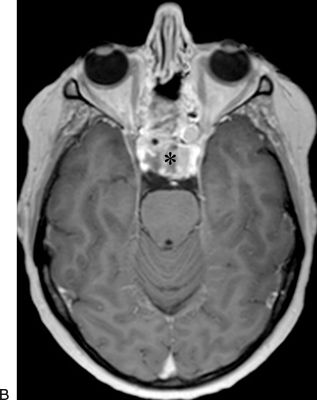
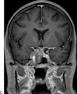
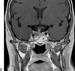
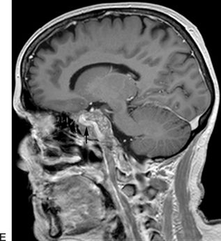
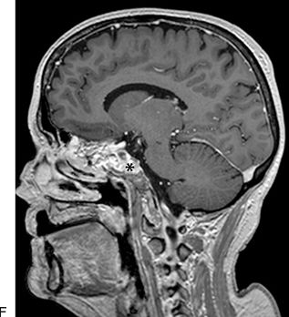
FIGURE 5.1 A–F. Preoperative (A,C,E) and postoperative (B,D,F) MRI of a patient with a prolactin-secreting pituitary macroadenoma extending laterally into the right cavernous sinus. The preoperative images demonstrate the extent of the tumor and the lateral displacement of the cavernous portion of the internal carotid artery (arrow). Given the size of the tumor and lack of response to medical treatment, the patient underwent a midline EEA. After removal of a portion of the lateral sphenoid sinus wall, medial to the ICA, the medial wall of the cavernous sinus was entered. The tumor was removed with angled instruments under visualization with a 30-degree-angled endoscope. The postoperative MRI demonstrates an excellent resection. The abdominal adipose tissue graft is demonstrated in the sphenoid sinus (asterisk). Following surgery, the patient’s prolactin level returned to well within normal range, indicating a biochemical cure.
PEARLS
Stay updated, free articles. Join our Telegram channel

Full access? Get Clinical Tree


