Dynamic Wound Healing


Dynamic Wound Healing


Gina D. Jefferson


INTRODUCTION

The otolaryngologist-head and neck surgeon encounters wound healing problems on a regular basis. An in-depth knowledge of the wound healing process and potential complicating factors is therefore imperative. There are several factors that may adversely affect the normal wound healing process including trauma, malignancy, history of radiation at the site of the wound, advanced age, metabolic diseases such as diabetes mellitus, and autoimmune disorders, just to name a few. Moreover, wounds are classified according to various criteria. Acute wounds follow a time course of healing within 30 days. The wounds repair themselves in an orderly fashion following the normal healing pathway. A chronic wound fails to progress through the normal phases of wound healing and is incomplete because it is often disturbed by various factors that impede the process.

The evolution in understanding wound healing began with single author publications in the late 1800s. These early reports were largely anecdotal experiences shared by individual surgeons. Various antiseptic agents were described. There were even reports of large rotational skin flaps utilized to prevent contraction scar deformities. In 1903, Cushing described nerve anastomoses for facial paralysis. Oschner reported in a symposium on the importance of septic techniques in 1904. In 1909, the surgeon John Stage Davis of Johns Hopkins Hospital published the first of many papers regarding wound closure, grafts, and flaps. Horsley discussed techniques for suturing blood vessels and recurrent laryngeal nerves in 1912. In 1919, Depage emphasized principles gleaned from experiences of World War I including antisepsis, delayed closure, and debridement with specific considerations for wounds of special areas. In 1925, Ravdin was the first to publish about the morbidity, mortality, and wound complication rate of a major surgical service (1).

During the 1930s, several papers appeared presenting observations of studies regarding wounds, wound healing, repair, and sepsis. The time course and various aspects of wound healing were discussed, and the Halstedian principles on operative technique were re-emphasized. In 1938, Ives and Hirshfeld relayed the importance of bacteria from a patient’s skin as a source of postoperative wound healing complications. During the late 1930s, the improvement in the course of wound healing was noted to occur with vitamin C and vitamin A supplementation (1).

During World War II, papers appeared espousing the use of penicillin to prophylax against surgical infection as well as papers regarding wound shock management, anesthesia, and wound pain. Jenkins described a new hemostatic agent, Gelfoam, in 1946 experimentally used for liver hemorrhage. Growth-promoting factors in tissues were reported in 1946 as well. In the 1950s, the role of nutrition was investigated with respect to wound healing. In 1956, Dunphy published regarding the role of vitamin C in collagen synthesis and the biochemical phases of wound healing. In 1964, factors influencing the incidence of wound infection led to the classification of wounds as clean, clean-contaminated, contaminated, and dirty. In the 1970s, Dunphy and others continued their research into the biochemistry of wound healing and the role and origin of various chemical mediators in the inflammatory process (1).

Recently, extensive research regarding adverse circumstances affecting wound healing has occurred. This research may lead to advanced treatment methodology directed at various phases of wound healing that targets specific substances or actually provide specific factors to improve the wound healing process.


THE NORMAL WOUND HEALING PROCESS

The biologic process of wound healing involves the sequential occurrence of four phases that are continuous and naturally overlap. One region of the wound may exhibit a different phase than the dominant phase occurring in another region of that same wound bed. These four phases of healing signal the introduction of specific cell types into the wound bed as depicted in Figure 6.1. Tissue injury incites the first phase, which is the coagulative and hemostatic phase of wound healing. The second inflammatory phase follows shortly thereafter. The body initiates wound repair with phase three, proliferation. Wound healing culminates with tissue remodeling. Importantly, all phases of wound healing depend upon the presence of various cytokines in order to accomplish the goal as briefly outlined in Table 6.1.


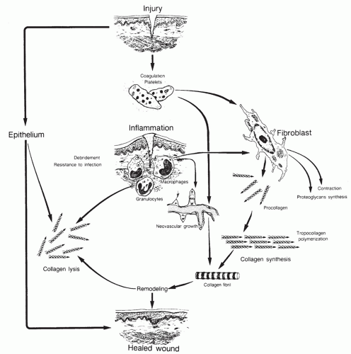
Figure 6.1 Brief depiction of the complexity of wound healing. (Modified and reprinted from Feinberg SE, Larson PE. Healing of traumatic injuries. In: Fonseca RJ, Walker RV, eds. Oral and maxillofacial trauma. Philadelphia, PA: WB Saunders, 1991, with permission.)


TABLE 6.1 CYTOKINES INVOLVED IN WOUND HEALING


Cytokine


Abbreviation


Source


Function


Human growth hormone


GH


Pituitary gland


Fibroblast proliferation; increases collagen content and tensile strength; anabolism; stimulates IGF-1


Epidermal growth factor


EGF


Platelets, bodily fluids (including saliva, urine, milk, and plasma)


Epithelial cell and fibroblast proliferation and migration; activates fibroblasts; angiogenic


Platelet-derived growth factor


PDGF


Platelets, macrophages, fibroblasts, endothelial cells, smooth muscle cells


Mitogenic for fibroblasts and smooth muscle cells; chemoattractant for neutrophils and macrophages; angiogenic


Fibroblast growth factors


FGF


Macrophages, brain, pituitary gland


Proliferation and migration of vascular endothelial cells; mitogenic and chemotactic for keratinocytes and fibroblasts


Transforming growth factors


TGF


Platelets, fibroblasts, neutrophils, macrophages, lymphocytes


Epithelial cell and fibroblast proliferation


Nerve growth factor


NGF


Schwann cell, muscle cells


Motoneurons, Schwann cells, muscle cells


Brain-derived neurotrophic factor


BDNF


Central nervous system, skeletal muscle, heart, lung


Support cranial and spinal motoneurons after axotomy


Ciliary neurotrophic factor


CNTF


Schwann cells


Promote survival and differentiation of neural and glial cells within the nervous system


Insulin-like growth factor 1


IGF-1


Fibroblasts, liver, plasma


Fibroblast proliferation, synthesis of proteoglycans and collagen


Tumor necrosis factor


TNF


Macrophages, mast cells, lymphocytes, other tissues and cells


Fibroblast proliferation


Interleukins


IL


Macrophages, lymphocytes, other tissues and cells


Fibroblast proliferation, neutrophil chemotaxis


Interferons


IFN


Fibroblasts, lymphocytes


Inhibition of fibroblast proliferation and collagen synthesis


Keratinocyte growth factors


KGF


Fibroblasts


Epithelial cell proliferation


Phase I: Coagulation and Hemostasis

The body’s immediate response to tissue injury is to protect the vascular system and prevent exsanguination. In addition, this phase provides cells that serve as a matrix for other cells that eventually appear in the wound in the later stages of wound healing. Microvascular injury results in extravasation of blood into the wound. A reflex vasoconstriction occurs due to vascular smooth muscle contraction. This rapid reflex response can temporarily halt blood loss. After a few minutes, this environment becomes hypoxic and acidotic triggering vascular smooth muscle relaxation with the resumption of blood loss. Exsanguination is thwarted however by activation of the coagulation cascade.

The coagulation cascade (Fig. 6.2) assembles a platelet plug and clot via extrinsic and intrinsic pathways. Cellular injury extrinsically exposes a tissue factor lipoprotein ubiquitously present on cell membranes that leads to conversion of factor VII to an active protease and ultimately to the common pathway of the coagulation cascade. Likewise, the contact or intrinsic coagulation pathway creates a complex on exposed subendothelial collagen with factor XII, high molecular weight kininogen, and prekallikrein. These factors convert to active proteases after binding and together activate factor XI, which leads to the final common coagulation pathway via factor X activation. Factor X is activated by the calcium-dependent complex formed between factors VIII, IX, and X or directly by an activated factor VII. Finally, in the presence of calcium and phospholipid, factor V becomes activated, and in concert with activated factor X, prothrombin is converted to thrombin. Fibrinogen in turn is converted to fibrin, which ultimately cross-links and forms a stable platelet plug.

The platelet plug contains platelet-derived growth factor (PDGF), transforming growth factor-beta (TGF-β), epidermal growth factor (EGF), and insulin-like growth factor (IGF). These growth factors promote the activation and
chemotaxis of neutrophils, macrophages, endothelial cells, and fibroblasts. Platelets also contain vasoactive amines like serotonin that cause vasodilation and increased vascular permeability. Fluid extravasation occurs contributing the edema observed during the second phase of wound healing, the inflammatory phase (2,3).


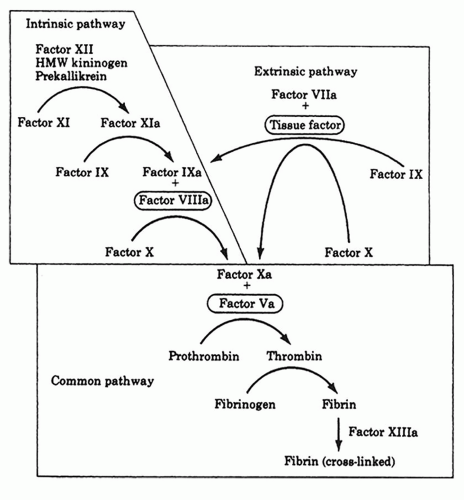
Figure 6.2 The coagulation cascade. (From Klingensmith ME. Washington manual of surgery, 5th ed. Lippincott Williams & Wilkins, 2005, with permission.)


Phase II: Inflammation

The inflammatory phase of wound healing functions to establish an immune barrier against invading bacteria. This phase is divided into an early phase and a late phase.


Early Inflammatory Phase

The early inflammatory phase of wound healing ensues during the late phase of coagulation during the first 24 to 36 hours after wounding occurs. This phase activates the complement cascade, which elaborates various chemoattractive factors such as C3a and C5a. These complement components synergistically act with local chemoattractive factors released from the activated platelets such as TGF-β to encourage neutrophil migration to the site of injury. Neutrophils are critical in performing phagocytosis to eliminate bacteria, foreign particles, and injured tissue components for the wound healing process to continue. Modification of surface adhesion molecules confers a sticky quality to neutrophils enabling the process of margination. Margination allows neutrophils to adhere to the endothelial cells of venules surrounding the wound bed. The endothelial cells secrete chemokines that activate a stronger adhesion system that is regulated by integrins. Through diapedesis, neutrophils migrate between endothelial cells to continue phagocytosis. Once neutrophils have completed their function of eliminating wound bacteria and foreign material, the products are extruded via apoptosis to the wound surface producing an exudate. This exudate is then further phagocytosed via macrophages (2,3).


Late Inflammatory Phase

The late inflammatory phase is heralded 48 to 72 hours of wounding with the appearance of macrophages. Macrophages originate as blood monocytes that undergo phenotypic changes upon arrival to the wound to become tissue macrophages. Macrophages have a longer lifespan than neutrophils and continue the function of phagocytosis of bacteria and foreign material within the wound bed. The macrophages are attracted to the wound by elaborated clotting factors, complement by-products, cytokines such as PDGF, TGF-β, leukotriene B4, and platelet factor IV plus elastin and collagen breakdown products. Additional growth factors essential for the later phases of wound healing are released by tissue macrophages to influence the growth and behavior of fibroblasts, assist in wound revascularization, and produce factors to modulate proteins comprising the extracellular matrix (ECM) scaffold. These factors include transforming growth factor alpha (TGF-α), heparin-binding EGF, fibroblast growth factor (FGF),
and collagenase. Activated keratinocytes, fibroblasts, and endothelial cells also play a role (4).

The next cells to arrive at the site of injury are lymphocytes. After 72 hours, interleukin-1 (IL-1), complement components, and immunoglobulin G breakdown products all attract lymphocytes to the wound bed. Lymphocytes regulate collagenase, which is later needed for collagen remodeling and to assist in producing ECM components such as fibrin and fibronectin, as well as participate in their degradation (2,3).


Phase III: Proliferation

The proliferative phase of wound healing begins on day 3 after injury once hemostasis is achieved and an adequate immune response has occurred. This phase lasts for about 2 weeks. Fibroblasts originate from nearby connective tissue cells after growth factors released from tissue macrophages stimulate the mitosis and proliferation of fibroblasts for collagen synthesis. Fibroblasts migrate into the wound and synthesize a new ECM replacing the original fibrin-fibronectin matrix scaffold thereby providing the lattice for collagen synthesis. Granulation tissue represents this phase of wound healing. Importantly, unwounded dermis contains 80% type I collagen and 25% type III collagen. Early in wound healing, granulation tissue however contains 40% type III collagen rendering a lower tensile strength. Later in wound healing, this type III collagen is largely replaced by type I collagen; however, the maximum tensile strength achieved is 80% of the previously unwounded tissue (5).

Angiogenesis is also a component of proliferation. Angiogenesis is critical for the restoration of blood flow into a wound, for reoxygenation of the wound surface, and for the continuation of wound healing through all of its phases. The local tissue response to hypoxia is to increase the production of vascular endothelial growth factor (VEGF). In addition to VEGF, other angiogenic factors including FGF, PDGF, angiogenin, TGF-α, and TGF-β promote the proliferation and growth of endothelial cells. VEGF is secreted by macrophages and binds to endothelial cell receptor tyrosine kinases VEGFR1 and VEGFR2. Binding to these receptors initiates an intracellular signaling cascade to promote angiogenesis. The endothelial cells sprout capillary buds at the wound surface. Ultimately, these capillary buds form a network that join other networks sprouting from the wound bed (2,3).

Cellular migration is imperative for both angiogenesis and continued collagen formation and is dependent upon three processes: (a) protrusion at the cell front, (b) adhesion for the actin cytoskeleton to attach to the substratum, and (c) traction, which propels the trailing cytoplasm forward (2,3).

The cytoskeleton is anchored by cell-cell junctions and cell-ECM adhesions. The internal actin network reorganizes at the leading edge pushing the plasma membrane outward and forming a filopodia. The cyclic assembly and disassembly of actin filaments at the leading plasma membrane edge and behind the edge, respectively, permits unidirectional movement toward other capillary sprouting matrices and other forming ECM frameworks (2,3).

Integrins serve as the primary receptors for ECM proteins and are therefore necessary for cellular migration. Integrins also function in signal transduction and in the regulation and stimulation of cell migration. Increasing adhesion optimizes the rate of cellular migration; however, mobility declines with further attachment. After attachment to the underlying ECM, integrins signal a change in cellular morphology from an oval, spindle shape to an irregular, flat shape. This occurs initially at the leading migratory edge of the plasma membrane. Migration is fastest immediately after injury and slows to a steady state maintained during the remainder of the wound healing process. Traction is effected by contractile forces transmitted through the integrin-cytoskeletal connections. These connections allow the cell to pull its cytoplasm forward. It is the interaction between myosin and actin fibers that pull the cell body forward (2,3).

Epithelialization also begins shortly after injury. This starts from the wound periphery as a single layer of cells forming over the defect. There is a marked increase in epithelial cell mitotic activity around the wound edges, and cells migrate across the wound by attaching to the underlying provisional matrix. When advancing epithelial cells come into contact with one another, migration ceases and basement membranes begin to form.


Phase IV: Remodeling

New epithelium forms in this phase of wound healing along with final scar tissue formation. This phase may last as long as 1 or 2 years. Normal wound healing occurs with the establishment of a balance between degradation and synthesis. The collagen fibers increase their diameter, while hyaluronic acid and fibronectin are degraded. There are three important stimuli inciting the deposition of collagen: (a) high lactate content in the wound, (b) adequate oxygen levels, and (c) growth factors, namely, TGF-β. The collagen fibers initially appear in a haphazard orientation within the wound but eventually become more oriented and cross-linked. The oriented, cross-linked collagen may regain about 80% of the original strength of the wounded area in comparison to the unwounded condition. The synthesis and breakdown of collagen and ECM components occur continuously and simultaneously, equilibrating during the third week postinjury as the rate of collagen synthesis is maximal during the first 2 weeks after injury. Matrix metalloproteinases produced by neutrophils, macrophages, and fibroblasts at the wound site carry out the task of degradation.

Further wound organization occurs during the final stages of the remodeling phase via wound contraction that began during the proliferative phase. The wound’s bed

Only gold members can continue reading. Log In or Register to continue

Stay updated, free articles. Join our Telegram channel

May 24, 2016 | Posted by in OTOLARYNGOLOGY | Comments Off on Dynamic Wound Healing

Full access? Get Clinical Tree

Get Clinical Tree app for offline access