Diseases of the Cornea


Diseases of the Cornea


Jagadesh C. Reddy

Christopher J. Rapuano


MANY DISEASES OF the cornea and anterior segment in children do not differ much from diseases in adults, with the exception of congenital and developmental abnormalities. Certain corneal disorders first appear in infancy and childhood, such that careful screening and examination at a young age would have elucidated the nature of disorders recognized later in adulthood.

This chapter delineates the common diseases of the earlier years of life that primarily affect the cornea but may also involve surrounding structures, such as the eyelids, conjunctiva, sclera, anterior chamber, iris, and lens.


EMBRYOLOGY AND DEVELOPMENTAL ABNORMALITIES

The anterior segment of the eye—the cornea, anterior chamber angle, iris, and lens—contains several anatomical and physiologic systems packed into a small space, so that its embryology and malformations are sometimes difficult to understand. The use of multiple names for each malformation further complicates the picture. We can reduce the confusion by reviewing the development of the anterior segment, observing how each abnormality might derive from arrested or aberrant growth, and classifying the abnormalities on a simple anatomical basis.

Congenital anomalies of the anterior segment result from abnormal induction, differentiation, or maturation of the tissues. Abnormalities can be due to inherited genetic defects, new genetic mutations and/or environmental factors. All cells of one individual begin with the same gene pool in their deoxyribonucleic acid (DNA) unless there is a chromosomal abnormality, but each differentiates to manifest a morphology controlled by only a portion of those genes—the genotype. Both internal and environmental influences regulate which genes will express themselves. During differentiation, different tissues are maximally susceptible to injury at different times, so that any agent that interferes during this sensitive period may produce an abnormality. Thus, similar malformations may result from abnormal genes, excessive or inadequate metabolites, viral or other infectious agents, exogenous toxins, hypoxia, or mechanical insults. Similarly, the same agents affecting the developing fetus at different time points produce different abnormalities, depending on which tissues are the most vulnerable at that particular moment. In some instances, such as congenital rubella, we know both the cause of the abnormalities and the approximate time of their development. In most instances, however, these factors are unknown, and we must fall back on more simple anatomical descriptions of the abnormalities, to which we often give eponyms, refining chronology and etiology as more information becomes available.


Developmental Variations in Limbal Anatomy

The limbus is a junctional zone where corneal epithelium and its basement membrane meet stem cells and conjunctival epithelium and its basement membrane. Corneal stroma juxtaposes sclera. Descemet’s membrane ends at Schwalbe’s ring and the trabecular meshwork. Corneal endothelium becomes continuous with the trabecular endothelium.

These transitional zones form a number of circular structures at the limbus that can be seen on slit-lamp examination (Fig. 10.1), and variations in these structures are common in congenital malformations of the anterior segment (1).


Epithelium

Corneal and conjunctival epithelia are continuous over the limbus. Corneal epithelium, a five- to eight-cell-layered stratified squamous epithelium, attaches to a smooth basement membrane that is supported by the acellular, fibrillar felt-work of Bowman’s layer. The conjunctival epithelium, a stratified cuboidal layer that contains goblet cells, lies on an undulating basement membrane, supported by irregular vascular connective tissue. At the limbus, the conjunctival epithelium forms a series of radially arranged extensions into the subepithelial connective tissue, each extension flanked by vascular connective tissue pegs that protrude into the epithelium. Pigmentation of the basal layers makes these epithelial extensions, the limbal palisades of Vogt, visible between the white connective tissue spaces (Figs. 10.1 and 10.2). These limbal palisades have tongue-like epithelial projections into the corneal stroma termed rete ridges. These epithelial rete ridges serve as a repository for corneal epithelial precursor cells.


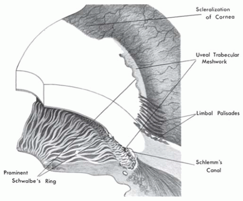
FIGURE 10.1. Developmental variations in limbal anatomy. This diagrammatic representation of limbal structures illustrates the appearance of each structure on the anterior surface and demonstrates its location in cross section.


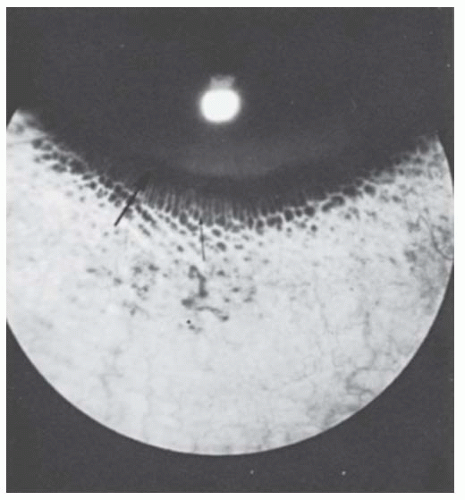
FIGURE 10.2. Limbal palisades of Vogt. This circle of white, fingerlike projections (small arrow) that breaks up the limbal pigment ring (large arrow) results from subepithelial connective tissue papillae pushing up near the surface, interrupting the pigmentation of the basal conjunctival epithelium.


Corneoscleral Junction

The components of both corneal stroma and sclera are the same: collagen fibrils, proteoglycans (acid mucopolysaccharides), water, and fibrocytes. The tissues appear different because the corneal collagen fibers have a uniform diameter and arrangement, and the proteoglycans are dehydrated, whereas scleral fibrils vary in size, are randomly oriented, and remain hydrated. At the limbus, the cornea inserts into the sclera as a wedge, like a watch crystal into its casing. Normally, the superficial rim of sclera extends 0.5 mm centrally over the wedge of cornea superiorly and inferiorly as a white, vascularized crescent (Figs. 10.1 and 10.3), so that the vertical corneal diameter is about 1 mm less than the horizontal diameter when measured on the anterior surface. Thus, deeper limbal structures, such as a prominent Schwalbe’s ring or uveal trabecular meshwork, are usually visible only medially and laterally. When this scleral tissue extends farther centrally, or when it is present for 360 degrees, the abnormality is called scleralization or scleral overriding.


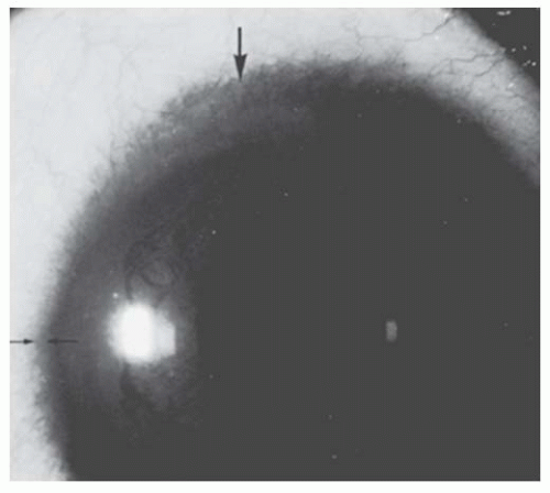
FIGURE 10.3. Scleralization of the cornea. At 12 o’clock position, the sclera and its vessels extend superficially into the cornea, hiding underlying iris and angle details (area between two large arrows). Compare the normal extent of sclera over the limbus at 9 o’clock position (area between two small arrows).


Trabecular Meshwork

A thin, translucent, gray band of uveal trabecular meshwork may extend up to and past Schwalbe’s line to form a gray arc on the posterior cornea, one sometimes accentuated by a pigmented epithelial ring and a prominent Schwalbe’s ring (Figs. 10.4 and 10.5).


Schwalbe’s Ring

The corneal endothelium and Descemet’s membrane meet the uveal trabecular meshwork at a junction designated as the anterior border ring of Schwalbe, or Schwalbe’s line. This ring, part of the uveal meshwork, has a structure similar to a trabeculum, that is, a collagen-proteoglycan core surrounded by thin leaves of the terminal portion of Descemet’s membrane and covered on its inner surface by endothelium. With a gonioscope, the clinician sees it as a change from the refractile corneal endothelium to the reticulated translucent trabecular meshwork.


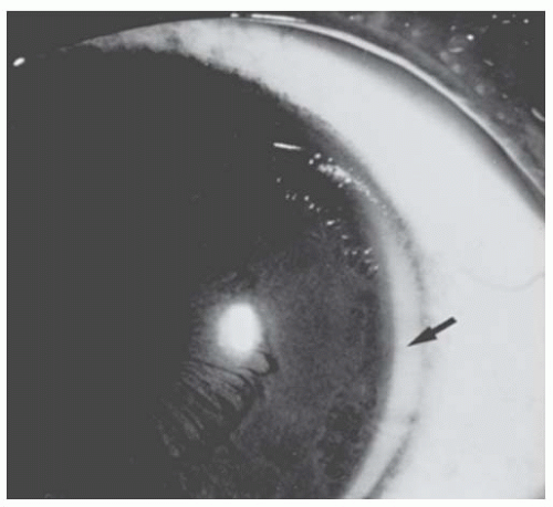
FIGURE 10.4. Prominent uveal trabecular meshwork. The limbus of the eye is demarcated by a pigment ring. Sclera extends up to but not beyond the ring. The light tissue lying central to the pigment ring (arrow) is the uveal trabecular meshwork.

Schwalbe’s ring may be thickened and positioned centrally, making it visible at the slit lamp as an irregular, refractile, white line lying concentric to the limbus and gonioscopically as a ridge protruding into the anterior chamber. It may be broken or continuous and frequently has pigment spots on its inner surface that represent previous attachment of iris strands. This centrally located prominent Schwalbe’s ring is commonly called a posterior embryotoxon (Greek, embryon [“embryo”] plus toxon [“bow”]). It appears in 8% to 15% of normal eyes but is seen this frequently only by those who specifically look for it.


Congenital Anomalies of the Cornea

The clinician confronted with a child who has a developmental anomaly of the ocular anterior segment often has difficulty classifying and naming the disorder. This is an activity of more than academic value, because a precise diagnosis is necessary before the ophthalmologist can predict the natural history of the disorder, look for specific associated ocular or systemic abnormalities, provide genetic counseling, and begin appropriate medical or surgical therapy. It is easiest to describe these abnormalities in terms of their anatomical components. Once clinicians have made a thorough description of the anatomical abnormalities, they can more precisely label the disorder, which may be aided by a simple classification system (Fig. 10.6).


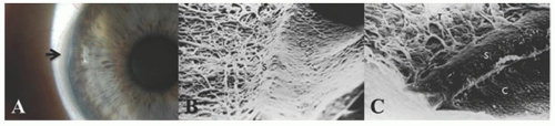
FIGURE 10.5. A: Prominent Schwalbe’s ring (posterior embryotoxon). The limbal pigment ring and prominent uveal trabecular meshwork are present. The distinct white ring demarcating the uveal meshwork centrally is the enlarged, displaced Schwalbe’s ring (arrow), which may be seen in about 10% of normal eyes. B: Scanning electron micrograph of normal Schwalbe’s ring (S), cornea (C), and uveal trabecular meshwork (T) about at this junction. C: Scanning electron micrograph of prominent Schwalbe’s ring (posterior embryotoxon). Endothelium covers both cornea (C) and elevated Schwalbe’s ring (S). (Scanning electron micrographs, courtesy of Morton Smith, MD.)

This anatomical approach makes sense embryologically, because it is the neural crest-derived mesenchymal tissue that differentiates into the cornea (except the epithelium), the angle structures, and the iris stroma, and because this mesenchymal tissue may have an inductive effect on the optic cup that determines the size and shape of the pupil and ciliary ring. This approach also makes sense clinically, because many of the abnormalities so easily described in isolation occur in combination with other anomalies. For example, Rieger’s anomaly (prominent Schwalbe’s ring, iris strands to Schwalbe’s ring, and hypoplasia of the anterior iris stroma) is accompanied by megalocornea in about 25% of cases, scleralization of cornea in about 80% of cases, juvenile glaucoma without buphthalmos in about 25% of cases, and one form of Peters’ anomaly in occasional cases.


Congenital Corneal Diseases without Corneal Opacification


Abnormalities of Corneal Size and Shape

Megalocornea. The newborn cornea measures approximately 10 mm in horizontal diameter and reaches the average adult measurement of 11.8 mm by age 2. Megalocornea is present if the horizontal diameter of a newborn cornea is 12 mm or more and if an adult cornea is 13 mm or more (Fig. 10.7).

An enlarged cornea occurs in three patterns: (a) megalocornea unassociated with other ocular abnormalities, usually inherited as an autosomal dominant trait; (2,3) (b) X-linked
megalocornea or anterior megalophthalmos, an X-linked recessive trait that consists of megalocornea, iris and angle abnormalities, and lens subluxation with early cataract formation; and (c) buphthalmos in infantile glaucoma (3). In keratoglobus, the protuberant thin cornea appears enlarged clinically but usually has a normal diameter. There seems to be no entity of “megaloglobus” in which the entire globe is congenitally enlarged with a normal intraocular pressure.


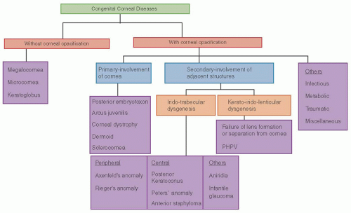
FIGURE 10.6. Flow chart showing clinical classification of congenital corneal diseases.


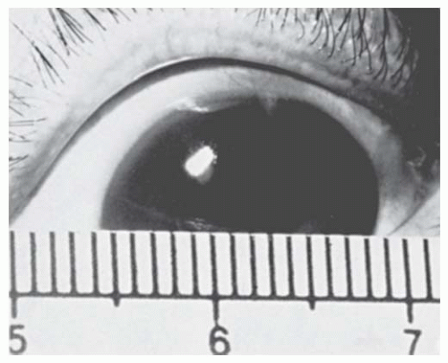
FIGURE 10.7. Anterior megalophthalmos. This cornea measures 15 mm in diameter and has normal thickness and clarity. The disorder is accompanied by transillumination defects in the iris and is associated with lens subluxation and cataract development in the fourth decade.

Simple Megalocornea. If bilateral clear corneas of normal thickness measure 13 mm or more in diameter without associated ocular abnormalities, the nonprogressive disorder of simple megalocornea exists, and once the diagnosis is clearly made, no other follow-up is necessary (3,4).

X-Linked Megalocornea (Anterior Megalophthalmos). X-linked megalocornea, a recessive disorder, is due to mutation of CHRDL1 gene at locus Xq23. It is the most common type of megalocornea. X-linked megalocornea occurs if the embryonic relationship (bell shaped optic cup) between the diameter of the anterior opening of the cup and the equatorial diameter persists, resulting in an increase in the relative diameter of the anterior segment of the eye compared to the posterior segment (3). It manifests as bilateral, symmetrically enlarged corneas that remain stable throughout life and sometimes contain a stromal mosaic pattern and arcus juvenilis (3,5). The deep anterior chamber occurs because the normal-sized lens, which is too small for the enlarged ciliary ring, subluxates. The iridocorneal angle is open but contains excess mesenchymal tissue, whereas the iris manifests a hypoplastic anterior stroma, transillumination defects, and pigment dispersion. The pupil is occasionally ectopic.

The two associations that threaten vision are the frequently elevated intraocular pressure (often due to pigmentary
glaucoma, spherophakia and/or lens subluxation), which requires lifelong annual examinations for early detection, and cataracts, which often appear in the fourth decade and may require the use of vitrectomy-type instruments during extraction, because the lenses are subluxated or dislocated (6,7). Due to enlarged anterior segment, custom-designed intraocular lenses may be required for lens stability and visual rehabilitation (8).

The clinician may have difficulty distinguishing among isolated megalocornea, anterior megalophthalmos, and infantile glaucoma in a young child with an enlarged cornea. Table 10.1 presents the distinctive features of these three disorders.


Table 10.1 DIFFERENTIAL DIAGNOSIS OF ENLARGED CORNEA


Simple Megalocornea


Anterior Megalophthalmos


Primary Infantile Glaucoma with Buphthalmos


Inheritance


Autosomal dominant (?)


X-linked recessive (male preponderance)


Sporadic


Time of appearance


Congenital


Congenital


First year of life


Bilaterality


Bilateral


Bilateral


Unilateral or bilateral


Symmetry


Symmetrical


Symmetrical


Asymmetrical


Natural history


Nonprogressive


Nonprogressive


Progressive


Symptoms


None


None


Photophobia, epiphora


Corneal clarity


Clear


Clear or mosaic dystrophy


Diffuse edema, tears in Descemet’s membrane


Intraocular pressure


Normal


Elevated in some adults


Elevated


Corneal diameter


13-18 mm


13-18 mm


13-18 mm


Corneal thickness


Normal


Normal


Thick


Keratometry


Normal


Normal/steep (cornea globosa); ↑ astigmatism (with the rule)


Flat


Gonioscopy


Normal


Excessive mesenchymal tissue


Excessive mesenchymal tissue


Globe diameter (A-scan)


Normal


Normal/increased


Increased and progressive


Anterior chamber depth


(A-scan)


Presumably normal (3 mm)


Approximately 5 mm


Approximately 4 mm


Vitreous length


Normal


Decreased


Increased


Major ocular complications


None


Lens dislocation, cataract <40 y, secondary glaucoma


Optic disc damage, late corneal edema


Associated systemic disorders


None


Occasionally Marfan’s and other skeletal abnormalities


None consistent


Keratoglobus. Keratoglobus is a distinct, rare entity that is characterized by generalized thinning and anterior bulging of the cornea. The thinning is greatest in the midperiphery of the cornea (Fig. 10.8A). Keratoglobus may occur as an autosomal recessive disorder that is part of the Ehlers-Danlos syndrome type VIA, in which it is accompanied by hyperextensible joints, blue sclerae, and neurosensory hearing loss. It has also been reported as an acquired condition, associated with various disorders, including vernal keratoconjunctivitis and thyroid ophthalmopathy. In keratoglobus the cornea is one-third normal thickness, usually has a normal diameter, and arcs highly over the iris, creating a very deep anterior chamber. Acute spontaneous breaks in Descemet’s membrane
may produce focal stromal edema (acute hydrops) and heal spontaneously in weeks to months (Fig. 10.8B). Minor blunt trauma to the eye or to the head may rupture the thin cornea and sclera, so the ophthalmologist must counsel the parents of these children to provide a safe environment and protective spectacles or eye guards. Amblyopia is often severe because of the high myopia, a problem diminished by carefully fitted spectacles or contact lenses. A scleral lens may be tried if contact lenses are not successful. In patients with severe thinning and anterior bulging of the cornea, surgical treatment may be contemplated. Given the diffuse corneal thinning to the limbus, surgical repair is problematic at best. A large limbus-to-limbus onlay lamellar keratoplasty or epikeratoplasty to both reinforce the corneal integrity and provide a more normal curvature can be performed. A “tuck-in” lamellar keratoplasty (central lamellar keratoplasty with intrastromal tucking of the peripheral flange) is another technique that can be performed in these patients. If the central cornea is scarred, typically from a previous episode of hydrops, a subsequent central visual penetrating keratoplasty can be performed (9,10,11,12,13,14).


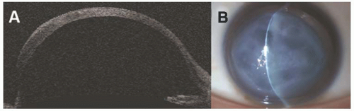
FIGURE 10.8. Keratoglobus A: Anterior segment optical coherence tomography showing ectasia of the entire cornea and midperipheral thinning. B: Slit-lamp photography demonstrating corneal edema due to corneal hydrops in a patient with keratoglobus.

Microcornea. A cornea 7 to 10 mm in diameter occurs in a variety of clinical settings, making classification difficult (Fig. 10.9). Both autosomal dominant and autosomal recessive patterns of inheritance occur, but microcornea may also appear sporadically. Although the exact cause is not known, it is assumed to be due to arrest of the growth of the cornea after differentiation is complete.

Microcornea may be an isolated abnormality in an otherwise normal eye (15,16). It may be associated with nanophthalmos (also called simple microphthalmos), a small, anatomically normal globe; (17,18,19) or it may be part of microphthalmos (also called complex microphthalmos), a small globe with multiple anomalies (20). A-scan ultrasonography can help distinguish isolated microcornea with normal axial length from microphthalmos with short axial length of the globe (17). All microphthalmic eyes have microcornea but the reverse is not true. Microcornea has syndromic associations with Ehlers-Danlos syndrome (mesodermal), Waardenburg syndrome (craniofacial), Norrie syndrome (neurologic), and Nance-Horan syndrome (osseous).

Management of eyes with microphthalmos varies according to the associated abnormalities. Hyperopia is commonly seen due to the flat cornea (cornea plana) but other refractive errors are also seen due to variation in the curvature. Early refraction may help prevent amblyopia. Lifelong examinations will detect intraocular pressure elevation, which occurs more commonly in eyes with microcornea due to associated anomalies of the angle and enlarging lens crowding the small anterior segment. Microcornea is commonly associated with congenital cataracts and is shown to be associated with mutations in various genes. These cataracts should be removed, using special care in eyes manifesting other anomalies (21).


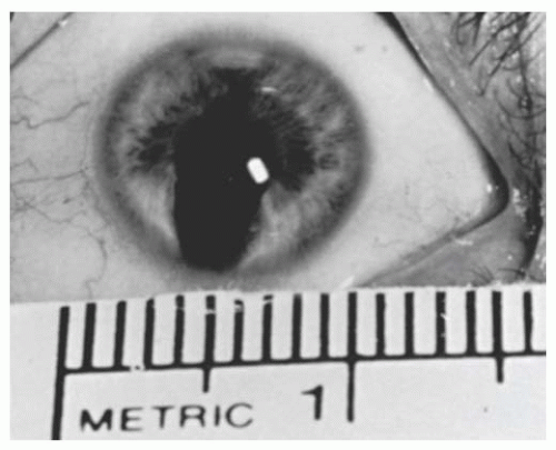
FIGURE 10.9. Microcornea, measuring 9 mm in diameter and accompanied by atypical iris coloboma and congenital cataract. The disorder was inherited as an autosomal dominant trait in this family.


Congenital Corneal Diseases with Corneal Opacification


Anterior Segment Dysgenesis

Abnormal development of the anterior segment structures (cornea, iris, ciliary body and lens) is called anterior segment dysgenesis. It is often difficult to separate abnormalities into individual disease entities. Anterior segment dysgenesis should be considered a heterogeneous clinical spectrum. The terms mesodermal dysgenesis of the iris and stoma (iris and stroma are neuroectoderm elements) and anterior chamber cleavage syndrome (no cleavage plane is formed during the anterior segment development) have been used, but these do
not seem to be appropriate in the description of abnormalities that occur during embryogenesis and are of neural crest differentiation (22,23,24,25).

Many abnormalities of the cornea, angle, and iris can be classified in an anatomical stepladder fashion, which builds from basic to more complex combinations (26). This approach simplifies our understanding of these anomalies, because the clinician or pathologist needs only to describe the anatomical findings, rather than worry about the proper eponyms or obscure Latin phrases. Unusual anomalies that do not fit into preestablished categories (27) can be inserted into this tabular classification on the basis of their anatomical components. Figure 10.10 represents this classification and includes commonly used eponyms.


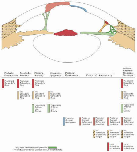
FIGURE 10.10. Composite illustration of the anatomical findings in the anterior chamber cleavage syndrome. The stepladder table demonstrates the spectrum of anatomical combinations and the terms by which they are commonly known. The colored markers in the table indicate the corresponding anatomical component in the illustration. (From Waring GO, Rodrigues M, Laibson PR, et al. Anterior chamber cleavage syndrome: a stepladder classification. Surv Ophthalmol 1975;20:5, with permission.)


These malformations conveniently fall into three groups: (a) peripheral (prominent Schwalbe’s ring, iris strands to Schwalbe’s ring, and hypoplasia of the anterior iris stroma); (b) central (central posterior corneal defect, central iridocorneal adhesions, corneolenticular approximation); and (c) combinations of the peripheral and central components.


Peripheral Anterior Segment Dysgeneses


Prominent Schwalbe’s Ring with Attached Iris Strands (Axenfeld’s Anomaly)

Iris strands that span the angle to insert on the prominent Schwalbe’s ring (Fig. 10.11) display variable morphology: fine threadlike filaments with a terminal knob, broad conical bands, or a confluent, fenestrated, lattice-like membrane. Prominent Schwalbe’s ring is usually only partly seen on slit-lamp biomicroscopy but may be seen 360 degrees on gonioscopy. In some cases the pupil is distorted. Axenfeld’s syndrome is defined as Axenfeld’s anomaly plus glaucoma (26).


Prominent Schwalbe’s Ring with Attached Iris Strands and Hypoplastic Anterior Iris Stroma (Rieger’s Anomaly)

Eyes with Rieger’s anomaly (Figs. 10.12, 10.13, 10.14, 10.15, 10.16 and 10.17) lack some superficial iris stroma, so that instead of crypts, furrows, and a collarette, the iris manifests a stringy appearance because the delicate radial fibrils of the posterior stroma show through. Abnormally shaped pupils (displaced in the direction of thickened iris band) occur commonly: slit-shaped, pear-shaped, round, ectopic, part of an atypical coloboma, or very large pupils, as in a partial aniridia. In rare cases, the iris atrophy progresses. Contracture of the primordial endothelial layer on the anterior surface of iris leads to these pupillary abnormalities. Rieger’s syndrome occurs when systemic anomalies are also present.

Axenfeld’s anomaly and syndrome, and Rieger’s anomaly and syndrome represent a spectrum of developmental dysgenesis and hence a single diagnostic entity of Axenfeld-Rieger syndrome has been proposed (28,29). Glaucoma is seen in 50% of cases and the onset is delayed compared to infantile glaucoma, and thus lifetime surveillance is required. The pathogenesis of glaucoma is due to improper migration of primordial tissue derived from the neural crest leading to persistence of endothelial layer on the angle and anterior insertion of peripheral iris onto trabecular meshwork leading to impaired aqueous outflow (30). It is inherited as autosomal dominant pattern (70%) with variable expressivity, but sporadic cases are also reported (31). Mutations at locus 4q25 of PITX2 gene and at locus 6p25 of FOXC1 gene are commonly implicated in the pathogenesis of Axenfeld-Rieger syndrome. Corneal endothelium, stroma, iris, ciliary body and sclera express PITX2. Mutations in PITX2 are commonly associated with extraocular systemic abnormalities of Axenfeld-Rieger syndrome. Mutations in FOXC1 are commonly associated with only ocular abnormalities and higher risk for glaucoma (32). The most common systemic condition associated with Axenfeld-Rieger syndrome is cardiovascular outflow tract abnormalities, but craniofacial and skeletal abnormalities are also seen (33,34,35).


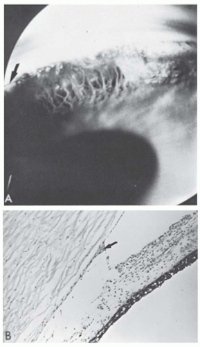
FIGURE 10.11. Axenfeld’s anomaly. A: Gonioscopic view shows the angle recess filled with dense iris processes (persistent mesenchymal tissue) that extend to a prominent Schwalbe’s ring (arrow). This configuration may exist alone or as a part of a variety of iridocorneal dysgeneses (Courtesy of Robinson D. Harley, MD). B: Histologic section showing the prominent centrally displaced Schwalbe’s ring (arrow) with iris processes extending to it and across the angle recess (×64). (Courtesy of Merlyn Rodrigues, MD.)


Iris Strands in Angle and Hypoplasia of Anterior Iris Stroma (Iridogoniodysgenesis)

This abnormality, inherited in an autosomal dominant pattern, resembles Rieger’s anomaly without the prominent Schwalbe’s ring (36). Affected individuals very commonly have juvenile glaucoma.


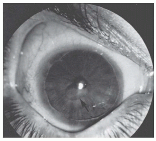
FIGURE 10.12. Rieger’s anomaly with central posterior corneal defect. This right eye of a 23-year-old dwarf demonstrates a prominent Schwalbe’s ring with the iris process extending to it, an atrophic anterior iris stroma, and a central posterior corneal defect (posterior keratoconus) (arrow). Intraocular pressure was normal. The left eye appeared similar.


Infantile Glaucoma

If this classification is extended, infantile glaucoma (with or without buphthalmos) can be added, on the basis that it represents a mesenchymal goniodysgenesis. In fact, some authors believe that the megalocornea seen in infantile glaucoma is a result of a primary keratodysgenesis, rather than a result of stretching from increased intraocular pressure (37).


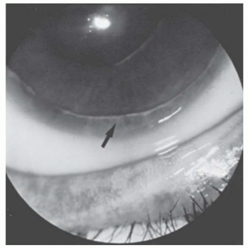
FIGURE 10.13. Rieger’s anomaly. Close-up of 6 o’clock limbus of eye in Figure 10.12. Iris processes (arrow) extend to the irregular prominent Schwalbe’s ring.


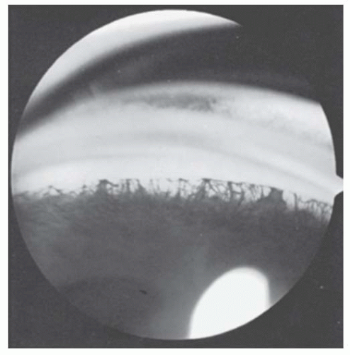
FIGURE 10.14. Angle in Rieger’s anomaly. Gonioscopic appearance of eye in Figures 10.12 and 10.13, showing iris processes extending to the prominent Schwalbe’s ring.

The management of infantile and juvenile glaucoma is discussed in Chapter 12.


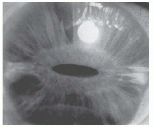
FIGURE 10.15. Rieger’s anomaly. The right eye shows marked hypoplasia of the anterior iris stroma. The deep stroma is thin and fibrillary, revealing the underlying iris epithelium and pupillary sphincter. The pupil is slit-shaped and central. The prominent Schwalbe’s ring is poorly illustrated. (Courtesy of George L. Spaeth, MD.)


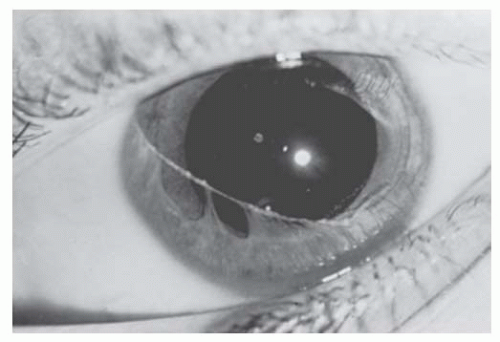
FIGURE 10.16. Rieger’s anomaly. This 10-year-old white girl has a centrally displaced prominent Schwalbe’s ring with iris processes extending to it from the angle recess and the collarette. Anterior iris stroma is absent at 11 o’clock position. The configuration is accentuated by the dilated pupil. Intraocular pressure was normal. No other ocular anomalies existed. (Courtesy of Harold Koller, MD.)


Central Anterior Segment Dysgenesis: General Features

The basic abnormality in this group is a focal attenuation or absence of the corneal endothelium and Descemet’s membrane, usually associated with an overlying corneal opacity. In contrast with the peripheral abnormalities, the central disorders have two separate etiologies, primary dysgenesis and secondary to inflammation, but the clinical and histopathologic distinction between the two is difficult. Presumably, if the cornea is avascular and there are no signs of inflammation, one can assume that the disorder is a primary dysgenesis; however, if the cornea is opaque and vascularized, intrauterine inflammation may have been present. Therefore, these entities are discussed on the basis of their anatomical findings alone, rather than their pathogenesis. Glaucoma is present in about half the cases, usually appearing as a nonbuphthalmic infantile form.


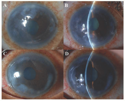
FIGURE 10.17. Rieger’s anomaly. A, B: The right eye demonstrating a peripheral corneal opacity and edema with iris strands anteriorly with peaking of pupil toward 1 o’clock. C, D: The left eye demonstrating peripheral corneal opacity and edema, prominent Schwalbe’s ring nasally with iris processes extending to it, atrophic anterior iris stroma (12-3 o’clock) and also inferiorly on gonioscopy. Intraocular pressure was normal.

Postnatally, the corneal opacity may clear somewhat, particularly if it is avascular, central, and consists mostly of edema. On the other hand, the opacity may progressively vascularize, particularly if the cornea is ectatic and the anterior segment derangement is severe.


Posterior Corneal Depression (Central Posterior Keratoconus)

This focal, discrete, posterior corneal indentation has a faint overlying stromal haze and is usually central, unilateral, and nonprogressive (Fig. 10.18) (38). In some instances, a ring of pigment clumps surrounds the depression, indicating previous iris contact (39). The anterior corneal curvature is not dramatically irregular, and the disorder is unrelated to the more common form of acquired progressive anterior keratoconus. Visual acuity is only moderately reduced, presumably because of the irregular astigmatism resulting in mild amblyopia. Some authors described a total posterior keratoconus, which may be a variant of the more common progressive
degenerative type (40). Histopathologically, abnormalities include an irregularly thickened epithelial basement membrane, focal disruption of Bowman’s layer, stromal irregularity, and a multilaminar Descemet’s membrane that contains wide-spacing material and focal excrescences (41). Scanning electron microscopy was used to evaluate a cornea with posterior keratoconus, revealing no excrescences of Descemet’s membrane or endothelial tags (42). Given these findings, Al-Hazzaa et al. (42) suggest there may be a subset of posterior keratoconus patients who do not fall into the category of anterior segment dysgenesis abnormalities.


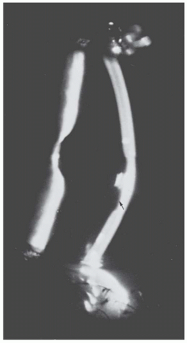
FIGURE 10.18. Posterior keratoconus. Slit-lamp view of the eye in Figure 10.12, showing the depression in the posterior corneal surface (arrow). The cornea overlying it is clear, and no iris processes extend to its margin.

The anterior corneal curvature in eyes with posterior keratoconus has generally been described as essentially normal. One problem is that most methods used to evaluate corneal curvature, including the keratometer and keratoscope, do not examine the central few millimeters of cornea well at all. With the advent of computerized corneal topography/tomography, central as well as midperipheral anterior corneal curvature can be effectively analyzed. Computerized corneal videokeratography was performed on an eye with posterior keratoconus, revealing a central steepened “cone” associated with the area of posterior corneal thinning (43).


Posterior Corneal Defect with Overlying Leukoma (Peters’ Anomaly)

Peters’ anomaly represents a range of features from a simple defect in the posterior cornea producing an overlying opacity to severe ocular and systemic malformations. If the opacity is dense enough, it may obscure the more normal anterior segment anatomy.


Posterior Corneal Defect with Stromal Opacity and Adherent Iris Strands (Peters’ Anomaly Type I)

The size and density of the corneal opacity and the depth of the posterior defect can vary widely, from a small, central, focal, ground-glass opacity (Fig. 10.19A); to a dense, round leukoma (Fig. 10.19B); to total corneal vascularization and scarring with an elevated mass (see Fig. 10.21A). The lens is clear and in normal position. The configurations of the iris strands that extend from the collarette to the margin of the posterior defect are as diversified as the opacity and include fine filaments (Fig. 10.19A), broad bands, and fenestrated sheets.

The histopathologic findings are equally varied but usually include thickening or fragmentation of Bowman’s layer, disorganization of stromal architecture, central absence of Descemet’s membrane and endothelium (both of which are present peripherally), and central iridocorneal adhesions (Figs. 10.20 and 10.21C,D).


Posterior Corneal Defect with Stromal Opacity, Adherent Iris Strands, and Corneolenticular Contact or Cataract (Peters’ Anomaly Type II)

In this variant of Peters’ anomaly, a variety of lens abnormalities occur (44), including adhesion of lens cortex to the corneal stroma at the site of the posterior defect (Figs. 10.22 and 10.23) (45), approximation to the back of the cornea with an intact lens capsule, displacement into the anterior chamber or into the pupil, or a central cataract with maintenance of a normal position (46). These lens abnormalities occur due to faulty separation of the lens vesicle from the surface ectoderm. It is also associated with other ocular abnormalities such as aniridia, microcornea, microphthalmos (47,48).


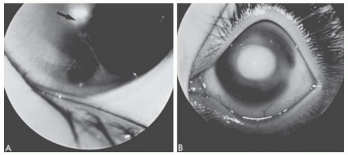
FIGURE 10.19. A: Peters’ anomaly. A mild form showing attenuated iris adhesions to the border of a small corneal opacity (arrow). This was present bilaterally in this 9-month-old white girl. B: Peters’ anomaly. This 10-month-old white girl had bilateral congenital central corneal opacities. During penetrating keratoplasty, iris adhesions were found extending from the pupillary margin to the borders of the opacity. An anterior polar cataract was present. (Courtesy of Harold Koller, MD.) The histopathologic findings are equally varied but usually include thickening or fragmentation of Bowman’s layer, disorganization of stromal architecture, central absence of Descemet’s membrane and endothelium (both of which are present peripherally), and central iridocorneal adhesions (Figs. 10.20 and 10.21C, D).

Peters’ anomaly associated with systemic abnormalities such as short stature, developmental delay, and cleft lip/palate is termed as Peters Plus syndrome. Peters’ anomaly is usually bilateral. Bilateral Peters’ anomaly is associated with more systemic malformations compared to unilateral cases. Peters’ anomaly is usually sporadic or autosomal recessive but autosomal dominant patterns have been reported. Peters’ anomaly is associated with mutations of homeobox genes PAX6 (locus-11p13), PITX2 (locus-4q25), and FOXC1 (6p25). Peters Plus syndrome is associated with mutation of the gene B3GALTL at locus 13q12.3 (48,49).


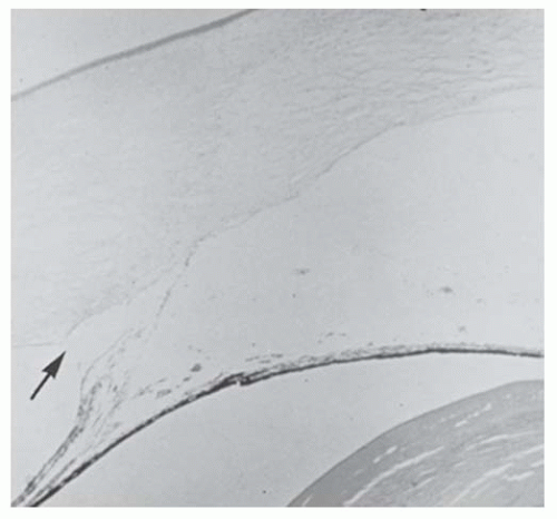
FIGURE 10.20. Peters’ anomaly. Histologic section demonstrates iris adhesions that extend from the collarette to the margin of a central posterior corneal defect. The overlying cornea is edematous. Descemet’s membrane ends abruptly at the margin of the central defect (arrow) (×6). (Courtesy of Robert D’Amico, MD.)


Corneal Staphyloma

In this most severe form of posterior corneal defect, the ectatic, thin, scarred, vascularized cornea is lined by uveal tissue and may protrude between the eyelids (Fig. 10.24) (50). The ectasia may be present at birth but usually becomes worse in the first week of life. Intraocular pressure is usually elevated, and the lens is incorporated into the scarred ectatic cornea. In rare instances, the cornea develops a hypertrophic keloid scar (51).


Pathogenesis of Posterior Corneal Defects

There are four pathogenic theories (44,52,53): (a) intrauterine keratitis, leaving a posterior defect commonly called the internal corneal ulcer of von Hippel; (b) incomplete central migration of the neural crest mesenchymal waves that form the corneal endothelium and stroma; (c) improper separation of the lens vesicle from the surface ectoderm, which may produce the central defect by blocking the ingrowth of the neural crest mesenchymal tissue and may result in a persistent keratolenticular adhesion without an intact lens capsule; and (d) secondary anterior displacement of the lens by a vitreoretinal-mass-like persistent hyperplastic primary vitreous or pupillary block from a persistent pupillary membrane. Because none of these four theories adequately explains all the clinical or histopathologic findings, and because there is experimental evidence supporting each one, these congenital anomalies must be regarded as a heterogeneous group with a similar clinical appearance.


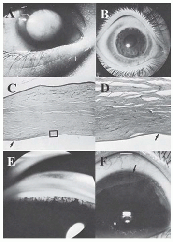
FIGURE 10.21. Anterior chamber cleavage syndrome. A: This 7-month-old boy had bilateral megalocornea (13 mm in diameter), (A-D) a central posterior corneal defect with corneal leukoma in the right eye (Peters’ anomaly), and (E, F) Rieger’s anomaly of the left eye (Courtesy of Turgut Hamdi, MD). B: Keratoplasty for central posterior corneal defect. A penetrating keratoplasty was performed in the right eye at age 22 months, and the graft remained clear for 5 months until graft rejection occurred. No iris processes extended to the corneal leukoma. An anterior polar cataract was discovered postoperatively. C: Central posterior corneal defect with scarring. Histopathologically, the corneal button shows superficial fibrovascular invasion and deep stromal edema. In this area, Bowman’s layer and Descemet’s membrane are absent (box). The margin of the button (left side) shows more normal cornea with edematous stroma. Descemet’s membrane is present in this area (arrow) (Periodic acid-Schiff (PAS) ×25). D: Central posterior corneal defect. The area in the box shows the transition (arrow) from intact Descemet’s membrane peripherally to its replacement by fibrous tissue centrally. Only fragments of endothelium were seen (PAS ×250). E: Megalocornea and Rieger’s anomaly. The left eye of this patient exhibited a 13-mm diameter cornea, a prominent Schwalbe’s ring (arrow) with iris processes extending to it, and a hypoplastic iris stroma. F: Iris processes in Rieger’s anomaly. The angle filled with delicate iris processes and mesenchymal tissue extending up to the prominent Schwalbe’s ring.

The descriptive anatomical classification is especially helpful in the central-peripheral combinations, because it allows one to see the exact components in each case, instead of resorting to a combination of eponyms.


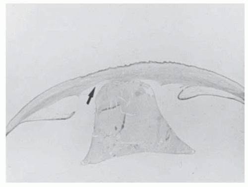
FIGURE 10.22. Congenital lens-corneal adhesion (Peters’ anomaly). The eye of this newborn demonstrates irregular and thickened corneal epithelium and stroma, central absence of Bowman’s and Descemet’s membranes, a central posterior corneal defect (arrow) with a lens-corneal adhesion, a conical cataractous lens, and malformation of the anterior chamber angles with adhesion of the iris to the cornea. (PAS ×3) (Courtesy of Charles G. Steinmetz, MD.)


Corneal Keloid

Corneal keloid is a gray-white elevated mass that may present as a localized solitary nodule or diffusely involving the entire cornea (Fig. 10.25). They are congenital or more typically secondary to trauma. Congenital corneal keloids are due to failure of normal differentiation of corneal tissue during embryogenesis. Intrauterine trauma due to amniocentesis may be a cause, but fortunately the incidence of complications due to amniocentesis has decreased due to more advanced ultrasound-guided techniques (54). In posttraumatic cases, a mechanical stimulus triggers a cellular inflammatory response (vasodilation and recruitment of immature fibroblasts). Subsequently, there is regression of blood vessels, myofibroblast proliferation and scar retraction. At times, this response leads to a vigorous fibrocytic response causing formation of an exuberant glistening mass. Diagnosis is confirmed based on histopathologic presence of hyalinized collagen, activated fibroblasts, and myofibroblasts. Congenital keloid may be associated with Lowe syndrome and Rubinstein-Taybi syndrome. Various surgical modalities such as superficial lamellar keratectomy, lamellar keratoplasty or full-thickness keratoplasty have been reported with variable success rates (55,56,57,58,59).


Aniridia

Aniridia is a bilateral congenital disorder associated with panocular abnormalities affecting not only the iris but also the cornea, anterior chamber angle, lens, retina, and optic nerve. It is inherited in an autosomal dominant pattern in most cases, but sporadic and rarely autosomal recessive
inheritance is also seen. Patients with sporadic aniridia are at risk for WAGR syndrome (Wilms tumor, aniridia, genitourinary abnormalities, mental retardation) and should undergo routine surveillance for kidney disorders. Aniridia is due to mutation at locus 11p13 of the paired box gene 6 (PAX6). It is usually associated with keratopathy, cataract, glaucoma, foveal hypoplasia and strabismus. Dental, musculoskeletal and developmental delays are systemic abnormalities frequently associated with aniridia. Keratopathy is thought to be due to an abnormally differentiated epithelium, abnormal cell adhesion, impaired healing response and limbal stemcell deficiency leading to conjunctivalization of cornea. It begins as vascularized thickening of the cornea at the periphery, which gradually advances centrally. Recurrent corneal erosions lead to subepithelial fibrosis causing corneal opacification. Corneal opacification that occurs due to recurrent erosions is caused by deficiency in matrix metalloproteinase 9 (regulated by PAX6), which is responsible for normal cell remodeling and wound healing. Penetrating keratoplasty for visual rehabilitation is generally unsuccessful because of recurrent surface breakdown. Keratolimbal allograft and Boston keratoprosthesis have proven to be effective for long-term visual rehabilitation (Fig. 10.26) (60,61,62).


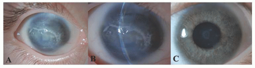
FIGURE 10.23. Peters’ anomaly A, B: The right eye of this child showing central corneal opacity with relatively clear periphery and iris adhesions to the midperiphery and central cornea. C: The left eye of the same patient demonstrating a corneal posterior opacity with an anterior lenticular opacity suggesting a partial dysgenesis.


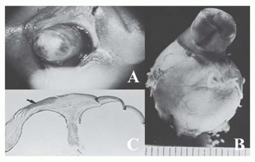
FIGURE 10.24. Congenital corneal staphyloma. A: This 5-day-old infant was born with a flat opaque right cornea. By age 2 days, the cornea had become blue and ectatic, as shown here. The left eye was normal, except for persistent pupillary membrane (Courtesy of Joseph H. Calhoun, MD). B: Gross appearance of the globe. The ectatic area is limited to the cornea (Courtesy of Merlyn Rodrigues, MD). C: Histologic section of globe. Areas of the cornea are thin and ectatic. A superficial corneal abscess from exposure is present (arrow). Bowman’s and Descemet’s membranes are absent. A rudimentary lens is adherent to the central posterior cornea, blending with stromal tissue. Uveal tissue is firmly adherent to the posterior cornea, sweeping down along the lens rudiment (Hematoxylin-eosin ×3). (Courtesy of Merlyn Rodrigues, MD.)


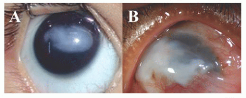
FIGURE 10.25. Congenital corneal keloid. A: 3-month-old boy with corneal keloid and normal intraocular structures, B: Corneal keloid with vascularization in a child associated with Lowe’s syndrome.


Neonatal Corneal Opacities


Differential Diagnosis

The ophthalmologist often feels stumped when confronted by a child with a neonatal corneal opacity. These feelings provide an apt acronym for the causes of neonatal corneal opacities: STUMPED (Table 10.2) (63).


Sclerocornea (Stumped)

Clinicians often use the term “sclerocornea” as a nonspecific description for any congenitally opaque, vascularized cornea. Too broad a use of the term, however, obscures valuable distinctions. Clinically, sclerocornea denotes a congenital peripheral white vascularized opacity that blends with the sclera, due to anterior displacement of limbal arcades, obliterating the corneoscleral limbus and scleral sulcus.

Previously proposed by Waring and Rodrigues, but recently reclassified by Nischal into three groups (64,65):

1. Isolated sclerocornea (Fig. 10.27A). Patients in this group have no other ocular abnormalities and show either exaggerated scleral extension (scleralization, scleral overriding) or more extensive peripheral corneal opacification and vascularization. It may be associated
with cornea plana (<38D) and a slightly shallow anterior chamber, which may be a risk factor for the development of secondary glaucoma (66). It should be differentiated from arcus juvenilis, which is devoid of vessels and has a clear lucid interval between the limbus and the corneal opacification.


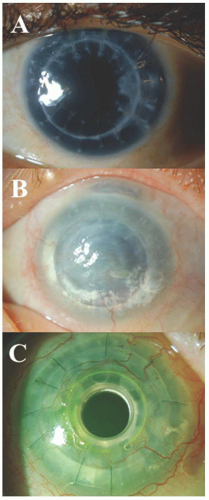
FIGURE 10.26. Aniridia. A: Central clear penetrating keratoplasty in a patient with aniridia. B: Failed penetrating keratoplasty due to aniridiainduced keratopathy. C: Boston keratoprosthesis in a patient with multiple failed grafts. The visual acuity remained 20/400 due to amblyopia and nystagmus.

2. Complex sclerocornea. It is usually associated with other ocular abnormalities such as microphthalmos, cataract and infantile glaucoma. The central cornea is relatively clear and the corneal thickness is normal or increased (67).

3. Total sclerocornea (Fig. 10.27B). When the cornea is opaque enough to prohibit visualization of the iris and lens, a precise clinical diagnosis is difficult. In some cases, the lens and iris may remain unseparated from the cornea. Limbal anlage that is well defined by the 10th week of gestation distinguishes the cornea and sclera by providing a ring of stability. The absence of this ring leads to corneal curvature similar to that in sclera.

Histopathologically, sclerocornea shows an irregular epithelium with variably thick basement membrane, a fragmented or absent Bowman’s layer, and disorganized spindles of vascularized stromal collagenous tissue that contain collagen fibrils. The collagen fibers are of larger diameter in the superficial stroma compared to the deep stroma, similar to the structure of sclera. The random structure of these large fibrils and their attendant blood vessels scatter light and give the cornea its white clinical appearance. Descemet’s membrane is abnormal, either being present as a thin irregular layer with collagenous tissue behind it or showing focal dehiscences that may contain fibrous tissue. The endothelium is usually damaged, precluding detailed description (68,69). Syndromic abnormalities inconsistently accompany sclerocornea (70,71).


Tears in Endothelium and Descemet’s Membrane (sTumped)

Birth trauma is discussed later in this chapter. Infantile glaucoma is discussed in Chapter 21.


Ulcers (stUmped)

Corneal ulceration in the neonate is extremely rare. It can occur due to a variety of causes including infectious and inflammatory reasons. We have observed one patient with a congenital sensory neuropathy of unknown type who was born with bilateral, shallow, central corneal ulcers. Corneal melting persisted, and in spite of therapy with tarsorrhaphies, soft contact lenses, and keratoplasties, both eyes were finally enucleated.

Viral infections of the cornea are discussed later in this chapter.


Table 10.2 STUMPED: DIFFERENTIAL DIAGNOSIS OF NEONATAL CORNEAL OPACITIES


Diagnosis


Laterality


Opacity


Ocular Pressure


Other Ocular Abnormalities


Natural History


Inheritance


S-Sclerocornea


Unilateral or bilateral


Vascularized, blends with sclera, clearer centrally/total scleralization of the cornea


Normal or elevated


Cornea plana


Nonprogressive


Sporadic


T-Tears in endothelium and Descemet’s membrane


Birth trauma


Unilateral


Diffuse edema


Normal, possibly elevated


Possible hyphema, periorbital ecchymoses


Spontaneous improvement in 1 month


Sporadic


Infantile glaucoma


Bilateral


Diffuse edema


Elevated


Megalocornea, photophobia and tearing, abnormal angle


Progressive unless treated


Autosomal recessive


U-Ulcers


Herpes simplex keratitis


Unilateral


Diffuse with dendritic or geographical epithelial defect


Normal or elevated


None


Often recurrent


Sporadic


Congenital rubella


Bilateral


Disciform or diffuse edema, no frank ulceration


Normal or elevated


Microphthalmos, cataract, pigment epithelial mottling


Stable, may be clear


Sporadic


Neurotrophic or exposure


Unilateral or bilateral


Central ulcer


Normal


Eyelid anomalies, congenital sensory neuropathy


Progressive unless treated


Sporadic


M-Metabolic (rarely present at birth) (all mucopolysaccharidoses except II, III; mucolipidosis Type IV)*


Bilateral


Diffuse haze, denser peripherally


Normal


Few


Progressive


Generally autosomal recessive or X-linked recessive


P-Posterior corneal defect (Peters’ anomaly)


Unilateral or bilateral


Central, diffuse haze or vascularized leukoma


Normal or elevated


Anterior segment dysgenesis


Stable; sometimes early clearing or vascularization


Sporadic, autosomal recessive


E-Endothelial dystrophy


Congenital hereditary endothelial dystrophy


Bilateral


Diffuse corneal edema, marked corneal thickening


Normal


None


Stable


Autosomal dominant or recessive


Posterior polymorphous corneal dystrophy


Bilateral


Diffuse haze, normal to moderate corneal thickening


Normal or elevated


Occasional peripheral anterior synechiae


Slowly progressive


Autosomal dominant


Congenital stromal corneal dystrophy (more stromal than endothelial)


Bilateral


Flaky, feathery stromal opacities; increased corneal thickness


Normal


None


Stable


Autosomal dominant


D-Dermoid


Unilateral or bilateral


White vascularized mass, hair, lipid arc


Normal


None


Stable


Sporadic


* Mucopolysaccharidosis II (Hunter’s syndrome); mucopolysaccharidosis III (Sanfilippo’s syndrome).


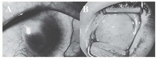
FIGURE 10.27. Sclerocornea. A: Scleral tissue extends in a geographical pattern toward the central cornea. Some clear cornea remains centrally. B: Total replacement of the cornea by sclera. Penetrating keratoplasty was unsuccessful. The iris and lens were grossly malformed. (Courtesy of Joseph Calhoun, MD.)


Metabolic (stuMped)

Because the fetus has access to maternal enzymes, systemic metabolic disorders, such as the mucopolysaccharidoses, mucolipidosis, and tyrosinosis that later develop corneal opacities, are rarely present at birth. A consistent exception to this is mucolipidosis type IV (ganglioside neuraminidase deficiency). These metabolic disorders are discussed in more detail in Chapter 21.


Posterior Corneal Defect, Peters’ Anomaly (stumPed)

These central corneal opacities are discussed earlier in this chapter.


Endothelial Dystrophies (stumpEd)

These dystrophies are discussed later in this chapter.


Dermoid (stumpeD)

A corneal dermoid tumor, classified as a choristoma (Fig. 10.28), is a solid, congenital, rounded mass consisting of keratinized epithelium overlying fibrofatty tissue that contains hair follicles, sebaceous glands, and sweat glands (72,73). Rarely, ectopic lacrimal gland, another choristoma, can appear similar to a dermoid. A dermoid is usually a single unilateral pink-white-gray mass, 1 to 5 mm in diameter, which straddles the limbus inferotemporally. They are usually sporadic but
rarely its occurrence in families has been reported. The clinical picture is highly variable, however. The masses may be multiple, bilateral, confined to the cornea alone, minutely small, or large enough to obscure the entire cornea (Fig. 10.29). The dermoid extends into the corneal stroma and sclera but seldom occupies the full thickness and only rarely grows into the angle. Hair is not always present on the surface. Dermoids have been classified into three grades: Grade-1, most frequent type and is small (up to 5 mm) and isolated. Grade-2, is much larger and may cover the entire corneal surface and may extend into deeper layers of the cornea. Grade-3, most severe and rare, it replaces the entire anterior segment (74).


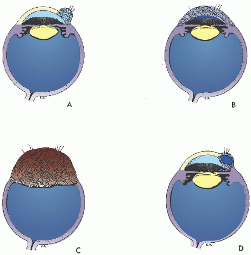
FIGURE 10.28. Corneal dermoids, schematics. A: Limbal dermoid tumor. B: Dermoid tumor replacing the entire cornea. C: Dermoid tumor replacing the entire anterior segment. D: Dermoid cyst of cornea (After Ida Mann).

Dermoids may enlarge slowly, especially at puberty or after trauma or irritation. A limbal dermoid may leave visual acuity unaffected, but if it grows over the visual axis or produces significant corneal astigmatism, amblyopia will likely result. Dermoids contain considerable fatty tissue, and a white arcuate haze of lipoid material commonly extends into the corneal stroma in front of the tumor. This lipid may encroach on the visual axis and blur vision.


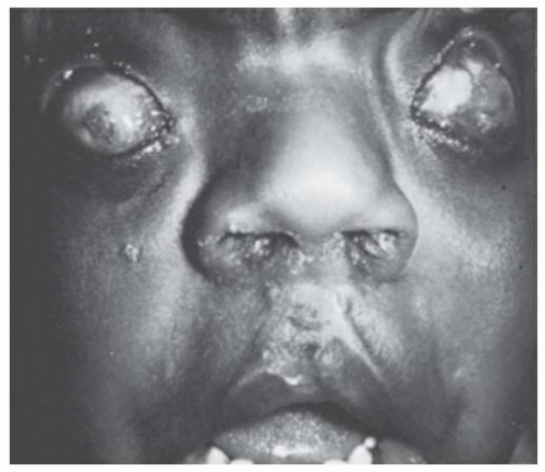
FIGURE 10.29. Bilateral dermoid tumors replacing the entire cornea. This 3-year-old boy was born with masses of vascularized tissue containing surface hair protruding grotesquely between his eyelids. He has had repair of cleft lip and palate. (Courtesy of Robison D. Harley, MD.)

Approximately one-third of patients with limbal dermoids have associated developmental anomalies. Among the most frequent is the constellation of epibulbar dermoids, preauricular appendages, and vertebral anomalies (Goldenhar syndrome [oculoauriculovertebral dysplasia]) (Figs. 10.30, 10.31, 10.32 and 10.33) (73,75).

In Goldenhar syndrome the epibulbar dermoid straddles the limbus in the inferotemporal quadrant. It is bilateral in about 25% of cases. A subconjunctival lipodermoid or dermolipoma (lipoma covered by keratinized or nonkeratinized epithelium with hair on the surface) is found in the superotemporal quadrant in about 50% of cases. This lipodermoid may blend with the epibulbar dermoid. A coloboma of the upper eyelid is present at the junction of the middle and inner third in about 25% of cases. Other associated ocular anomalies include Duane syndrome, lacrimal duct stenosis, and iris and choroidal colobomas.


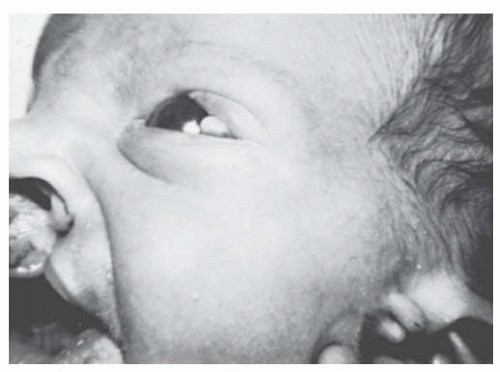
FIGURE 10.30. Goldenhar’s (oculoauriculovertebral dysplasia) syndrome. The limbal dermoid and preauricular skin tags are present, in addition to a cleft lip. (Courtesy of Robison D. Harley, MD.)


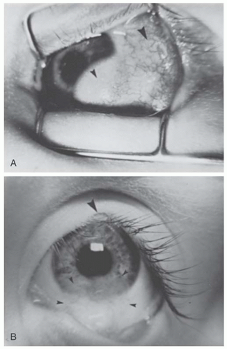
FIGURE 10.31. Goldenhar’s (oculoauriculovertebral dysplasia) syndrome. A: A lipodermoid of the conjunctiva (large arrow) and an epibulbar dermoid of the limbus (small arrow) are present concurrently in about half the cases. B: A coloboma of the upper eyelid at the junction of the middle and inner thirds is present in about one-fourth of cases (large arrow). The limbal dermoid tumor has been excised (small arrows). (Courtesy of Jules Baum, MD.)

Auricular anomalies—usually on the same side as the dermoid—include preauricular appendages, posteriorly placed ears, preauricular sinuses, and stenosis of the external auditory meatus. Vertebral anomalies occur in about two thirds of patients, including fused cervical vertebrae, hemivertebrae, spina bifida, and occipitalization of the atlas. Lumbosacral abnormalities also occur. Facial malformations include micrognathia, macrostomia, dental abnormalities, and facial asymmetry. The diagnosis of Goldenhar syndrome should lead to complete examination for associated
systemic abnormalities, especially cardiovascular, renal, genitourinary, and gastrointestinal defects. Goldenhar syndrome occurs sporadically.


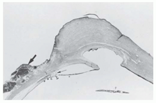
FIGURE 10.32. Corneal dermoid, posterior corneal defect, and Axenfeld’s anomaly. In the right eye, cornea is replaced by a mass of vascularized connective tissue. Ectopic lacrimal gland is present at the limbus (large arrow). In this area the angle is deep and contains a prominent Schwalbe’s ring with iris processes adherent to it (small arrow). A central posterior corneal defect is present. On one side, the iris stretches from the angle to the edge of the defect. Descemet’s membrane is present in this area. On the opposite side, iris lines the corneal defect and posterior cornea; Descemet’s membrane is absent in these areas (Hematoxylin-eosin ×3).


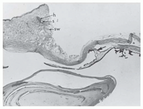
FIGURE 10.33. Corneal dermoid, central posterior corneal defect, and iris-corneal adhesion. The left eye of the patient shown in Figure 10.32. Anterior cornea is replaced by vascularized connective tissue containing hair follicle (h), sebaceous gland (s), and sweat gland (sw). A biopsy has been taken for diagnostic purposes, leaving a defect. Descemet’s membrane is present peripherally but absent centrally. An iris adhesion (arrow) is present centrally. Angle structures are disorganized (Hematoxylin ×4).

Jun 20, 2016 | Posted by in OPHTHALMOLOGY | Comments Off on Diseases of the Cornea

Full access? Get Clinical Tree

Get Clinical Tree app for offline access