Purpose
To determine the developmental sequence of retinal layers to provide information on where in utero pathologic events might affect retinal development.
Design
Qualitative and quantitative descriptive research.
Methods
A histology collection of human eyes from fetal week (Fwk) 8 to postnatal (P) 10 weeks was analyzed. The length of the nasal and temporal retina was measured along the horizontal meridian in 20 eyes. The location of the inner plexiform layer (IPL) and outer plexiform layer (OPL) was identified at each age, and its length measured.
Results
The human eye retinal length increased from 5.19 mm at Fwk 8 to 20.92 mm at midgestation to 32.88 mm just after birth. The IPL appeared in the presumptive fovea at Fwk 8, reached the eccentricity of the optic nerve by Fwk 12, and was present to both nasal and temporal peripheral edges by Fwk 18–21. By contrast, the OPL developed slowly. A short OPL was first present in the Fwk 11 fovea and did not reach the eccentricity of the optic nerve until midgestation. The OPL reached the retinal edges by Fwk 30. Laminar development of both IPL and OPL occurred before vascular formation.
Conclusions
In human fetal retina, the IPL reached the far peripheral edge of the retina by midgestation and the OPL by late gestation. Only very early in utero events could affect IPL lamination in the central retina, but events occurring after Fwk 20 in the peripheral retina would overlap OPL laminar development in outer retina.
The primate retina develops over many months both in utero and postnatally. Moreover, it has a prominent foveal-to-peripheral gradient such that points on the retina only 2 mm apart may be at strikingly different stages of development. This marked gradient is not mentioned or not recognized in many older papers, making it difficult to interpret the data presented. Moreover, having accurate timelines of human retinal development at known retinal loci is important for medicolegal issues as well as for knowledge of what regions and layers of the retina may be impacted by in utero events. The widespread use of optical coherence tomography to visualize the prenatal and postnatal human retina has greatly expanded our knowledge of foveal development, but there is little systematic information on laminar development outside of the fovea in humans. We have analyzed a large collection of human retinas from embryonic to infant, which provides well-preserved material from which such an assessment can be made morphologically.
Methods
Qualitative and quantitative descriptive analysis was done on 2 groups of human eyes. These were obtained with Human Subjects approval (0447-E/A07). Eyes from fetal week (Fwk) 6–22 were sourced from aborted fetuses obtained after consent by the Human Tissue Laboratory, University of Washington (UW), Seattle. Fetuses containing obvious abnormalities were excluded. Eyes from Fwk 24–40 and postnatal (P) infants were obtained through the support of the UW Neonatal Intensive Care Nursery and the Lions Eye Bank, Seattle. Infants containing obvious abnormalities or congenital conditions that might affect the eye, or eyes that had poor retinal structure, were not used in this study. Fetal age was determined by crown-rump and foot length, and should be taken to be ±1 week.
Fetal eyes <Fwk 16 were fixed whole by immersion in 4% paraformaldehyde in phosphate buffer pH 7.4; in older eyes, the cornea and lens were removed before fixation. Eyes were fixed overnight, washed in phosphate buffer, and measured using micrometer calipers for axial length and diameter. For the best morphology, the horizontal meridian containing fovea and optic nerve was embedded in glycol methacrylate, and serially sectioned at 2 μm using glass knives. For frozen sections to be used in other studies, the horizontal meridian was cryoprotected in 30% sucrose in phosphate buffer, and serially frozen sectioned at 12 μm. In both series, every 10th slide was stained with 1% azure II/methylene blue in pH 10.5 borax buffer to identify the fovea, and then additional sections within the fovea were stained for analysis.
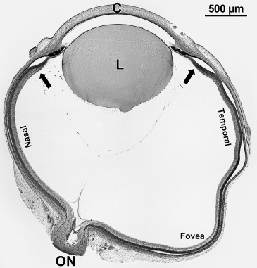
A stained section was selected from 1 well-preserved eye of infants from Fwk 8 to P 1 week; an example is shown in Figure 1 . The section contained optic nerve, fovea, and the entire retina, including both nasal and temporal far peripheral edges. The far peripheral edge of the retina is called “edge” in the text and is indicated by arrows in Figure 1 . The length of the nasal retina from optic nerve to edge; the nasal retina from optic nerve to foveal center; and the temporal retina from foveal center to edge was measured using a microscope ocular micrometer ( Table 1 ). In selected sections, the inner plexiform layer (IPL) containing bipolar cell and amacrine synapses and outer plexiform layer (OPL) containing cone and rod synapses were identified and the distance from the center of the developing fovea was measured ( Table 2 ). The amount of retina containing IPL and OPL was divided by the total length and expressed as “% coverage” for a given age ( Table 2 ). Selected regions of well-preserved retinas were imaged digitally using a Nikon E1000 wide-field digital microscope. These images were processed in Adobe Photoshop CS5 for size, color balance, sharpness, and contrast.
| Individual Eyes | Average of Age Groups | ||||||||
|---|---|---|---|---|---|---|---|---|---|
| Age Fetal Week | Nasal ON to Edge a (mm) | Nasal ON to Fovea (mm) | Temporal Fovea to Edge a (mm) | Total Length (mm) | Age Fetal Week | Nasal ON to Edge a (mm) | Nasal ON to Fovea (mm) | Temporal Fovea to Edge a (mm) | Total Length (mm) |
| 8 | 1.9 | 2.98 | 4.88 | ||||||
| 8.4 | 2.3 | 3.2 | 5.5 | 8.2 | 2.1 | 3.09 | 5.19 | ||
| 11.4 | 3.25 | 2.52 | 2.42 | 8.19 | |||||
| 11.4 | 5.05 | 3.35 | 3.24 | 11.64 | 11.4 | 2.95 | 2.66 | 3.40 | 9.02 |
| 12 | 3.70 | 2.75 | 4.45 | 10.9 | |||||
| 13 | 3.75 | 2.72 | 4.17 | 10.64 | 12.5 | 3.73 | 2.74 | 4.31 | 10.78 |
| 14 | 5.75 | 3.52 | 7.48 | 16.45 | — | — | — | — | — |
| 15 | 6.16 | 3.90 | 7.17 | 17.23 | |||||
| 15 | 6.01 | 3.24 | 6.70 | 15.95 | 15 | 5.97 | 3.55 | 7.17 | 16.64 |
| 18 | 7.18 | 3.35 | 8.95 | 19.48 | |||||
| 18 | 8.13 | 4.18 | 9.06 | 21.37 | 18 | 7.66 | 3.77 | 9.01 | 20.43 |
| 21 | 8.66 | 4.36 | 8.25 | 21.27 | |||||
| 21 | 8.26 | 4.2 | 8.1 | 20.56 | 21 | 8.46 | 4.28 | 8.18 | 20.92 |
| 25 | 11.7 | 4.13 | 11.87 | 27.7 | |||||
| 26 | 12.23 | 3.17 | 13.41 | 28.81 | 25.5 | 11.97 | 3.65 | 12.64 | 28.26 |
| 35 | 13.36 | 3.16 | 14.07 | 30.59 | |||||
| 37 | 12.54 | 3.16 | 14.74 | 30.44 | 36 | 12.95 | 3.16 | 14.41 | 30.52 |
| 40 | 14.28 | 3.10 | 15.94 | 33.32 | |||||
| 41 | 13.8 | 2.93 | 15.71 | 32.24 | 40.5 | 14.04 | 3.02 | 15.83 | 32.88 |
a Edge refers to far peripheral retinal edge ( Figure 1 , arrows).
| Age Fetal Week | Total Length Retina (mm) | Inner Plexiform Layer | Outer Plexiform Layer | ||||||||
|---|---|---|---|---|---|---|---|---|---|---|---|
| Nasal ON to Edge a (mm) | Nasal ON to Fovea (mm) | Temporal Fovea to Edge a (mm) | Length IPL (mm) | % Coverage b IPL | Nasal ON to Edge a (mm) | Nasal ON to Fovea (mm) | Temporal Fovea to Edge a (mm) | Length OPL (mm) | % Coverage b OPL | ||
| 8 | 4.88 | — | 1.24 | 0.5 | 1.74 | 36 | — | — | — | — | — |
| 8.4 | 5.5 | — | 1.11 | 0.4 | 1.51 | 28 | — | — | — | — | — |
| 11.4 | 8.19 | 0.65 | 2.3 | 0.55 | 3.5 | 43 | — | 0.4 | 0.3 | 0.7 | 9 |
| 12 | 10.64 | 0.47 | 2.25 | 0.91 | 3.63 | 34 | — | 0.31 | 0.44 | 0.75 | 7 |
| 13 | 10.9 | 1.65 | 2.75 | 3.11 | 7.51 | 69 | — | 1.55 | 1.26 | 2.81 | 26 |
| 15 | 17.23 | 4.91 | 3.9 | 6.52 | 15.33 | 89 | — | 2.4 | 2.89 | 5.29 | 31 |
| 15 | 15.95 | 5.51 | 3.24 | 5.92 | 14.67 | 92 | 5.97 | 1.95 | 1.97 | 9.88 | 62 |
| 18 | 19.48 | 7.18 | 3.35 | 7.75 | 18.28 | 94 | 1.6 | 3.11 | 3.84 | 8.55 | 44 |
| 18 | 21.37 | 6.53 | 4.18 | 8.3 | 19.41 | 89 | 0.35 | 3.27 | 4.09 | 7.71 | 36 |
| 21 | 22.56 | 8.26 | 4.2 | 8.1 | 20.56 | 91 | 0.5 | 3.1 | 3.38 | 6.98 | 31 |
| 25 | 27.7 | 10 | 4.13 | 11.67 | 25.8 | 93 | 11.5 | 4.13 | 12.41 | 27.3 | 98 |
| 26 | 28.81 | 12.23 | 3.17 | 13.41 | 28.11 | 98 | 11.4 | 3.65 | 11.2 | 26.25 | 92 |
Stay updated, free articles. Join our Telegram channel

Full access? Get Clinical Tree


