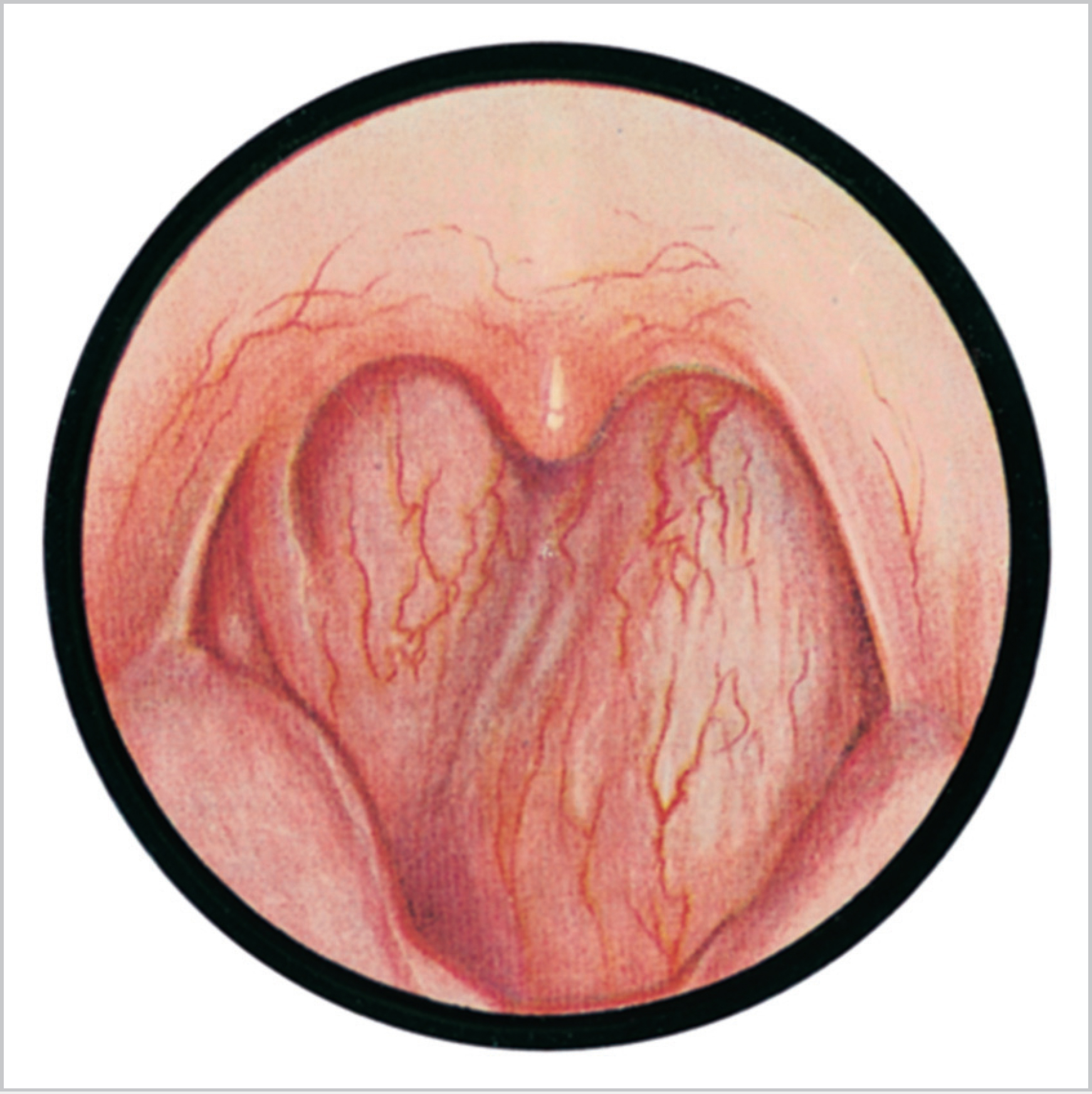
55 Deep Neck Space Infection • Retropharynx • Parapharyngeal • Anterior deep neck • Fascial layers: – Superficial: investing – Middle: visceral – Deep: prevertebral, alar • Spaces * All 3 contribute to carotid sheath • 2nd most common neck abscess in children (peritonsillar abscess/quinsy most common) • Retropharyngeal lymph nodes atrophy with age • Aetiology: • Presentation: • Management: • Ludwig angina; floor of mouth cellulitis • Anatomy Fig. 55.1 Retropharyngeal abscess. • Features • Aetiology • Management – Sublingual space only—intra-oral – Submaxillary space—external • Anatomy • Presentation • Aetiology • Treatment
55.1 Sites
55.2 Anatomy
 Fascial compartments are potential spaces between fascial planes
 Superficial and deep layers
 Deep,* divided into:
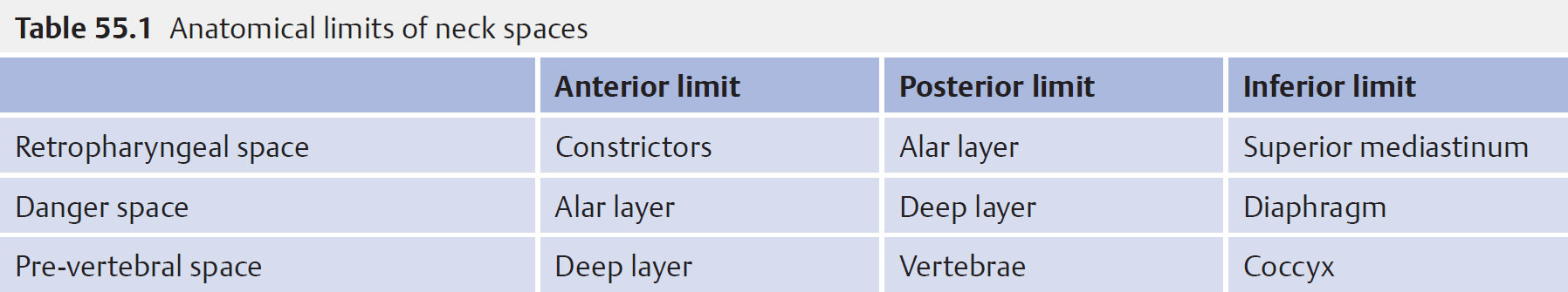
 See Table 55.1
55.3 Retropharyngeal Abscess (Fig. 55.1)
 Suppurative adenoiditis
 Trauma
 Vertebral osteomyelitis
 Pyrexia*
 Neck pain*
 Airway obstruction
 Drooling
 Torticollis
 Age of presentation is typically 6 months to 6 years (mean 3–5 years)
 Airway is the priority
 Transoral aspiration/drainage if abscess medial to great vessels unless other neck spaces involved
 Specimen pus urgent Gram stain and culture and sensitivity and abscess wall
 Intravenous antibiotics (discuss with microbiology common regimes include: clindamycin/piperacillin or penicillin/gentamicin + metronidazole) ± steroids
55.4 Anterior Deep Neck
 Submandibular space extends from hyoid to mandible divided by mylohyoid muscle
 Sublingual space superior to muscle
 Submaxillary space inferior
 Mouth pain
 Drooling
 Dysphagia
 Neck pain
 Swelling in floor of mouth
 Superior tongue displacement
 80% dental—typically lower 3rd molar
 20% soft tissue/tonsil infection
 Airway is priority consider HDU for observation
 Timely surgical intervention with appropriate IV ABx
 Surgical drainage
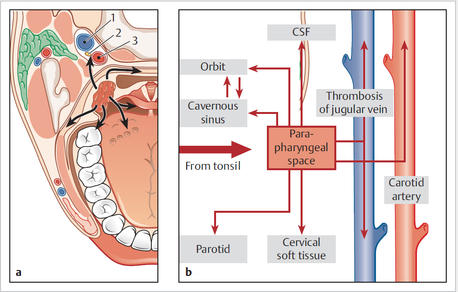
55.5 Parapharyngeal Abscess
 Parapharyngeal space 2 compartments: pre- (anterior) and post-styloid
 Communicates anterior with submaxillary space via buccopharyngeal gap created by styloglossus
 Communicates posteriorly with the retropharyngeal and danger spaces
 Airway obstruction—stridor/stertor
 Pain
Pain
 Dysphagia/dysphonia
 Trismus (20%)
 Snoring/OSA
 Neck swelling/fullness/increasing erythema
 Tonsillitis (particularly immunocompromised) (Fig. 55.2)
 Dental
 IV drug abusers
 Epiglottitis
 Parotitis
 Foreign body
 Branchial cleft cysts (if recur)
 Extension from petrous apex/mastoid tip (Citelli abscess)
 Ensure airway stable, may need tracheostomy
 May require ICU admission
 Preop CT scan for surgical planning (if airway stable), and cardiothoracic referral if mediastinum involved
 Incision guided by imaging and clinical examination
 Urgent microbiology inc. Gram stain and culture
 Multiple neck spaces may need opening, finger dissection useful at breaking down loculations and entering planes
![]()
Stay updated, free articles. Join our Telegram channel

Full access? Get Clinical Tree


