 Conjunctival Neural, Xanthomatous, Fibrous, Myxomatous, and Lipomatous Tumors
Conjunctival Neuroma and Neurofibroma
Several related soft tissue tumors appear to derive from neural, histiocytic, fibrous myxomatous, or lipomatous elements of the conjunctiva. These can have distinct features or overlap considerably in their cellular constituents. They are discussed briefly, beginning with neuroma and neurofibroma.
General Considerations
Neural tumors like simple neuroma and neurofibroma can occur in the conjunctiva (1,2,3,4,5,6,7,8,9,10,11,12,13). The best known simple neuromas are the soft mucosal neural tumors that appear in the conjunctiva and other mucous membranes in patients with multiple endocrine neoplasia type 2b (10,11,12,13). Such patients have prominent corneal nerves in 100% of cases. Because of the high association with life-threatening medullary thyroid carcinoma, ophthalmologists should be familiar with these conjunctival lesions. These benign neural tumors are generally asymptomatic and usually require no treatment. We have seen a patient with this uncommon disorder but have not been able to obtain acceptable photographs.
Neurofibroma is a peripheral nerve sheath tumor that can occur in the conjunctiva as a solitary mass or as a diffuse or plexiform variety associated with von Recklinghausen’s neurofibromatosis (NF-1; 1,2,3,4,5,6,7,8,9). Conjunctival and orbital neurofibromas can be divided into solitary, diffuse, and plexiform types (5). The solitary type is not usually associated with NF-1, the diffuse type is sometimes associated with NF-1, and the plexiform type is generally considered to be almost pathognomonic of NF-1. There was only one neurofibroma in the authors’ clinical series of 1,643 conjunctival tumors.
Clinical Features
Solitary conjunctival neurofibroma appears as a yellow-gray sessile or dome-shaped mass located in the conjunctival stroma. The sessile variant can have poorly defined margins. The plexiform variant is an ill-defined, firm, irregular mass that has been likened to a bag of worms. The conjunctival plexiform neurofibroma is often in continuity with the same lesion of the eyelid and orbit.
Pathology
Histopathologically, diffuse and plexiform neurofibromas are composed of bundles of enlarged nerves with proliferation of Schwann cells and endoneural fibroblasts in a mucoid matrix. A distinct perineural sheath defines the individual tumor cores. The localized neurofibroma lacks a perineural sheath and is encapsulated. It can sometimes be difficult to differentiate from other spindle cell tumors and special stains for axons may help make the diagnosis in such cases (4,5,6).
Management
Solitary tumors appear as slowly enlarging elevated stromal masses that can be managed by complete surgical resection. The plexiform type can be extremely difficult to remove intact and debulking procedures are often necessary. This can result in extensive scarring. The systemic prognosis is good; malignant transformation is extremely rare.
Selected References
1. Shields JA. Peripheral nerve tumors of the orbit. Neurofibroma. In: Shields JA, ed. Diagnosis and Management of Orbital Tumors. Philadelphia: WB Saunders; 1989:149-152.
2. Shields CL, Shields JA. Tumors of the conjunctiva and cornea. Surv Ophthalmol 2004;49:3-24.
3. Shields CL, Demirci H, Karatza E, et al. Clinical survey of 1643 melanocytic and nonmelanocytic tumors of the conjunctiva. Ophthalmology 2004;111: 1747-1754.
4. Brownstein S, Little JM. Ocular neurofibromatosis. Ophthalmology 1983; 91:1595-1599.
5. Krohel GB, Rosenberg PN, Wright J, et al. Localized orbital neurofibromas. Am J Ophthalmol 1985;100:458-464.
6. Kobrin JL, Blodi FC, Weingiest TA, et al. Ocular and orbital manifestations of neurofibromatosis. Surv Ophthalmol 1979;24:45-51.
7. Perry HD. Isolated episcleral neurofibroma. Ophthalmology 1982;89:1095-1098.
8. Dabezies OH Jr, Penner RJ. Neurofibroma or neurilemoma of the bulbar conjunctiva. Arch Ophthalmol 1961;66:73-75.
9. Kalina PH, Bartley GB, Campbell RJ, et al. Isolated neurofibromas of the conjunctiva. Am J Ophthalmol 1991;111:694-698.
10. Jacobs JM, Hawes MJ. From eyelid bumps to thyroid lumps: report or a MEN type IIb family and review of the literature. Ophthal Plast Reconstr Surg 2001;17:195-201.
11. Riley FC Jr, Robertson DM. Ocular histopathology in multiple endocrine neoplasia type 2b. Am J Ophthalmol 1981;91:57-64.
12. Robertson DM, Sizemore GW, Gordon H. Thickened corneal nerves as a manifestation of multiple endocrine neoplasia. Trans Sect Ophthalmol Am Acad Ophthalmol Otolaryngol 1975;79:772-787.
13. Shields JA, Shields CL, Perez N. Choroidal metastasis from medullary thyroid carcinoma in multiple endocrine neoplasia. Am J Ophthalmol 2002;134:607-609.
Conjunctival Neurofibroma
Perry HD. Isolated episcleral neurofibrom a. Ophthalm ology 1982;89:1095-1098.
 Figure 21.1. Very subtle diffuse neurofibroma of the inferior bulbar conjunctiva in a young girl with NF-1. |
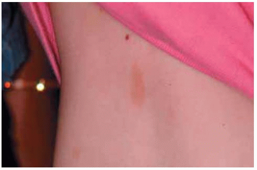 Figure 21.2. Cutaneous café-au-lait spoton patient shown in Figure 21.1. |
 Figure 21.3. Involvement of superior aspect of conjunctiva with plexiform neurofibrom a in a 4-year-girl with NF-1. (Courtesy of Frederick Blodi, MD.) |
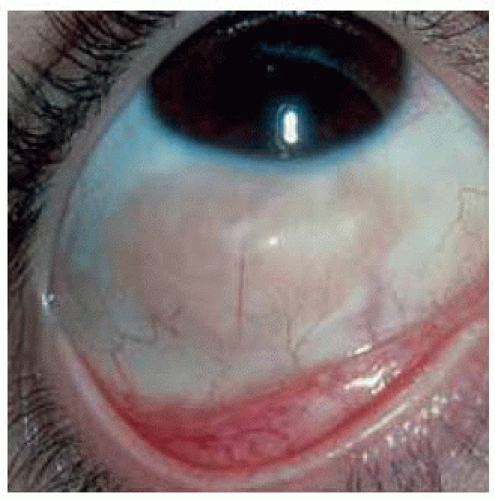 Figure 21.4. Circum scribed episcleral neurofibrom a in a 22-year-old woman. Although initially reported as being unassociated with neurofibrom atosis, the patient eventually developed NF-2 (personal communication, Henry D. Perry M D). (Courtesy of Henry Perry, M D.) |
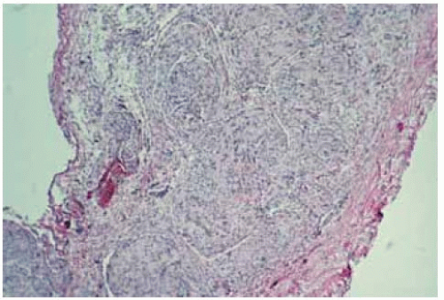 Figure 21.5. Histopathology of lesion shown in Figure 21.4, showing closely compact spindle cells. (Hematoxylin-eosin 10.) |
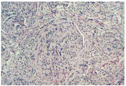 Figure 21.6. Slightly higher magnification photom icrograph of lesion shown in Figure 21.5. Note with enlarged nerve bundle with numerous spindle-shaped cells. (Hematoxylin-eosin 30.) |
Conjunctival Schwannoma and Granular Cell Tumor
Conjunctival Schwannoma
General Considerations
Clinical Features
In the conjunctiva, schwannoma presents as a light pink-yellow, elevated mass that generally lies in the stroma of the bulbar conjunctiva or episcleral tissues. It is a slow-growing lesion that may have mildly dilated conjunctival or episcleral nutrient vessels.
Pathology
Histopathologically, it is composed of a typical arrangement of spindle cells, usually forming an Antoni A or B pattern and Verocay bodies (1). Ultrastructurally, the cytoplasm of the cells contain areas of wide-spacing collagen, a typical feature of Schwann cells.
Management
The best management of conjunctival schwannoma is complete surgical resection. As for orbital schwannoma, it is important to completely excise the lesion within its capsule, because of the possibility of recurrence after incomplete excision. Malignant peripheral nerve sheath tumor (malignant schwannoma) has been known to arise in the orbit (1), but, to our knowledge, has not been reported in the conjunctiva.
Conjunctival Granular Cell Tumor
General Considerations
Clinical Features
In the conjunctiva, like in the orbit, granular cell tumor is clinically indistinguishable from most other well-circumscribed, nonpigmented conjunctival neoplasms.
Pathology
Microscopically, granular cell tumor consists of cords and lobules of round, benign cells with a pronounced granular cytoplasm. Pseudoepitheliomatous hyperplasia of the overlying conjunctival epithelium is a recognized feature of this tumor. Based on electron microscopic studies, it has been suggested that the cells may be modified Schwann cells, although the precise histogenesis of the tumor is still disputed. A malignant variation of this tumor may be indistinguishable from alveolar soft-part sarcoma. Granular cell tumor is rarely diagnosed clinically.
Management
Like other slowly progressive, circumscribed, benign tumors, the best management is complete surgical excision. The prognosis is good.
Selected References
1. Shields JA. Peripheral nerve tumors of the orbit. Neurilemoma. In: Shields JA, ed. Diagnosis and Management of Orbital Tumors. Philadelphia: WB Saunders; 1989:149-169.
2. Charles NC, Fox DM, Avendano JA, et al. Conjunctival neurilemoma. Report of 3 cases. Arch Ophthalmol 1997;115:547-549.
3. Le Marc’hadour F, Romanet JP, Fdili A, et al. Schwannoma of the bulbar conjunctiva. Arch Ophthalmol 1996;114:1258-1260.
4. Dabezies OH Jr, Penner RJ. Neurofibroma or neurilemoma of the bulbar conjunctiva. Arch Ophthalmol 1961;66:73-75.
5. Vincent NJ, Cleasby GW. Schwannoma of the bulbar conjunctiva. Arch Ophthalmol 1968;80:641-642.
6. Ferry AP. Granular cell tumor (myoblastoma) of the palpebral conjunctiva causing pseudoepitheliomatous hyperplasia of the conjunctival epithelium. Am J Ophthalmol 1981;91:234-237.
7. Charles NC, Fox DM, Glasberg SS, et al. Epibulbar granular cell tumor. Report of a case and review of the literature. Ophthalmology 1977;104:1454-1456.
Conjunctival Schwannoma and Granular Cell Tumor
Conjunctival tumors of Schwann cell origin and presumed Schwann cell origin include schwannoma (neurilemoma) and granular cell tumor, respectively.
1. Le Marc’hadour F, Romanet JP, Fdili A, et al. Schwannoma of the bulbar conjunctiva. Arch Ophthalmol 1996;114:1258-1260.
2. Charles NC, Fox DM, Glasberg SS, et al. Epibulbar granular cell tumor. Report of a case and review of the literature. Ophthalmology 1977;104:1454-1456.
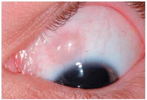 Figure 21.7. Epibulbar schwannoma in a 15-year-old boy. The lesion appeared and enlarged slowly over several weeks. |
 Figure 21.8. Histopathology of lesion shown in Figure 21.7 demonstrating mostly Antoni A pattern of schwannoma. (Hematoxylin-eosin 80.)
Stay updated, free articles. Join our Telegram channel
Full access? Get Clinical Tree
 Get Clinical Tree app for offline access

|