Ultraviolet exposure
 Chronic dryness and hot, dusty environment
 These factors lead to elastotic degeneration of the substantia propria of the conjunctiva, resulting in subepithelial proliferation of fibrovascular tissue, initially on the conjunctiva and then on the cornea.
Symptoms
 Irritation, redness nasally or temporally, tearing, occasionally decreased vision
 Occasionally, contact lens intolerance
 Rarely, pain if inflamed
Signs
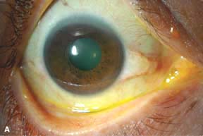
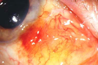
 Pinguecula: yellow-white, often triangular, slightly elevated conjunctival lesion adjacent to the nasal or temporal side of the limbus (Fig. 2-1A). They may become mildly to moderately inflamed (Fig. 2-1B).
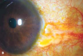
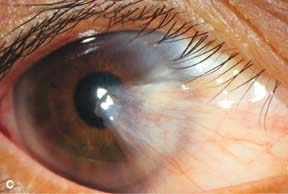
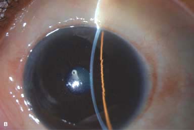
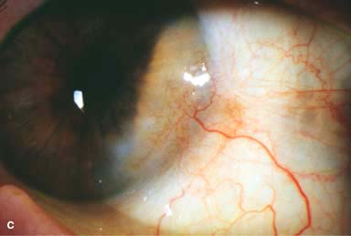
 Pterygium: triangular, wing-shaped fibrovascular sheet of tissue extending onto the cornea at the 3 and 9 o’clock positions (Fig. 2-1C). An iron line (Stocker’s line) in the corneal epithelium may be present central to the apex of the pterygium.
 An area of thinning due to desiccation (delle) may be present in the cornea adjacent to an elevated lesion.
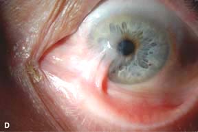
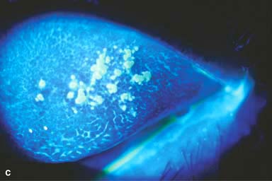
 Large or recurrent pterygia can cause symblepharon formation and even restrict ocular motility (Fig. 2-1D).
Differential Diagnosis
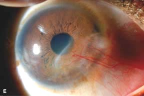
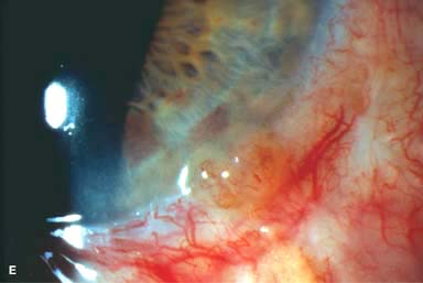
 Pseudopterygium: An adhesion of conjunctiva onto the corneal surface after corneal injury (Fig. 2-1E). Unlike a true pterygium, the adhesion is only at the apex and not throughout the underlying surface. It is typically unilateral and often not at the 3 and 9 o’clock positions.
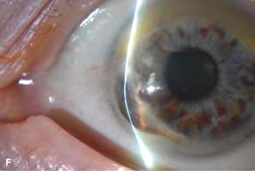
 Fuchs’ marginal keratitis: Associated with mild to severe peripheral corneal thinning (Fig. 2-1F).
 Conjunctival papilloma, nevus, intraepithelial neoplasia, or squamous cell carcinoma: If not typical for a pterygium or pinguecula, consider a conjunctival biopsy.
Diagnostic Evaluation
 Slit-lamp examination to look for unusual features suspicious of other diagnoses. Pingueculae and pterygia have classic appearances.
 Excisional biopsy in cases suspicious for malignancy
Treatment
 Avoid excessive sunlight exposure and wear good-quality ultraviolet-blocking sunglasses.
 Artificial tears to prevent dry eyes
 Topical antihistamines (e.g., emedastine, levocabastine, naphazoline, antazoline), nonsteroidal anti-inflammatory agents (e.g., ketorolac, bromfenac, nepafenac), or, rarely, corticosteroids (e.g., loteprednol 0.2%, fluorometholone 0.1%) q.d. to q.i.d. to reduce redness or inflammation.
 Surgical excision is indicated if there is excessive irritation, difficulty with contact lens wear, for cosmetic reasons, or when there is progression toward the visual axis. The recurrence rate is much lower when excision is combined with a conjunctival autograft. If inadequate conjunctival tissue is available, an amniotic membrane graft can be used to cover the bare sclera.
 Intraoperative application of mitomycin C and postoperative use of beta radiation may decrease the recurrence rate, but both are associated with an increased risk of corneoscleral necrosis and are usually not necessary when a conjunctival autograft is performed.
Prognosis
 Good to very good, depending on severity
 Pterygia can recur in about 10% to 15% of patients, occasionally worse than the original pterygium.
Figure 2-1. Pinguecula. A. A triangular, creamy-white elevated conjunctival mass is seen at the nasal limbus from the 3 to the 4:30 clock position. B. This pinguecula, also located between the 3 to 4:30 positions, is slightly inflamed, as demonstrated by mild surrounding conjunctival injection. Pterygium. C. A nasal wing-shaped fibrovascular growth is apparent in this patient’s right eye with a classic nasal pterygium. This pterygium is reaching into the visual axis. Pterygium. D. This large recurrent nasal pterygium has resulted in significant symblepharon formation and restriction of ocular motility. Pseudopterygium. E. This large nasal pseudopterygium developed as part of the healing process after an episode of peripheral ulcerative keratitis. Some residual peripheral scarring is seen inferotemporally. Fuchs’ marginal keratitis. F. This eye has a history of unilateral chronic nasal peripheral corneal inflammation, thinning, and neovascularization due to Fuchs’ marginal keratitis. Six years prior, it had developed a small Seidel-positive leak, which resolved with medical treatment. The eye has remained quiet on low-dose topical steroids. Note the severe corneal thinning inferonasally.
OTHER CONJUNCTIVAL DEGENERATIONS
AMYLOIDOSIS
 Amyloidosis is a degenerative condition in which the noncollagenous protein amyloid is deposited in the conjunctiva (Fig. 2-2AandB).
 It may be primary or secondary.
 It may be localized to the conjunctiva or be related to a systemic disorder such as amyloidosis, plasma cell dyscrasias, or, rarely, lymphoma.
 Primary localized amyloidosis is the most common form. Primary systemic amyloidosis involves amyloid deposition throughout the eye and eyelids and can affect the heart and kidneys.
 Rule out systemic amyloid conditions.
CALCIUM CONCRETIONS
 Calcium concretions are yellow-white calcium deposits that are embedded in the upper and/or lower palpebral conjunctiva.
 Generally they are located below the surface of the conjunctiva and do not cause any symptoms. Occasionally, the concretions erode through the surface of the conjunctiva, stain with fluorescein dye, and cause foreign-body symptoms (Fig. 2-2C).
 If they are mild, the symptoms can be treated with topical lubrication; if they are severe, the concretions can be removed, but they often recur.
Figure 2-2. Conjunctival amyloidosis. A. An elevated yellow-colored conjunctival mass is noted in this elderly patient. It was mobile over the sclera, and it did not have the classic appearance of a pterygium or the papillomatous pattern of a squamous cell tumor. Conjunctival biopsy revealed amyloidosis. There is often associated subconjunctival hemorrhage, as seen in this case. Conjunctival amyloidosis. B. A creamy white, rubbery conjunctival lesion covered the superior and nasal bulbar conjunctiva of this left eye. Conjunctival biopsy revealed amyloidosis. Conjunctival calcium concretions. C. Multiple white calcium concretions have eroded through the conjunctival epithelial surface of the upper eyelid, resulting in fluorescein staining, seen upon eyelid eversion. These exposed concretions cause a chronic foreign-body sensation and require removal.
MELANOCYTIC CONJUNCTIVAL LESIONS
CONJUNCTIVAL EPITHELIAL MELANOSIS (RACIAL MELANOSIS)
 Common in pigmented races, usually bilateral, but may have asymmetric ocular involvement
 Becomes more pronounced during puberty.
 Flat, patchy, brownish pigmentation scattered over the conjunctiva, but frequently involves the perilimbal regions (Fig. 2-3A).
 Mobile over the sclera. May be perforated by anterior ciliary arteries or nerves.
 No malignant potential.
OCULODERMAL MELANOSIS (NEVUS OF OTA)
 A congenital condition characterized by blue-gray hyperpigmentation of skin and mucous membranes in the distribution of the fifth cranial nerve
 Almost always unilateral.
 Three variants are seen: dermal, ocular, and oculodermal melanoses.
 Involves the dermis of the skin and episclera of the eye, thus the lesion does not move over the sclera.
 May affect ipsilateral uveal tissues, orbit, and central nervous system.
 Malignant transformation, uveal melanoma, and glaucoma can develop, and patients should be followed up regularly.
NEVUS
 Develops during puberty or early adulthood.
 Most are subepithelial or compound nevi.
 Appears as a well-demarcated, flat or slightly elevated lesion, usually in the interpalpebral areas. It is usually solitary, and has a predilection for the limbus, plica, caruncle, and eyelid margin. Cystic spaces within the nevus are common and are the key to diagnosis. The degree of pigmentation may vary and may increase at puberty (Fig. 2-3B).
 Enlargement can occur but may be a sign of malignant transformation. Nevi involving the cornea, tarsal, or forniceal conjunctivae are extremely rare and should be excised for histopathologic evaluation.
 Periodic photographic documentation of the lesion may be helpful for follow-up.
PRIMARY ACQUIRED MELANOSIS
 This is an uncommon, unilateral, premalignant condition that is usually seen in middle-aged to elderly white patients.
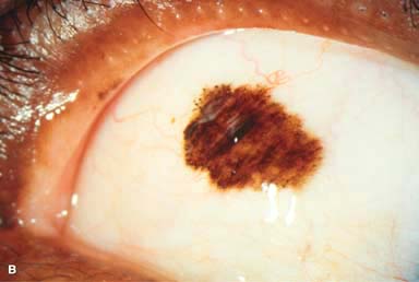
 Unifocal or multifocal flat patches with indistinct margins that may involve any part of the conjunctiva. Cystic spaces are absent (Fig. 2-3C).
 Follow-up with clinical documentation (e.g., slit-lamp photography) should be performed every 6 months. Malignant change should be suspected if the patches become nodular.
 Local wide excision with cryotherapy is often performed for suspicious lesions. Postoperative topical chemotherapy (e.g., with mitomycin C) may be beneficial. Incomplete excision and/or recurrence is common, requiring more aggressive treatment (e.g., local radiation therapy or topical chemotherapy). Topical mitomycin C has also been used successfully to treat primary acquired melanosis without wide excision.
SECONDARY ACQUIRED MELANOSIS
 Causes include:
 Adrenochrome deposits: discrete clumps of melanin on tarsal and forniceal conjunctiva associated with long-term use of topical epinephrine—becoming rare
 Alkaptonuria: interpalpebral, bluish-gray or black pigmentation of the conjunctiva, episclera, sclera, and tendons of horizontal rectus muscles due to accumulation of homogentisic acid
 Mascara deposits
 Age-related
 Addison disease
 Hemochromatosis
 Argyrosis: As a result of long-term use of drops containing silver, becoming rare
 Dark foreign bodies
MALIGNANT MELANOMA
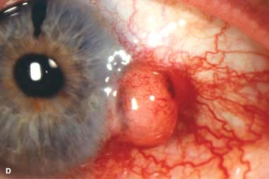
 Malignant melanoma is an uncommon, malignant tumor that may be pigmented or nonpigmented. It may arise de novo, from preexisting primary acquired melanosis, or from a nevus.
 Elevated nodule that can affect any part of the conjunctiva, but has a predilection for the limbus and may extend onto the cornea. Feeder vessels may be seen (Fig. 2-3D and E). Advanced melanomas may invade the eyelids and orbit.
 Treatment is local excision with cryotherapy. Local radiation therapy may also be beneficial. Exenteration may be necessary for orbital involvement. Use palliation with chemotherapy if metastasis is present (lymph nodes, central nervous system, liver, etc.).
Figure 2-3. Conjunctival epithelial (racial) melanosis. A. An area of poorly demarcated conjunctival epithelial melanin pigment is seen in this African American patient. These lesions have minimal to no malignant potential. Conjunctival nevus.B. A pigmented patch can be seen in the superior conjunctiva of this African American woman. It is well demarcated, has not changed in size, and has numerous microcysts, all pointing to the diagnosis of a nevus. Primary acquired melanosis. C. An area of flat conjunctival pigmentation is seen at the limbus from the 3 to 5 o’clock positions. There is a mild increase in vascularization. This lesion is suspicious for malignant transformation. Malignant melanoma of the conjunctiva.D. Biopsy of this large, solid conjunctival mass revealed malignant melanoma. It is relatively amelanotic, but pigmented areas can be seen at the 3 and 9 o’clock aspects of the lesion. There is also significant surrounding vascularization, indicating an aggressive process. E. A small recurrent conjunctival malignant melanoma is seen at the limbus at the 5 o’clock position after surgical excision. It was reexcised and treated with a radioactive plaque.

Stay updated, free articles. Join our Telegram channel

Full access? Get Clinical Tree


