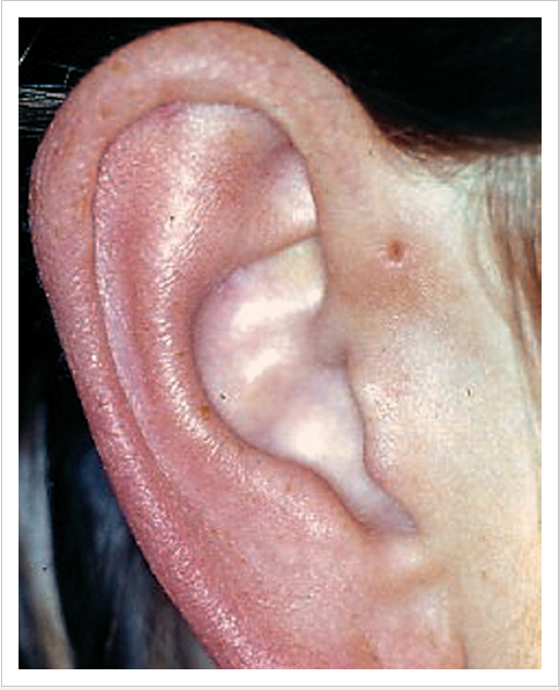
62 Congenital Malformations of the Ear • Common in those with SNHL; may involve membranous, bony, or both parts of labyrinth (former probably most common but do not show on imaging) • Various classification systems—Jackler et al (1987) based on their occurrence in embryogenesis; most likely to arise between 4–8/40 • Michel aplasia: arrest of inner ear development before 4/40—complete aplasia of all inner ear structures; associated with thalidomide exposure, anencephaly, Klippel–Feil syndrome; total SNHL, not candidate for CI • Common cavity: failure at 4/40, with membranous labyrinth poorly differentiated in a large common cavity; severe to profound HL; can try CI (risk CSF gusher) • Cochlear aplasia: failure at 5/40; rare; normal vestibular development; profound SNHL, unlikely candidate for CI • Cochlear hypoplasia: failure at 6/40 with variable HL • Mondini dysplasia; failure at 7/40, with incomplete partition of cochlea so only basal turn cochlea present (most common cochlear malformation seen on imaging) with variable HL (some high-tone residual hearing possible); may be associated with widened vestibular aqueduct, some associated with stapes footplate anomalies, Waardenburg, DiGeorge, Pendred syndromes • Widened vestibular aqueduct: definition varies, e.g., ≥1.5 mm; may be associated with Pendred and branchio-otorenal syndrome, cochlear dysplasias, or stapes fixation; often normal hearing at birth with progressive SNHL, sometimes sudden after mild HI; avoid HI/pressure changes (flights, diving); 30% have vestibular disturbance; autosomal recessive • Aplasia or dysplasia of lateral Scc most common (because develops later than other Sccs); may be associated with CHARGE (coloboma, heart defects, atresia of nasal choanae, retardation of growth, genital and/or urinary abnormalities, ear abnormalities and deafness) syndrome (which also has stenotic cochlear aperture, underdeveloped vestibule, incomplete partition of cochlea) • Occur in isolation or with bony labyrinthine anomalies (because do not show on imaging, only determined histologically) • Numerous ossicular abnormalities; malleus head fixation most common (due to incomplete pneumatization of epitympanum) • Persistent stapedial artery: 2nd arch remnant, should regress at 10/40; if it does not, passes over stapes footplate; may be associated with aberrant course of ICA • High jugular bulb • Facial nerve: aberrant course most likely in presence of other congenital abnormalities • Microtia – 1st degree dysplasia: most structures of normal auricle recognizable – 2nd degree: some recognizable structures but additional skin/cartilage required for reconstruction – 3rd degree: no normal structures seen so total reconstruction required • Pre-auricular tags and accessory auricles • Pre-auricular pits and sinuses (Fig. 62.1) • Prominent ears – Marking of desired site for antihelical fold with methylene blue dye – Cartilage weakening, e.g., by anterior scoring (Converse technique); as lateral surface of auricle involved in procedure, risk of poor cosmesis – Stay sutures techniques (e.g., Mustardé) – Diamond burr drilling of groove on medial surface – Conchomastoid sutures and cartilage excision may help reduce deep conchal bowl appearance – Can also excise medial part of conchal bowl ± soft tissue over mastoid • Canal atresia – I: TM present – II: atretic plate, normal tympanic cavity – III: hypoplastic tympanic cavity
62.1 Inner Ear
62.1.1 Cochlear Anomalies
62.1.2 Vestibular Anomalies
62.1.3 Membranous Anomalies
 Bing–Siebenmann malformation: isolated membranous malformation within well-formed bony capsule; profound SNHL; associated with Usher and Jervell–Lange-Nielsen syndromes
 Scheibe malformation: most common membranous inner ear malformation—malformed organ of Corti and saccule; severe to profound SNHL; associated with Usher, Jervell–Lange-Nielsen, Refsum, Waardenburg, trisomy 18; autosomal recessive inheritance
 Alexander malformation: least severe—dysplastic basal turn of cochlea; high-frequency SNHL
62.2 Middle Ear
62.3 External Ear
 1:10,000, M>F, R>L-hand side; 4:1 unilateral: bilateral
 Association with other congenital abnormalities in ~50% cases (e.g., Treacher–Collins, Goldenhar, hemifacial microsomia) and teratogens (e.g., thalidomide)
 Weerda classification:
 Rx: do nothing; minor malformations can be excised/repaired with flaps; consider autogenous reconstruction (e.g., using rib cartilage) or prosthesis; bone conduction hearing aid
 Association with canal atresia (especially 3rd degree cases)
 Usually anterior to tragus
 If cartilage present = accessory auricle
 Often bilateral; may be part of a syndrome (e.g., Goldenhar)
 Beware excision in children as VII n may be involved in root of accessory auricle
 Related to incomplete fusion of the hillocks of His and 1st branchial arch formation
 Anterior to anterior crus of helix
 Pits may extend down to cartilage; sinuses extend to tympanic ring; possible relationship to facial nerve
 Autosomal inheritance with incomplete penetrance
 Usually asymptomatic but recurrent infection possible
 Complete excision usually requires removal of a small oval of helical cartilage at base of tract
 Pinna stops growing by ~5 years of age and children become “body aware” by ~7 years
 5% population have protruding ears
 Usually a failure of folding of the antihelix or hyperplasia of the conchal bowl
 Normal auriculotemporal angle ~30°; >40° is protruding
 In newborns may be corrected by taping
 Surgical repair: various techniques used to create an antihelical fold
 Altman classification:
 Jahrsdorfer grading system assesses CT findings to determine suitability for surgery
 BAHA often best option rather than canal reconstruction
![]()
Stay updated, free articles. Join our Telegram channel

Full access? Get Clinical Tree


