Complications of Rhinosinusitis


Complications of Rhinosinusitis


Carla M. Giannoni


Acute bacterial sinusitis occurs commonly, usually as a sequela of an upper respiratory infection. Symptoms include cough, purulent nasal discharge, headache, facial pain and pressure, nasal congestion, fetid breath, fever, malaise, and lethargy. Spread of infection outside the sinuses results in complicated sinusitis. The incidence of complications from both acute and chronic sinusitis has decreased fourfold since the 1950s but appear to have stabilized in the past several decades (1,2,3). This has been attributed to the widespread use of antibiotics for rhinosinusitis but clinical data are lacking in this area (see “Prevention of Complications for Sinusitis” later in this chapter for further discussion). Complications of sinusitis can be divided into three categories: orbital, intracranial, and bony. This chapter reviews each of these complications in detail, describing the pathophysiology, evaluation, microbiology, and medical and surgical treatments.


PRESEPTAL CELLULITIS

Preseptal infections are far more common than postseptal infections. Preseptal cellulitis can be a complication of ethmoid sinusitis but it can also occur as a result of infection of the eyelids and orbital adnexa, trauma (including insect

bites), or a foreign body (6,7). In contrast to other orbital complications, medical comorbidities such as cystic fibrosis, HIV, leukemia, diabetes, and other immune deficiencies are risk factors for preseptal cellulitis (7). Preseptal cellulitis manifests as eyelid swelling, erythema, and tenderness. Occasionally these may progress to an eyelid abscess (Fig. 38.2) and can also be associated with edema of the orbital (postseptal) contents. There are no limitations of extraocular movements and no impairment of visual acuity. Sinonasal infections cause periorbital swelling due to impaired venous drainage of the ethmoidal vessels that are obstructed by inflammation and pressure.


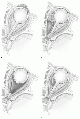
Figure 38.1 Chandler classification of orbital complications of sinusitis. A: Preseptal cellulitis (eyelid edema), (B) Subperiosteal abscess, (C) Orbital cellulitis, (D) Orbital abscess,


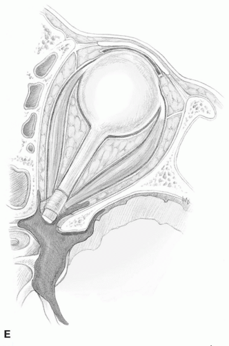
Figure 38.1 (Continued) (E) Cavernous sinus thrombosis.


TABLE 38.1 ORBITAL COMPLICATIONS OF SINUSITIS


Typical Findings


Treatment


Preseptal cellulitis


Edematous, erythematous eyelids


Extraocular muscles (EOM) intact


Normal vision


Medical therapy (Rarely, drainage of secondary abscess)


Orbital cellulitis


More diffuse orbital edema ± Impaired EOM


Usually normal vision until later in disease course


Medical therapy


± Sinus drainagea


Subperiosteal abscess


Edematous, erythematous eyelids; Proptosis


Impaired EOM


Usually normal vision, esp in the case of small abscesses


Visual changes more likely with larger abscesses


Medical therapy


± Sinus drainage


± Abscess drainage


Orbital abscess


Severe exophthalmos, chemosis,


Ophthalmoplegia, common


Visual impairment, common


Medical therapy


Sinus drainage, often


Abscess drainage, usually


Cavernous sinus thrombophlebitis


Bilateral orbital pain, chemosis, proptosis


Ophthalmoplegia


CN III, IV, V1, V2, V3, VI can be affected


Medical therapy


Sinus drainage, often


± Anticoagulation (controversial)


a Surgical sinus drainage may be limited to maxillary sinus aspiration or may include endoscopic or open sinus surgery; its necessity depends on severity of symptoms, physical examination, duration of medial treatment and need for cultures to direct antibiotic therapy.


SUBPERIOSTEAL ABSCESS

With subperiosteal abscess, a collection of pus forms at the medial aspect of the orbit between the periorbita and the lamina papyracea (Fig. 38.3). This is the second most common orbital complication of sinusitis. In response to inflammation in the ethmoid sinus, an inflammatory tissue collection beneath the orbital periosteum can develop and become a subperiosteal phlegmon (9). This subperiosteal phlegmon can then progress into a discrete abscess. A subperiosteal phlegmon or abscess can displace the orbital contents and globe downward and laterally with normal mobility in the early stages. An abscess may occasionally rupture through the orbital septum and present in the eyelids.

Typically, younger children develop isolated orbital complications, especially medial subperiosteal abscesses associated with acute ethmoiditis. Interestingly, a second, notable subset of patients has been identified: teenage males, who develop simultaneous orbital and intracranial complications (10,11,12,13,14). This phenomenon is likely related to the agedependent development of the frontal and sphenoid sinuses.


ORBITAL CELLULITIS

Orbital cellulitis is a postseptal infection that manifests as diffuse edema of the orbital contents without a discrete abscess (Fig. 38.4). There is eyelid edema and erythema, proptosis, and chemosis with limited or no impairment of extraocular movements and normal visual acuity early in the disease process. Visual changes and ophthalmoplegia
indicating optic neuritis and/or ischemia can occur as the disease progresses; these are prognostically worrisome findings. Not all cases of orbital cellulitis are due to sinusitis, however. Patients presenting with pain and diplopia and a history of recent orbital trauma or dental surgery should be assessed for orbital cellulitis. In diabetic patients with ketoacidosis and immunocompromised patients, more severe infections such as invasive fungal sinusitis should be considered. Orbital cellulitis is more concerning than preseptal cellulitis because it can evolve into an orbital abscess.


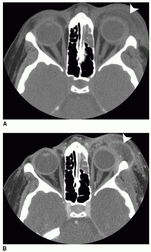
Figure 38.2 Preseptal abscess secondary to preseptal cellulitis. A: Axial CT scan taken on admission showing the patient with left ethmoid sinusitis and left preseptal cellulitis (arrow); patient also had frontal sinusitis (not pictured). B: Axial CT scan after 4 days of IV antibiotic treatment showing progression of infection to left preseptal abscess (arrow). The patient also developed an orbital subperiosteal abscess, scalp abscess, and small epidural abscess.


ORBITAL ABSCESS

An orbital abscess occurs when orbital cellulitis coalesces into a discrete collection of pus within the orbital tissues. This is a serious complication that can be associated with severe exophthalmos and chemosis, complete ophthalmoplegia and visual impairment with a risk for progression to irreversible blindness. On rare occasions there is spontaneous drainage of purulent material through the eyelid.


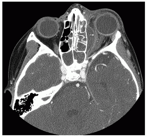
Figure 38.3 Axial CT scan demonstrating a left subperiosteal orbital abscess. The abscess is immediately adjacent to the lamina papyracea (the arrow is inside the abscess and identifies the lateral edge of the abscess); note the proptosis and the displacement of the orbital periosteum and medial rectus muscle.


CAVERNOUS SINUS THROMBOSIS

Cavernous sinus thrombophlebitis or thrombosis is a complication that can be considered an orbital as well as an intracranial complication of sinusitis. Venous congestion
in the orbit results in orbital pain, chemosis, proptosis, and ophthalmoplegia. Cranial nerves III, IV, V1, V2, V3, and VI traverse the sinus and can all be affected. Extension of the phlebitis posteriorly into the cavernous sinus results in progression of symptoms in the opposite eye. This contralateral involvement is a distinguishing feature of cavernous sinus thrombosis. It can be associated with sepsis and meningismus or frank meningitis may be present.


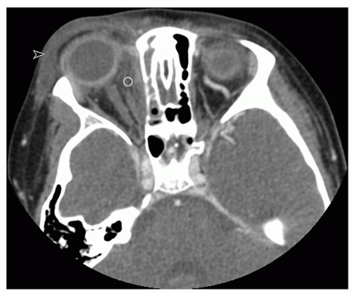
Figure 38.4 Axial CT scan showing right orbital cellulitis with diffuse orbital inflammatory changes that are intra- and extraconal (open circle); there is concurrent preseptal edema and inflammatory changes (arrow).