and Yi Ning J. Strube2
(1)
Wright Foundation for Pediatric Ophthalmology and Adult Strabismus Medical Center, Los Angeles, CA, USA
(2)
Queen’s University, Kingston, Ontario, Canada
Keywords
Duane’s syndromeSynergistic divergenceCongenital fibrosis syndromeDVDDHDThyroid strabismusBrown’s syndromeMonocular elevation deficit syndromeOrbital floor fractureHigh myopia and esotropiaHeavy eye syndrome7.1 Duane’s Syndrome
The pathophysiology is congenital agenesis of the sixth nerve nucleus and innervational misdirection of part of the medial rectus nerve to the lateral rectus muscle. The result is limited abduction and co-contraction of the medial and lateral recti on adduction. The co-contraction causes the globe to retract, producing lid fissure narrowing on adduction. If the medial and lateral recti receive the same reciprocal innervation, the eye will rest in primary position (equal). The result is a stand-off with equal medial and lateral forces, so there is limited abduction and adduction or Duane’s type 3 syndrome. In cases where the lateral rectus muscle receives more innervation, an exotropia Duane’s type 3 syndrome occurs. In most cases, the medial rectus muscle gets most of the medial rectus nerve, so the resting eye position is in adduction. This is esotropia Duane’s syndrome type 1 (limited abduction, intact adduction). Duane’s syndrome type 2, characterized as good abduction with poor adduction, is very rare (if it exists at all), and most cases are actually Duane’s type 3. Synergistic divergence is a rare type of Duane’s syndrome with abduction on attempted adduction. The lateral rectus muscle receives almost all the innervation from the medial rectus nerve, and there is agenesis of the sixth nerve nucleus [1]. Duane’s syndrome is usually sporadic without systemic manifestations, but it can be familial or associated with systemic disease (Goldenhar syndrome, Klippel-Feil syndrome, and intrauterine thalidomide exposure).
7.1.1 Duane’s Syndrome Type 1 (Esotropia)
7.1.1.1 Clinical Features
Ipsilateral face turn
Esotropia in primary position
Abduction deficit
Lid fissure narrowing in adduction, widening in abduction
7.1.1.2 Treatment
The treatment of Duane’s syndrome differs from a sixth nerve palsy because in Duane’s syndrome the lateral rectus muscle is innervated, albeit by the medial rectus nerve. Thus there is tonic lateral rectus muscle tone in Duane’s syndrome, but not in sixth nerve palsy. The presence of lateral rectus tone is why a simple medial rectus recession works well to hold the eye in primary position in patients with esotropia type 1 Duane’s syndrome. Virtually all patients with esotropia type 1 Duane’s syndrome will have a tight medial rectus muscle. Thus recession of the medial rectus muscle is of prime importance. The indication for surgery in esotropia Duane’s syndrome type 1 is a significant face turn. Figure 7.1 shows preoperative and postoperative photographs of a typical case of esotropia type 1 Duane’s syndrome.
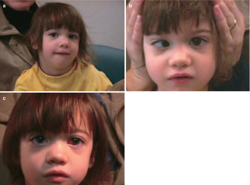
Fig. 7.1
Left esotropia Duane’s syndrome type 1 with face turn to the left, eyes right to obtain binocular fusion. The left lid fissure is narrow when in adduction (a), and lid fissure widening (b) occurs on attempted abduction (poor abduction of left eye). (c) Postoperative photograph (left medial rectus recession 6.0 mm) shows no face turn and orthotropia in the primary position
7.1.1.3 Surgery for Type 1 Esotropia Duane’s Syndrome (Authors’ Choice)
The procedure is recession of the ipsilateral medial rectus muscle:
Moderate face turn (15°–20°): 5.5–6.5 mm
Large face turn (25°–40°): 6.5–7.5 mm
For a residual esotropia and face turn after previous medial rectus recession, consider a re-recession ipsilateral MR of 3–4 mm. A residual esotropia usually persists if the previous MR recession was less than 6 mm, so the surgeon can re-recess the ipsilateral MR an additional 3 mm.
7.1.1.4 Other Treatment Options
Some suggest using the transposition surgery as a primary procedure [2]. Disadvantages of transposition surgery for Duane’s syndrome include induced hypertropia (10–15 %), a high rate of undercorrection requiring a second operation, disruption of anterior ciliary circulation, and significantly more scarring than a simple medial rectus recession. The proposed advantage is improved abduction, but this has not been our experience.
Bilateral medial rectus recessions have been advocated to stimulate the Duane’s eye to abduct through Hering’s law of yoke muscles. (The medial and lateral recti are yoke muscles.) Recessing the contralateral medial rectus muscle does not improve abduction, however [3]. In Duane’s syndrome, the lateral rectus muscle is not innervated by the sixth nerve, so Hering’s law of yoke muscles does not apply.
7.1.2 Duane’s Syndrome Type 2
Duane’s syndrome type 2 is identified as good abduction with poor adduction. These cases are very rare (if they exist at all), and most cases are actually Duane’s type 3. (See Duane’s type 3 for treatment.)
7.1.3 Duane’s Syndrome Type 3 (Exotropia)
7.1.3.1 Clinical Features
Contralateral face turn
Exotropia in primary position
Abduction and adduction deficit (eye does not move much)
Lid fissure narrowing in adduction, widening in abduction
Upshoot and downshoot are frequent
7.1.3.2 Treatment
For exotropia Duane’s type 3 and contralateral face turn, perform an ipsilateral lateral rectus recession.
For Duane’s type 3 without a face turn (orthotropia in primary position), but severe co-contraction and lid fissure narrowing, perform an ipsilateral medial rectus recession and ipsilateral lateral rectus recession, recessing the muscles about the same amount.
If an upshoot and downshoot are present, add a Y-split procedure of the lateral rectus muscle. (See Chap. 15.)
7.1.4 Duane’s Syndrome with Upshoot and Downshoot
7.1.4.1 Clinical Features
Upshoot and downshoot on attempted adduction
Usually Duane’s type 3 with abduction and adduction deficit
Lid fissure narrowing on adduction, widening on abduction
These patients have severe co-contraction and globe retraction, usually with marked lid fissure narrowing on attempted adduction. The upshoot or downshoot of the Duane’s eye may be due to co-contraction of the lateral rectus muscle with slippage above and below the globe, or it may be due to aberrant innervation of the vertical muscles with part of the medial rectus nerve.
7.1.4.2 Treatment
For Duane’s type 3 with upshoot or downshoot without a face turn (orthotropia in primary position), perform an ipsilateral medial rectus recession and ipsilateral lateral rectus recession with a Y-split of the recessed lateral rectus. (See Chap. 15 for Y-split surgery.) Note that recession of the medial and lateral rectus muscles reduces the co-contraction, and the Y-split of the lateral rectus muscle reduces the upshoot and downshoot [4].
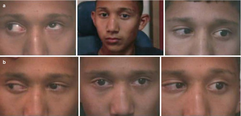
Fig. 7.2
(a) Left exotropia Duane’s type 3 syndrome with poor adduction and poor abduction. The center photograph shows a compensatory face turn to the right, eyes left. The left eye does not move horizontally, as it is fixed in abduction. There is an upshoot of the left eye on attempted adduction. (b) Postoperative photographs (left lateral rectus recession 4 mm with Y-split). Note the improved adduction and head position, and the absence of upshoot
7.1.5 Synergistic Divergence
Synergistic divergence is characterized by abrupt abduction on attempted adduction—so both the eyes abduct on gaze away from the Duane’s eye. Treatment consists of reducing the inappropriate abduction by eliminating ipsilateral lateral rectus function and tightening the medial rectus muscle with a large resection. Lateral rectus function can be greatly reduced by total tenotomy or by suturing the lateral rectus muscle to the lateral orbital wall. Simultaneous removal of the inferior and superior oblique muscles has been suggested to further reduce abduction forces [5].
7.2 Congenital Fibrosis Syndrome
Congenital fibrosis syndromes (CFS) are associated with tight, fibrotic muscles, with the medial and inferior rectus most often involved. CFS is usually inherited as an autosomal dominant trait. There has been evidence that maldevelopment of the oculomotor (III), trochlear (IV), and abducens (VI) nuclei is associated with congenital fibrosis [6]. In rare cases, there may be hypoplasia or aplasia of the inferior rectus muscles associated with muscle fibrosis without craniofacial dysostosis syndromes or neurofibromatosis [7].
7.2.1 Treatment
Treatment consists of recessing the tight muscles to improve ductions. If both inferior rectus muscles are tight and there is a chin elevation, recess both inferior recti. If there is a hypotropia, perform asymmetric inferior rectus muscle recessions with a larger recession on the hypotropic side [8]. Esotropia can usually be treated by bilateral medial rectus recessions.
7.3 A-Patterns, V-Patterns, and Oblique Overaction
7.4 Dissociated Strabismus Complex (DVD and DHD)
Dissociated strabismus complex occurs in approximately 40 % of patients with infantile esotropia, usually presenting after 2 years of age. Dissociated strabismus can, however, occur primarily or secondarily with any disorder that disrupts normal binocular visual development. Two types have been described: dissociated vertical deviation (DVD) and dissociated horizontal deviation (DHD), with DVD being the most commonly recognized form. Dissociated strabismus has three components: elevation, abduction, and extorsion. The vertical component is predominant with DVD, and the horizontal component is predominant with DHD. Dissociated strabismus is typically latent and is manifest when one eye is occluded for several seconds. It can also become manifest spontaneously, often occurring when the patient is fatigued or is daydreaming.
DVD is characterized by a slow drift of one eye up and out with slight extorsion. It is almost always bilateral but is often asymmetric. DVD can be distinguished from a true hypertropia by the lack of a corresponding hypotropia in the contralateral eye when the ipsilateral hypertropic eye returns to primary position (Fig. 7.3).
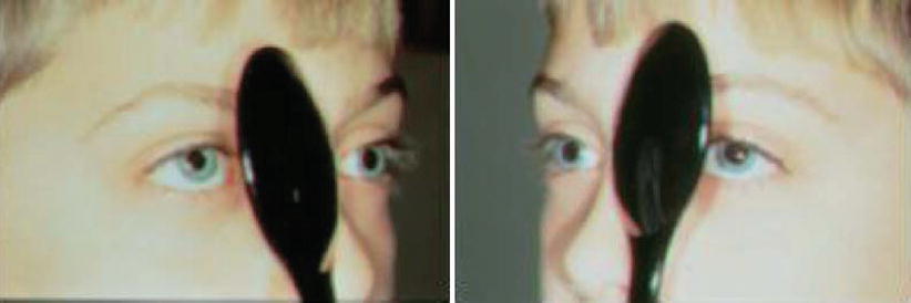
Fig. 7.3
Bilateral DVD, with a right hypertropia with the right eye covered and a left hypertropia with the left eye covered
The appearance of DHD resembles consecutive exotropia, with an eye that will intermittently drift outward, especially when the child is tired or daydreaming. In unilateral cases, covering the DHD eye induces an exodrift, but there is no drift when the fellow eye is covered. DHD can be unilateral, bilateral, or asymmetric. In cases where there is a small residual esotropia plus a unilateral DHD, alternate cover testing will disclose an exo-shift (inward movement) from the DHD eye, while the fellow eye will show an eso-shift (outward movement). If the DHD is larger than 10–15 PD, it can be symptomatic and may need surgery.
7.4.1 Treatment
Surgery is indicated if the deviation is obvious and bothersome to the patient or parents, or if the deviation is increasing in frequency. The indication to operate is subjective.
DHD: Ipsilateral lateral rectus recession (usually 4–6 mm).
DVD: Ipsilateral large superior rectus recession (between 5 and 8 mm) using a fixed suture technique. Most cases require bilateral surgery; if the DVD is asymmetric, then perform asymmetric superior rectus recessions. Patients with amblyopia of two lines or more will not fixate with the amblyopic eye, and unilateral surgery (a superior rectus muscle recession of the amblyopic eye) is indicated for these patients.
DVD plus inferior oblique overaction: DVD and inferior oblique overaction often coexist. In these cases, an inferior oblique anteriorization procedure is indicated, as it will correct both the inferior oblique overaction and the DVD. Avoid combining inferior oblique anteriorization and superior rectus recession, as this will significantly limit up gaze. Severe DVD and mild inferior oblique overaction can be treated with a small inferior oblique recession and a superior rectus recession, but an inferior oblique anteriorization will be sufficient for most patients.
7.5 Thyroid Strabismus
Strabismus associated with thyroid eye disease occurs as inflamed muscles change to become fibrotic and stiff. All the extraocular muscles are affected, but the inferior rectus (IR) and medial rectus muscles are most severely involved. Asymmetric IR fibrosis will cause a hypotropia with limited elevation of both eyes, worse on the side of the hypotropic eye. Fibrosis of the medial rectus muscles results in an esotropia. A combination of hypotropia and esotropia is common. Involvement of the lateral rectus muscle is uncommon, however; if there is an exotropia, one must rule out a history of prior orbital decompression or coexistent myasthenia gravis. Surgery should be performed only after the acute inflammatory phase subsides, when the angle of strabismus is stable.
Stay updated, free articles. Join our Telegram channel

Full access? Get Clinical Tree


