Classification, Symptoms, and Signs of Uveitis


Classification, Symptoms, and Signs of Uveitis


Debra A. Goldstein

Sarju Patel

Howard H. Tessler


The term Uveitis is used to describe inflammation of the middle vascular tunic of the eye, the uvea. The uvea is comprised of the iris, ciliary body, and the choroid. The name is derived from the Latin word uva, meaning “grape,” because early anatomists compared the vascular coat of the eye to the inside of a purple grape skin.1 In practice, the term uveitis is often applied to any intraocular inflammation, even if the inflammation is not predominantly in the uvea. For example, infectious retinitis caused by cytomegalovirus typically falls under the rubric of uveitis.


CLASSIFICATION

There have been many classifications proposed for uveitis. The most recent and widely employed is the one set forth by the Standardization of Uveitis Nomenclature (SUN) Working Group, who classified uveitic terminology in regard to clinical presentation, inflammatory grading, clinical complications, and clinical outcomes.2 Standardization of terminology not only helps clinicians discuss individual cases, but allows for establishment of better diagnostic criteria for uveitic entities, as well as comparison of clinical research from around the world, enabling meta-analyses for rare conditions. Uveitis should be classified according to the anatomic location of the inflammation (anterior, intermediate, posterior, or panuveitis), disease onset (sudden or insidious), duration (limited or persistent), clinical course (acute, recurrent, or chronic), and clinical activity (worsening, improving, or in remission).

Anatomically, uveitis is divided into anterior, intermediate, posterior, and panuveitis, as set forth previously by the International Uveitis Study Group and endorsed by the SUN working group (Table 32.1).2,3 The classification of the anatomic location of the uveitis should be based on the site(s) of inflammation and not on the presence of structural complications. For example, a patient with dense vitreitis would be classified as having intermediate uveitis, even in the presence of vascular sheathing and/or macular edema. The term panuveitis should be reserved for situations where inflammation is present in the anterior chamber, vitreous, and retina or choroid, without a predominating site.

Presentation of uveitis should be described as sudden, typically presenting with acute symptoms of pain, redness or photophobia, or as insidious, usually presenting over an indeterminate period with symptoms such as cloudy vision or floaters (Table 32.2). Duration of disease is considered limited if it resolves in less than 3 months versus persistent if it lasts beyond 3 months. Together these terms can characterize the clinical course, such as acute uveitis, which is of sudden onset with limited duration. This classification helps to narrow the differential diagnoses and diagnostic work-up. Recurrent disease is characterized by repeated episodes, typically sudden in onset, with quiescence off all therapy for at least 3 months between exacerbations. Chronic uveitis is persistent disease with relapse within 3 months if therapy is stopped. Describing the clinical course of disease not only focuses the clinician’s differential, but also has important treatment implications.

The SUN Working Group has also clarified the use of various ambiguous terms. The term pars planitis should be used only for that subset of intermediate uveitis where there is formation of snowbanks or snowballs, occurring in the absence of an associated systemic disease, that is, it is idiopathic. The use of the term retinal vasculitis should be limited to those cases in which there is evidence of ocular inflammation and retinal vascular changes, such as vascular sheathing or leakage. The presence of occlusive retinal vasculopathy, for example, in the absence of visible inflammation (e.g., antiphospholipid antibody syndrome) should not be considered retinal vasculitis.

When attempting to categorize uveitis, it is also important to note whether the inflammation is unilateral or bilateral. Certain conditions, such as HLA-B27-associated iridocyclitis, tend to be recurrent, involving one eye at a time, often alternating between eyes. Other conditions, such as toxocariasis and Fuchs iridocyclitis, tend to remain unilateral. Still other entities, such as Vogt-Koyanagi-Harada (VKH) syndrome or tubulointerstitial nephritis and uveitis (TINU) syndrome, tend to present with simultaneous or nearly simultaneous bilateral involvement.


TABLE 32-1 Anatomic Classification of Uveitis


Type


Primary Site of Inflammationa


Includes


Anterior uveitis


Anterior chamber


Iritis


Iridocyclitis Anterior cyclitis


Intermediate uveitis


Vitreous


Pars planitis Posterior cyclitis Hyalitis


Posterior uveitis


Retina or choroid


Focal, multifocal, or diffuse choroiditis Chorioretinitis Retinochoroiditis Retinitis Neuroretinitis


Panuveitis


Anterior chamber, vitreous, and retina or choroid


a As determined clinically.


Reprinted with permission from The Standardization of Uveitis Nomenclature (SUN) Working Group. Standardization of uveitis nomenclature for reporting clinical data. Results of the First International Workshop. Am J Ophthalmol. 2005;3:509-516.


Causation, if known, is used to modify the previously described classification. Patients such as those with sarcoidosis, Behçet disease, or HLA-B27-associated disease have this modifier added to their diagnosis. For example, using this schema, we may describe a patient as having bilateral chronic iridocyclitis associated with sarcoidosis or recurrent acute iridocyclitis associated with HLA-B27-positive ankylosing spondylitis.

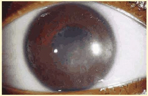
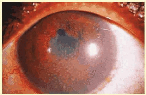
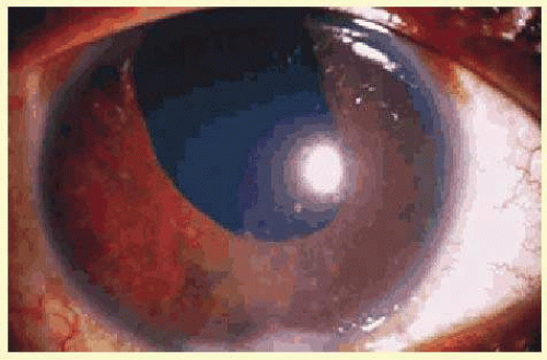
Another classification divides uveitis into granulomatous or nongranulomatous disease. This is a descriptive classification based on clinical examination, not histology, but it may help narrow the differential diagnosis. Uveitis is considered granulomatous if there are Busacca or Berlin nodules in the iris stroma, large greasy “mutton-fat” keratic precipitates, or choroidal granulomas. The presence of small Koeppe nodules at the pupillary margin does not imply that the disease process is granulomatous. Common causes of granulomatous inflammation include sarcoidosis, VKH, sympathetic ophthalmia, tuberculosis, and syphilis, although all of these entities may also present with nongranulomatous disease.


TABLE 32-2 Descriptors of Uveitis Onset, Duration, and Course


Category


Descriptor


Comment


Onset


Sudden


Insidious


Duration


Limited


<3 mo duration


Persistent


>3 mo duration


Course


Acute


Episode characterized by sudden onset and limited duration


Recurrent


Repeated episodes separated by periods of inactivity without treatment >3 mo in duration


Chronic


Persistent uveitis with relapse in <3 mo after discontinuing treatment


Reprinted with permission from The Standardization of Uveitis Nomenclature (SUN) Working Group. Standardization of uveitis nomenclature for reporting clinical data. Results of the First International Workshop. Am J Ophthalmol. 2005;3:509-516.


SYMPTOMS


Pain

The pain of iritis is primarily related to ciliary spasm. The ciliary body is innervated by the trigeminal nerve, and pain caused by its inflammation may therefore radiate to the periorbital region and to the eye. Because branches of the trigeminal nerve supply the cornea and iris, inflammation of these areas can cause a retrograde reflex with vascular dilation and swelling in the ciliary body, the so-called axon reflex. Cycloplegia is therefore useful in cases of iritis and in some cases of keratitis because paralysis of the ciliary muscle alleviates the pain of ciliary spasm.4


Photophobia

Photophobia (pain caused by light exposure) is often accompanied by tearing and blepharospasm. It must be distinguished from the photodysphoria or photoaversion in patients with achromatopsia, cone dystrophy, or retinitis pigmentosa and from glare caused by lenticular opacities. Photophobia is a prominent symptom in patients with inflammation of the iris or ciliary body, and in patients with keratitis and scleritis because of the axon reflex. Again, cycloplegia may lessen photophobia and pain.


Blurred Vision

Blurred vision may be caused by change in refraction or cloudy media, although vision is often surprisingly good in the presence of dense inflammatory reaction in the anterior chamber and vitreous. Floaters are a more frequent manifestation of cells and debris in the vitreous cavity. Macular edema, which frequently occurs with both anterior and posterior uveitis, can produce blurred vision, micropsia, and metamorphopsia.


SIGNS


Ciliary Injection

Ciliary injection, or “ciliary flush,” is manifest by a ring of dilated episcleral vessels radiating from the limbus. It should be distinguished from the deeper and more peripheral injection of scleritis and from the sectoral or diffuse injection of episcleritis. Overlying conjunctival injection may mask ciliary flush, but topically applied neosynephrine blanches the overlying conjunctiva, allowing visualization of deeper vessels.


Pupillary Miosis or Irregularity

The pupil is typically small in patients with iritis because iris inflammation results in the release of prostaglandins, which constrict the pupil5; exceptions include Posner-Schlossman syndrome and herpetic uveitis,6 which may present with a dilated pupil. The pupil in patients with anterior uveitis often becomes irregular and fixed because of the development of posterior synechiae.


Band Keratopathy

Long-standing chronic iridocyclitis, especially in children, may result in calcific band keratopathy, the deposition of calcium hydroxyapatite in the cornea at the level of Bowman’s membrane. Band keratopathy usually begins as grayish white opacities at the periphery of the interpalpebral region. The opacification may spread centrally and in time may form a complete band within the interpalpebral zone. A gap (lucid interval) is noted between the band and the limbus because Bowman’s layer does not extend to the absolute limbus.7 Small clear areas are noted in the opacity, representing the location where corneal nerves penetrate Bowman’s layer. These holes impart a “Swiss cheese” appearance to band keratopathy (Fig. 32.1). Band keratopathy should be distinguished from the Vogt limbal girdle and spheroidal degeneration. Occasionally, band keratopathy is atypical and starts centrally. Rarely, it forms a reticular pattern resembling lattice dystrophy, called superficial reticular degeneration of Koby.8


Keratic Precipitates

Clusters of inflammatory cells deposited on the endothelial surface of the cornea are known as keratic precipitates. The cells, which have been deposited from the aqueous humor, are often found inferiorly on the cornea in a linear vertical formation (Turk’s line) or in the form of a base-down triangle (Arlt’s triangle) (Fig. 32.2). The inferior corneal distribution results from convection currents within the anterior chamber that rise along the warm iris and fall along the cool cornea. Exceptions to the rule of inferior distribution of keratic precipitates include inflammation secondary to herpes simplex, Varicella zoster, cytomegalovirus, and Fuchs heterochromic iridocyclitis, which typically result in smaller stellate keratic precipitates, often throughout the whole cornea. Sarcoidosis and VKH may also result in keratic precipitates distributed above the midline, but these keratic precipitates tend to be larger and greasier in appearance than those associated with Fuchs or herpetic iridocyclitis.


FIG. 32.1 Band keratopathy in the interpalpebral area in a patient with juvenile idiopathic arthritis. Note the small holes in the opacity, which represent the location where corneal nerves penetrate Bowman’s layer. Also note the lucid interval between the band and the limbus.


FIG. 32.2 Chronic iridocyclitis. Note large greasy keratic precipitates in Arlt’s triangle. Also note extensive posterior synechiae and Busacca nodules.

The cells that precipitate on the corneal endothelium reflect the composition of those in the aqueous humor. In acute inflammation, polymorphonuclear leukocytes predominate, whereas in chronic inflammation, lymphocytes, plasma cells, and pigment are more common.9 Fine fibrin dusting may also be present, alone or among the keratic precipitates. Fibrin is a prominent feature of the keratic precipitates of Fuchs iridocyclitis, forming the bridges and stellate arms of the keratic precipitates.

In addition to size, shape, and distribution of keratic precipitates, color may be an important characteristic. Fresh keratic precipitates tend to be whiter and round, whereas older keratic precipitates may be heavily pigmented, faded, or have an irregular crenelated appearance. Descemet’s membrane may cover old keratic precipitates, imparting a glassy or ghostlike appearance (Fig. 32.3). Large greasy white keratic precipitates approaching 1 mm in diameter are often termed mutton-fat keratic precipitates and represent clusters of macrophages and epithelioid cells (Fig. 32.4). These are pathognomonic of granulomatous inflammation.

Although many keratic precipitates fade without sequelae, transient decompensation of the cornea may ensue with secondary corneal edema. The endothelium may have the appearance of having guttae (cornea pseudoguttata) secondary to endothelial cell swelling.10 Permanent opacification of the inferior corneal endothelium has also been described in chronic iridocyclitis, resulting from fibrous metaplasia of the endothelium due to the presence of keratic precipitates (Fig. 32.3).11 It is important to document the size, color, distribution, and number of keratic precipitates at each visit. For example, one should describe 30 small white keratic precipitates and five medium pigmented keratic precipitates all located on the inferior cornea, rather than simply noting that the cornea has keratic precipitates.


FIG. 32.3 Chronic iridocyclitis. Patient is aphakic. Note inferior corneal endothelial scarring, and old glassy “ghost” keratic precipitates inferiorly.


Anterior Chamber Flare

Normal aqueous humor is optically empty. If the slit lamp beam is seen in the anterior chamber, it is termed flare and represents breakdown of the blood-aqueous barrier with exudation of protein. Flare is graded on a 0 to 4+ scale in accordance with the SUN Working Group classification system (Table 32.3). The light intensity of the slit lamp is turned to maximum, and the ray of light is directed at an oblique angle to the plane of the iris. Flare may also be measured using a laser flare meter or flare-cell meter, which quantifies anterior chamber protein by measuring light scattering of a helium-neon laser beam in the anterior chamber.12,13 Flare is clinically relevant. For example, in chronic anterior uveitis in children, flare has been associated with increased vision loss and development of vision threatening complications, independent of anterior chamber cells.14

Only gold members can continue reading. Log In or Register to continue

Stay updated, free articles. Join our Telegram channel

Jul 11, 2016 | Posted by in OPHTHALMOLOGY | Comments Off on Classification, Symptoms, and Signs of Uveitis

Full access? Get Clinical Tree

Get Clinical Tree app for offline access