Fig. 6.1
Collar button design: front plate, back plate, and locking ring (Courtesy of Dr. Claes H. Dohlman)
There are two types of Boston Keratoprosthesis:
Type 1 (Fig. 6.2a, b): Used in non-cicatrizing disease like graft failures, aniridia, trauma, postinfections, etc.
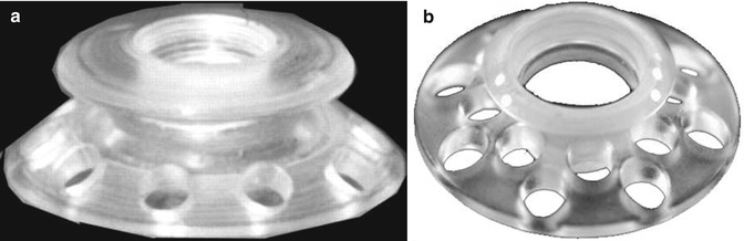
Fig. 6.2
(a, b) Type 1 Boston Keratoprosthesis assembled complex (Courtesy of Dr. Claes H. Dohlman)
Type 11 (Fig. 6.3): Used in cicatrizing diseases and severe dry eye conditions, like Stevens–Johnson’s syndrome, graft-versus-host disease, rheumatoid arthritis, etc.
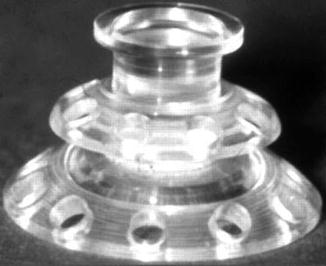
Fig. 6.3
Type 11 Boston Keratoprosthesis assembled complex (Courtesy of Dr. Claes H. Dohlman)
6.4.1 Front Plate Type 1 (Fig. 6.4)
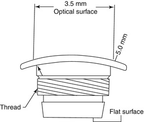
Fig. 6.4
Type 1 front plate (Courtesy of Dr. Claes H. Dohlman)
The front plate is made of PMMA. It has a main optical surface which is 3.7–3.5 mm in diameter, and the whole front plate is 5.0 mm in diameter. It is this optical surface where all the dioptric power of the KPro is machined in. The radius of curvature of this optical surface determines the power of the KPro. The Boston KPro can be made into either a pseudophakic or aphakic type depending upon the patient requirement. For an aphakic patient the eye’s axial length is required to determine the KPro power.
The edge of the front plate is machined to the minimum structurally feasible so as not to crack during manufacturing or handling. This extremely refined edge prevents the foreign body sensation that the patient may develop and also provides a smooth blend between the PMMA and the cornea. This PMMA/cornea interface is the most important area where infections can go inside the eye. And it has also been observed to be the most common site of initial corneal melt. The host corneal epithelium cannot grow over the PMMA; therefore, this PMMA/corneal junction always in principle remains open. The bandage contact lens that is placed in KPro patients keeps a certain amount of tear film intact over the KPro front surface and prevents Dellen formation, thus dryness, at this PMMA/corneal junction. This tear film and contact lens prevents the foreign body sensation and has in our experience reduced the corneal melt.
Stay updated, free articles. Join our Telegram channel

Full access? Get Clinical Tree


