(1)
Department of Ophthalmology and Visual Sciences, University of Iowa Hospitals and Clinics, Iowa City, IA, USA
Optic nerve head (ONH) circulatory disorders together constitute one of the most important ocular vascular disorders (e.g., anterior ischemic optic neuropathy, glaucomatous optic neuropathy, and optic disc vasculitis). To understand these disorders, a good basic understanding of the blood supply of the ONH is a first essential. I have discussed this at length elsewhere [1, 2], and the following is a brief account.
The ONH, from both the anatomical and the blood supply points of view, consists of the following four distinct regions from anterior to posterior aspect:
1.
The surface nerve fiber layer
2.
The prelaminar region
3.
The lamina cribrosa region
4.
The retrolaminar region
In the literature, the ophthalmoscopic term “optic disc” has been applied interchangeably either to the whole or a part of the anterior-most part of the ONH (i.e., the surface nerve fiber layer and the prelaminar region) or to the entire ONH. Similarly the term “papilla” has been used as a synonym for the optic disc or ONH. The term “papilla” was coined by Briggs [3] in 1676 based on an erroneous impression that the normal ONH was elevated like a papilla. Since the structure usually is not elevated above the level of the adjacent retina, but lies in the same plane as the retina and has in fact a central depression (i.e., the physiological cup) or may even be flat, the term “papilla” is a misnomer. Thus, the terms “papilledema” or “papillitis” are misleading.
Anatomy of the Optic Nerve Head
The Surface Nerve Fiber Layer
This is the most anterior layer of the ONH, containing the compact optic nerve fibers as they converge here from all over the retina and bend to run into the optic nerve (Figs. 5.1 and 5.2 ). It is separated from the vitreous by the inner limiting membrane of Elschnig [5, 6] which is composed of astrocytes [4, 7, 8]. In the region of the physiological cup, this membrane is often thick but is very thin when the cup is large.
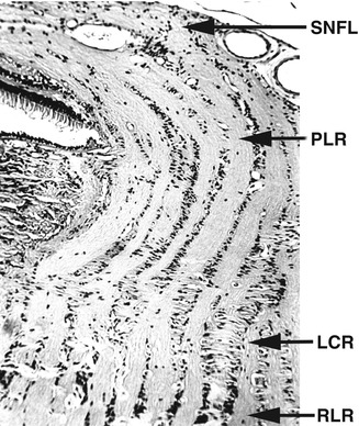
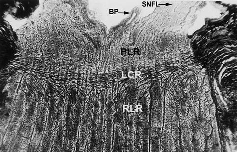
Fig. 5.1
Longitudinal section of a half of the normal human optic nerve showing the region of the optic nerve head and the retrolaminar optic nerve. LCR lamina cribrosa region, PLR prelaminar region, RLR retrolaminar region, SNFL surface nerve fiber layer (Reproduced from Hayreh [2])
Fig. 5.2
Longitudinal section of a normal rhesus monkey optic nerve showing the region of the optic nerve head and the retrolaminar optic nerve and Bergmeister’s papilla (BP). LCR lamina cribrosa region, PLR prelaminar region, RLR retrolaminar region, SNFL surface nerve fiber layer (Reproduced from Hayreh [2])
The region posterior to the surface nerve fiber layer is subdivided into three parts from front backward [4]: (1) pure glial part (prelaminar region), (2) mixed part – a transition zone between the prelaminar and the lamina cribrosa regions, and (3) connective tissue part (lamina cribrosa) (Fig. 5.3 ).
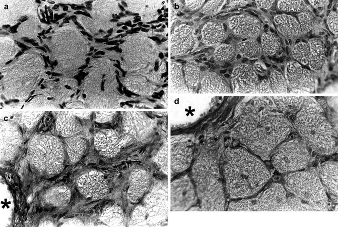
Fig. 5.3
Transverse sections of various parts of the ONH showing the arrangement of the septa. (a) Of the glial part of the prelaminar region. (b) Of the mixed part of the prelaminar region. (c) Of the lamina cribrosa region. (d) Of the retrolaminar region. Asterisk indicates central retinal artery
The Prelaminar Region
This consists almost entirely of glial tissue, and connective tissue fibers are seen only in connection with the vessels [4]. Glial fibers are much finer than connective tissue fibers, and they still retain a direction perpendicular to the nerve fiber bundles. Centrally, they remain attached to the connective tissue surrounding the central retinal vessels, and peripherally they are attached to the choroid as well as to the elastic membrane (Fig. 5.2 ). The number of glial cells in this part is enormous, and they are densely packed in transverse sheets (Fig. 5.3a). There may be many capillaries, surrounded by a glial limiting membrane, built up from the footplates of the glial cells.
The prelaminar region consists of optic nerve fibers arranged in bundles, surrounded by tubelike glial channels formed by specialized astrocytes (Figs. 5.1 , 5.2 , and 5.3a, b). The glial tissue between the nerve fiber bundles forms trabeculae (Fig. 5.3a, b), and capillaries are located within the glial septa. The glial cells in the prelaminar portion are loose, and the arrangement of the glial sheets is lost [4]. It would seem probable that this loose arrangement of the glial framework in the prelaminar portion might play a role in the pathologic swelling of the optic disc.
In the central part exists a central depression of varying degree corresponding to the physiological cup (Figs. 5.2 and 5.4 ). The bottom of this depression is separated from the vitreous by the inner limiting membrane of Elschnig (Figs. 5.2 and 5.4 ). The depression may or may not reach the lamina cribrosa. The prelaminar region at its edge is separated by several layers of astrocytes from the adjacent deeper layers of the retina (“Intermediate tissue of Kuhnt” [9]) and from the adjacent choroid (“Border tissue of Jacoby” [10]). These astrocytes not only form a column around the circumference of the chorioretinal canal and send processes internally among the nerve bundles but also invest the connective tissue of all vessels entering this portion of the ONH [11].
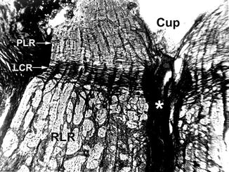
Fig. 5.4
Longitudinal section of the anterior part of the optic nerve in rhesus monkey showing a thick horizontal fibrous band of lamina cribrosa and the connective tissue sheath of the central retinal vessels centrally (*), which reaches the bottom of the optic disc cup (Hortega’s stain). LCR lamina cribrosa, PLR prelaminar region, RLR retrolaminar region (Reproduced from Hayreh [2])
The optic nerve fibers coming from the retina make a 90° bend in the surface layer of the ONH to run back in the optic nerve (Figs. 5.1 and 5.2 ), and as they make the bend, their main support is the glial tissue of the prelaminar region. The nerve fibers are arranged in bundles. Within the bundles, the axons are separated either from each other by an intercellular space measuring 150 Å or from an astrocyte process by a similar space [11]. The nerve fibers in this part, as in the surface nerve fiber layer and the retina, are unmyelinated and vary in diameter and are arranged in bundles; toward the retina the bundles become closely packed.
The Lamina Cribrosa Region
The lamina cribrosa is usually convex posteriorly and concave anteriorly (Figs. 5.1 , 5.2 , and 5.4 ). There is no sharp transition between the anterior prelaminar glial and posterior connective tissue parts, resulting in the transitional zone (mixed part – Fig. 5.3b) of varying size between the two.
The Connective Tissue Part of the Lamina Cribrosa
This extends transversely across the entire thickness of the ONH, bridging the scleral canal (Figs. 5.1 , 5.2 , and 5.4 ). At the periphery, the connective tissue part of the lamina cribrosa is anchored to the surrounding sclera by thick columns of the connective tissue with enlarged bases (Figs. 5.4 and 5.5 ); the same kind of anchorage is present centrally (Figs. 5.4 and 5.5 ), binding the lamina cribrosa firmly to the connective tissue envelope of the central retinal vessels.
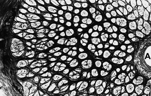
Fig. 5.5
Transverse sections of the lamina cribrosa, showing coarse connective tissue septa, rounded openings for the nerve bundles, and, in the periphery, glial fibers expand across the openings. A central retinal artery (Hortega’s stain) (Reproduced from Hayreh and Vrabec [4])
The connective tissue fibers of the lamina cribrosa are tightly packed together so that, in a longitudinal section through this region, it is seen as a dense compact band of the connective tissue bridging the scleral canal (Figs. 5.2 and 5.4 ). In cross sections of the lamina cribrosa, this compact connective tissue reveals many openings for the transmission of the optic nerve fiber bundles (Figs. 5.3c and 5.5 ). The lamina cribrosa is of a lamellar nature with collagen bundles alternating with glial sheets [12, 13]; posteriorly the collagen tissue sheets become increasingly prominent. The posterior part of this is not sharply delimited from the septal system of the retrolaminar optic nerve, so that the connective tissue septa of the latter are attached to the posterior surface of the lamina cribrosa (Figs. 5.2 and 5.4 ). A continuous glial membrane, similar to that seen in the retrolaminar part, separates the nerve fibers from the connective tissue in the region of the lamina cribrosa.
The lamina cribrosa, throughout its entire thickness, is pierced centrally by the central retinal vessels with their accompanying connective tissue (Figs. 5.4 and 5.5 ). The latter tissue forms a cylindrical sheath surrounding the vessels, within which numerous fine nerve fibers (autonomic) are seen running along the central vessels to the optic disc [4]. Some of these are closely attached to the vessel wall. In our study [4], in some sections we were able to follow larger trunks of such nerve fibers, representing the so-called nerve of Tiedemann.
The nerve fibers in this region are similar to those seen in the prelaminar region. The border tissue of Elschnig, much more strongly developed on the temporal than on the nasal side, separates the sclera from the nerve fibers and is composed of dense collagenous tissue with many glial and elastic fibers and some pigment [7, 14]. It continues forward to separate the choroid from the prelaminar region. The glial framework, formed by the astrocytes, extends throughout the entire ONH.
The Retrolaminar Region
This part of the optic nerve lies immediately behind the lamina cribrosa and is enclosed by dura, arachnoid, and pia. This area has coarse connective tissue septa (Figs. 5.2 , 5.3d, and 5.4 ), containing blood vessels. The septa run in all directions – longitudinal and transverse septa in the optic nerve are all attached to one another (Figs. 5.2 , 5.3d, and 5.4 ). In the central part of the optic nerve, near the central vessels, the main direction of the connective tissue fibers is longitudinal. The longitudinal fibrous septa of the retrolaminar part of the optic nerve are firmly anchored to the posterior surface of the lamina cribrosa (Figs. 5.2 and 5.4 ). The transverse septa at the periphery are attached to the pia on the surface (Figs. 5.2 and 5.4 ) and to the fibrous envelope around the central retinal vessels centrally (Figs. 5.3d and 5.4 ). Here the nerve fiber bundles lie in polygonal spaces formed by the connective tissue septa (Figs. 5.2 , 5.3d, and 5.4 ). The septa form a complicated intercommunicating tubular framework for the nerve fiber bundles. The nerve fibers in this part are myelinated but are unmyelinated anterior to that.
Blood Supply of the Optic Nerve Head
Arterial Supply of the ONH
The Surface Nerve Fiber Layer
This most anterior part of the ONH (surface of the optic disc) is typically supplied by the retinal arterioles (Figs. 5.6 and 5.7). In some cases, however, its temporal region may instead be supplied by the posterior ciliary artery (PCA) circulation from the deeper prelaminar region. The cilioretinal artery (rarely a tiny cilio-papillary artery), when present, usually supplies the corresponding sector of the surface layer (Fig. 5.7).
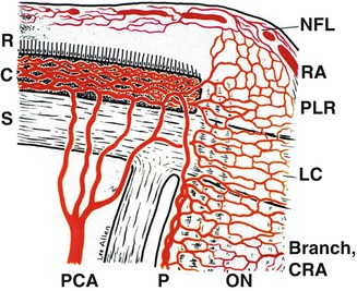
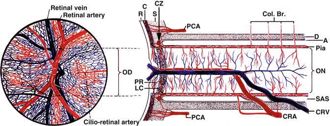
Fig. 5.6
Schematic representation of blood supply of the optic nerve head (Reproduced from Hayreh [15]). Abbreviations used: C choroid, CRA central retinal artery, LC lamina cribrosa, NFL surface nerve fiber layer of the disc, ON optic nerve, P pia, PCA posterior ciliary artery, PLR prelaminar region, R retina, RA retinal arteriole, S sclera
Fig. 5.7
Schematic representation of blood supply of the optic nerve and retina. Left half shows retinal appearance (Modified from Hayreh [16]). Abbreviations used: A arachnoid, C choroid, CRA central retinal artery, Col. Br. Collateral branches, CRV central retinal vein, CZ circle of Haller and Zinn, D dura, LC lamina cribrosa, OD optic disc, ON optic nerve, P pia mater, PCA posterior ciliary artery, PR prelaminar region, R retina, S sclera, SAS subarachnoid space
The Prelaminar Region
The peripapillary choroid is the main source of blood supply to this region (Figs. 5.6, 5.8, 5.9, 5.10, and 5.11). Fine centripetal branches from the peripapillary choroid go to the corresponding part of the prelaminar region, as demonstrated by many fluorescein angiographic (Figs. 5.8, 5.10, and 5.11) and morphologic studies [15, 17–51], the former providing the most convincing information. The peripapillary choriocapillaris (Fig. 5.12) and central retinal artery (Fig. 5.7) play no role in its supply. The blood supply in this region is sectoral (Figs. 5.9, 5.10, and 5.11), which agrees with the overall segmental distribution of the PCA circulation [39] (see Chap. 4), as well as with the segmental nature of visual loss in ONH ischemic disorders, e.g., anterior ischemic optic neuropathy.
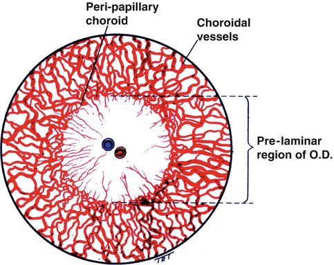
Fig. 5.8
Schematic representation of the choroidal and peripapillary arteries and their centripetal contribution to the prelaminar region of the optic nerve head. OD optic disc (Reproduced from Hayreh [17])