Bevacizumab for the Treatment of Exudative Age-Related Macular Degeneration
Lucienne C. Collet
Borja F. Corcóstegui
INTRODUCTION
Age-related macular degeneration (AMD) is the main cause of irreversible blindness in developing countries (1,2,3,4,5,6,7). In the United States, AMD causes 46% of the cases of severe visual loss (visual acuity [VA] 20/200 or worse) in patients older than 40 years (7), affecting approximately 1.75 million people (6). In Europe, AMD is also the leading cause of blindness, and its prevalence in patients aged 65 to 75 years is reported to range between 9% and 25% (8). Visual impairment secondary to AMD varies among the European countries. Studies have shown a prevalence of 40% in France (9), 39% in Germany (10), 36% in the Netherlands (11), 16% in the European North of Russia (12), and 14% in Bulgaria (13). In China, the prevalence of early AMD is 9.2% and late AMD 1.9%. The prevalence of early AMD increased from 5% in the 65- to 69-year group to 24.4% in patients older than 80 years and for late AMD, from 1% to 9% (14). Age is the most significant factor associated with AMD.
AMD is classified into nonexudative and exudative forms. The nonexudative form is characterized by the presence of drusen, retinal pigment epithelium (RPE) abnormalities (hypo- or hyperpigmentation), and geographic atrophy. The exudative or neovascular form is characterized by choroidal neovascularization (CNV), serous or hemorrhagic detachment of the retina or RPE, hard exudates, subretinal and sub-RPE fibrovascular proliferation, and disciform scar.
VASCULAR ENDOTHELIAL GROWTH FACTOR
During the last two decades, different treatment modalities have been used for the treatment of AMD. In the beginning, thermal laser photocoagulation was used to destroy CNV, but it caused irreversible visual loss due to retinal burns and had a recurrence rate of 50% (15). Next, photodynamic therapy (PDT) with verteporfin was introduced, and the majority of the patients reduced the risk of visual loss but few gained vision (16,17).
In 1989, Ferrara and Henzel (18) isolated and purified an endothelial cell mitogen, and they named it vascular permeability factor or vascular endothelial growth factor (VEGF). In the last 20 years, intensive research on VEGF-mediated disease has increased our knowledge of neovascular ocular pathology. VEGF-A is a homodimeric glycoprotein that functions as a growth factor specific for endothelial cells (19). It induces vascular permeability and is a regulator of vasculogenesis and angiogenesis. Blood vessel formation involves both vasculogenesis and angiogenesis.
Vasculogenesis takes place in the embryo and is characterized by the differentiation of endothelial cell precursors, while angiogenesis describes the formation of new blood vessels from existing vessels. The process of angiogenesis is responsible for new blood vessel formation in adults. This may take place in biologic or pathologic situations, such as AMD, cancer, proliferative retinopathies, diabetic maculopathy, and retinal vein occlusion among others (20).
Vasculogenesis takes place in the embryo and is characterized by the differentiation of endothelial cell precursors, while angiogenesis describes the formation of new blood vessels from existing vessels. The process of angiogenesis is responsible for new blood vessel formation in adults. This may take place in biologic or pathologic situations, such as AMD, cancer, proliferative retinopathies, diabetic maculopathy, and retinal vein occlusion among others (20).
There are factors that promote angiogenesis and factors that inhibit it. The factors that activate angiogenesis are VEGF, transforming growth factor α, transforming growth factor β, fibroblast growth factor, angiopoietin-1, angiopoietin-2 (21,22,23), and cysteine-rich 61 (Cyr61) (24).
VEGF can activate angiogenesis, lymphangiogenesis, vascular permeability, and prevents apoptosis promoting survival of endothelial cells. Without VEGF, endothelial cells in immature vessels could not survive, grow, or proliferate (19,23,25,26). The VEGF gene family includes VEGF-A, VEGF-B, VEGF-C, and VEGF-D (23). There are four active VEGF-A isoforms: VEGF121, VEGF165, VEGF189, and VEGF206. The most frequently expressed isoform is VEGF A165 (27).
VEGF receptors are located on the surface of endothelial cells. Three receptor tyrosine kinases have been identified for VEGF: VEGFR-1, VEGFR-2, and VEGFR-3. VEGFR-1 (fms-like tyrosine kinase-1) has both positive and negative angiogenic effects; VEGFR-2 (fetal liver kinase-1 and kinase insert domain-containing receptor) is the main mediator of angiogenic, mitogenic, and vascular permeability effects of VEGF A; VEGFR-3 promotes angiogenic effects on lymphatic vessels (19,28).
VEGF-A binds to receptors: VEGFR-1 and VEGFR-2. Studies on humans and primates have shown that VEGF-A is important in the pathogenesis of AMD and other eye diseases (29,30,31).
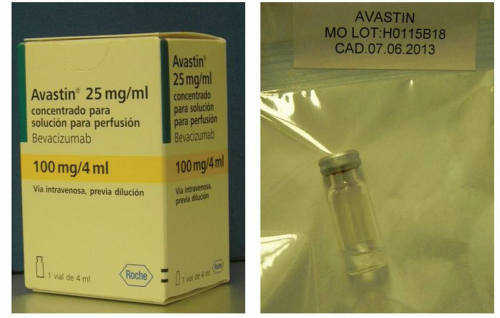 Figure 18.1 ▪ Bevacizumab (Avastin: Genentech, South San Francisco, CA) was first introduced and was the first antiangiogenic agent approved by the U.S. Food and Drug Administration (FDA) to inhibit tumor growth. |
In fact, increased concentrations of VEGF-A have been found in exudative AMD. VEGF-A has been found to be expressed in RPE cells, suggesting their involvement in new vessel formation (30). Additionally, high levels of VEGF-A, along with increased VEGF121 and VEGF165 expression, have been detected in CNV from AMD patients (31,32) as well as increased levels of VEGF in the vitreous humor of patients with CNV (33). VEGF-A is a target for the treatment of eye diseases in which angiogenesis is important, such as AMD. Treatment strategies are aimed at inhibition of VEGF.
THERAPIES TO INHIBIT VEGF
Therapies to inhibit VEGF include an aptamer (pegaptanib sodium), specific antibodies (bevacizumab and ranibizumab), the immunoglobulin G—VEGF receptor fusion protein (VEGF trap), and fragment antibodies of small interfering RNA (siRNA, bevasiranib) (34).
Bevacizumab
Bevacizumab is a full-length recombinant humanized monoclonal immunoglobulin-G that binds to all isoforms of VEGF and prevents VEGF from binding to its receptors (35). In 1997, bevacizumab (Avastin: Genentech, South San Francisco, CA) was first introduced and was the first antiangiogenic agent approved by the US Food and Drug Administration (FDA) to inhibit tumor growth (Fig. 18.1). In 2004, bevacizumab was approved for treatment of metastatic colorectal cancer.
The off-label use of intravenous bevacizumab for neovascular AMD showed promising results (36). Preclinical studies on pharmacokinetics of intravenous bevacizumab show a serum half-life of 1 to 2 weeks (37), and clinical studies in patients show a half-life of 21 days (38).
The off-label use of intravenous bevacizumab for neovascular AMD showed promising results (36). Preclinical studies on pharmacokinetics of intravenous bevacizumab show a serum half-life of 1 to 2 weeks (37), and clinical studies in patients show a half-life of 21 days (38).
Pharmacokinetics studies of intravitreal bevacizumab in rabbit models have shown that after an injection of 1.25 mg bevacizumab, a peak concentration of free bevacizumab (400 µg/mL) is achieved in the vitreous humor. Vitreous concentrations decline with a half-life of 4.32 days, but concentrations ≥10 µg/mL were maintained in the vitreous for 30 days. A peak concentration in the aqueous humor reached at day 3 after drug administration. Low concentrations were detected in the serum after intravitreal injection and in the aqueous humor of the fellow eye (39). In another study done in macaques, the aqueous humor and serum bevacizumab concentration were measured after intravitreal bevacizumab injection. At day 1, a peak in the aqueous humor was observed while the peak concentration in the serum was at 1 week. VEGF levels returned to preinjection concentrations after 42 days (40). In humans treated with intravitreal bevacizumab, a study showed decrease in VEGF concentration within 7 days of treatment (41).
SANA (Systemic Avastin for Neovascular AMD) was the first prospective study in patients with exudative AMD treated with intravenous bevacizumab. Eighteen patients with subfoveal CNV were enrolled in an open-label study in which bevacizumab 5 mg/kg was injected intravenously every 2 weeks for two or three infusions. VA improved from 54 letters (Snellen VA 20/80) at baseline to 68 letters (Snellen VA 20/40) at 24 weeks, an increase of 14 letters (P ≤ 0.001). Additionally, retinal thickness decreased from 392 to 280 µm at week 24. Adverse events occurred in 18.5% of the patients. At week 3, increased blood pressure was observed and controlled with antihypertensive drugs (36,42). Additionally, bevacizumab was also associated with increased risk of venous thromboembolism in cancer patients (43). Intravenous bevacizumab improved VA and decreased retinal thickness as early as 1 week after therapy; however, its potential systemic side effects outweighed its benefits.
The first anti-VEGF drug designed for intraocular use in the treatment of AMD was pegaptanib sodium (Macugen). Pegaptanib is a 28-base RNA oligonucleotide ligand or aptamer that binds to human VEGF165 with high specificity and affinity. Pegaptanib showed efficacy for the treatment of AMD; however, the VISION study concluded that intravitreal pegaptanib injection at 6-week intervals stabilized vision in 70% of cases and improved it in only 6% (44,45).
In 2006, phase III clinical trials, MARINA and ANCHOR, demonstrated that the use of anti-VEGF ranibizumab (Lucentis, Genentech, Inc. South San Francisco, CA) promoted VA improvement in patients with neovascular AMD, stabilizing vision in 95% of patients and improving VA between 7.2 and 11.3 letters in 30% to 40% of patients (46,47). While these trials were still in progress, Rosenfeld et al. (48) showed that bevacizumab could also lead to VA improvement at a considerably lower cost and became the first to report the use of intravitreal bevacizumab for the treatment of exudative AMD to minimize the risk of systemic treatment (48). Consequently, bevacizumab was and is used worldwide by clinicians for the treatment of neovascular AMD. Exudative AMD is the most common indication for intravitreal bevacizumab. However, the use of bevacizumab developed in the absence of formal protocols. Many papers, including retrospective and prospective studies, uncontrolled case series, and uncontrolled randomized trials, have shown improvement in VA as well as decrease in macular thickness after treatment.
Subsequently, intravitreal injection of bevacizumab achieved worldwide success because of its safety profile, availability, short-term effects, and low cost compared to other anti-VEGF drugs. Since ranibizumab and bevacizumab are derived from the same monoclonal antibody and have a similar mechanism of action, it was suggested that they have similar efficacy and safety in treating AMD. Despite the lack of large-scale clinical trials, bevacizumab is the most commonly used treatment in the United States for exudative AMD (49). In an analysis of 222,886 Medicare beneficiaries from 2008, 146,276 (64.4%) received bevacizumab and 80,929 (35.6%) received ranibizumab (49).
In the beginning, there were many issues to resolve, such as retinal penetration, toxicity, and schedule for retreatment. A question of particular interest was whether intravitreal bevacizumab could penetrate the retina given its large size, but some reports concluded full retina penetration after injection (50). In fact, an animal study using confocal immunohistochemistry demonstrated full retinal thickness penetration 24 hours after intravitreal injection (51). With regard to toxicity, several experimental animal studies confirmed no retinal toxicity even at doses of 5 mg of intravitreal bevacizumab (52,53). Electrophysiologic and histologic studies have not demonstrated toxicity as well (52,53,54,55,56). Studies in patients with AMD have shown improvement in mf-ERG responses and its correlation with improvement in VA. Specifically, improvement in P1 amplitude was observed and correlated with decreased leakage on fluorescein angiography. These results may explain an increase in neural activity in the retina.
By 2006, level 1 evidence has supported the safety and efficacy of Lucentis. Level 1 evidence is evidence obtained from properly conducted well-designed, randomized, controlled trials (46,47). In the case of bevacizumab, no level 1 evidence existed until 2011. During this time, data were limited to case series and randomized controlled trials with small numbers of patients or short follow-up periods. El-Mollayes et al. (57) reviewed 571 articles involving bevacizumab use and AMD between 1997 and 2010 (57). Of these articles, only eight included at least 30 patients with AMD treated with bevacizumab for at least 12 months of follow-up (58,59,60,61,62


Stay updated, free articles. Join our Telegram channel
Full access? Get Clinical Tree
