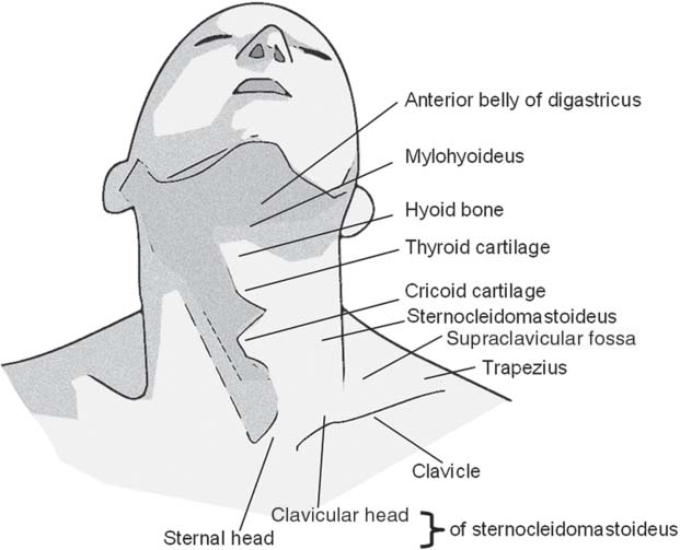
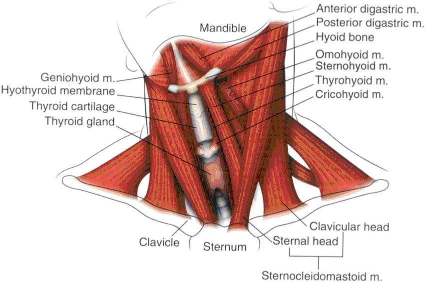
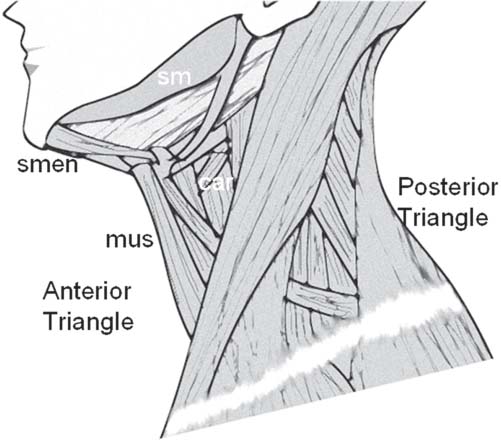
6 Applied Anatomy for Percutaneous Approaches to the Cervical Spine To understand the surgical technique of percutaneous endoscopic cervical diskectomy (PECD) and to produce successful outcomes, it is imperative to have a thorough knowledge of the regional anatomy of the neck. The approach to the cervical disk in PECD is always anterior; hence we have focused on the anterior triangle of the neck in describing the surgical anatomy in this chapter. Orientation to surface anatomy helps to locate the surgical level and proper needle trajectory to the disk space (Fig. 6.1). The sternocleidomastoid (SCM) muscle separates the anterior and posterior triangles of the neck. The landmarks are described below from the midline and beginning with the top of the neck and moving downward: • Thyroid cartilage — Most prominent midline structure, especially in post-pubertal males — Corresponds with C4-C5 level — Also corresponds with the carotid artery bifurcation into external and internal carotids • Hyoid bone — About 1.5 cm higher to the thyroid cartilage — Corresponds with the level of C3 vertebra • Cricoid cartilage — Located just below the thyroid cartilage — Corresponds to the C6 vertebral level • A horizontal plane approximately at the junction of the C6-C7 has the following associations: — Pharyngoesophageal junction — Laryngotracheal junction — Inferior thyroid artery, carotid sheath, and omohyoid muscle — Entrance of the inferior laryngeal nerve (recurrent nerve) into the larynx — Entrance of the vertebral artery into the transverse foramen of C6 — The thyroid isthmus and the greatest height of the thoracic duct are located at the C7 level. Fig. 6.1 Surface anatomy of the cervical region. Fig. 6.2 The surgical anatomy of the anterior triangle of the neck. sm, submandibular triangle; smen, submental triangle; mus, muscular triangle; car, carotid triangle. The neck, as already described, is divided into anterior and posterior triangles. The following description presents the surgical anatomy of the anterior triangle of the neck (Figs. 6.2 and 6.3). Fig. 6.3 The infrahyoid and suprahyoid muscles of the neck. • Lateral: SCM muscle • Superior: inferior border of the mandible • Medial: anterior midline of the neck — The anterior triangle is further subdivided into the following sections: — Submandibular — Submental — Carotid — Muscular • Superiorly: inferior border of the mandible • Inferiorly: anterior and posterior bellies of the digastric muscle • The submandibular gland is the largest structure of the triangle. • The roof is formed by skin, superficial fascia composing platysma, and the underlying mandibular and cervical branches of the fascial nerves. • Below the roof, from superficial to deep, lie the retromandibular vein, part of the facial artery, the submental branch of facial artery, the superficial layer of the submaxillary fascia (deep cervical fascia), the lymph nodes, the deep layer of the deep cervical fascia, and the hypoglossal nerve.
Surface Anatomy
Topographic Anatomy of the Cervical Spine
Boundaries of the Anterior Triangle
Submandibular Triangle
Boundaries
Content
Stay updated, free articles. Join our Telegram channel

Full access? Get Clinical Tree


