ANTERIOR SCLERA AND IRIS
EPISCLERITIS
Episcleritis is a benign, transient, recurrent, self-limited and usually nonspecific condition affecting young adults. It rarely progresses to scleritis, and should not be considered as a milder form of scleritis.
Etiology
• Idiopathic
• Collagen vascular diseases
• Rosacea
• Gout
• Herpes zoster, herpes simplex, syphilis
Symptoms
• Acute onset of redness, mild irritation, tearing
Signs
There are two forms: simple and nodular epis-cleritis.
Simple Episcleritis
• Sectorial or diffuse hyperemia, primarily involving the middle episcleral plexus, with some secondary involvement of the overlying conjunctival vessels (Figures 9-1A,B).
• The inflamed episcleral vessels have a straight, radial configuration.
• Topical phenylephrine 2.5% will cause blanching and enhance visualization of the normal deep vascular plexus over the sclera.
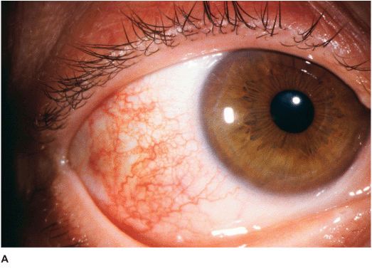
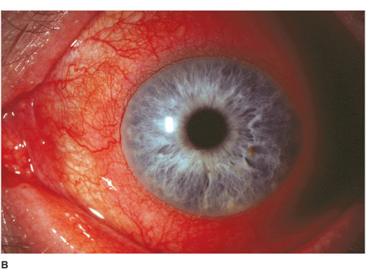
Figure 9-1 Episcleritis A. Localized injection of the episcleral vessels is evident in this patient with sectorial episcleritis. B. Diffuse injection of the episcleral vessels is present in this eye. Episcleritis can be difficult to differentiate from scleritis in certain patients. Episcleral vessels tend to blanch with topical phenylephrine 2.5% while scleral vessels do not.
Nodular Episcleritis
• Somewhat tender, usually solitary, localized injected nodule which can be moved slightly over the sclera.
• A localized area of corneal thinning secondary to desiccation (delle) may develop adjacent to the episcleral nodule.
Differential Diagnosis
• Conjunctivitis: usually associated with papillary or follicular response in the tarsal conjunctiva. May have a mucoid or purulent discharge.
• Scleritis: pain is deeper and more severe, sclera has a purple or bluish hue under natural light, injected scleral vessels have criss-crossing configuration, vessels are immobile over the globe, patients tend to be older.
• Inflamed pinguecula: located in the completely mobile conjunctiva.
Diagnosis
• Examination is made with the slit-lamp and under natural or room lights.
• Attempt to move the episcleral vessels over the sclera using a cotton-tipped stick under topical anesthesia.
• Apply phenylephrine 2.5% and observe for blanching of episcleral vessels after 15 minutes.
• Systemic work-up if history is suggestive of collagen vascular disease or gout.
Treatment
• Artificial tears and cool compresses qid.
• Consider a short course of topical antihistamine drops (e.g., tetrahydrozoline, naphazoline, or antazoline tid).
• Consider a short course of a topical non-steroidal anti-inflammatory agent (NSAID) (e.g., diclofenac, ketorolac, or ibuprofen qid).
• Oral NSAID (e.g., indomethacin 12.5 to 25 mg qd to tid or flurbiprofen 100 mg bid to tid) for moderate to severe cases.
• Consider a short course of topical cortico-steroids (e.g., fluorometholone 0.1 to 0.25%, loteprednol 0.2 to 0.5%) qid in recalcitrant cases.
Prognosis
• Good to very good. Episcleritis is often a recurrent condition.
ANTERIOR SCLERITIS
Scleritis is a severe, potentially sight-threatening ocular disorder that carries a totally different prognosis than episcleritis. It may be mild and benign or severe and destructive. Females are affected more often than males and the condition is frequently bilateral. The majority of scleritis affects the anterior sclera. Scleritis can be further divided into the following clinical forms:
NONNECROTIZING SCLERITIS
• Diffuse Diffuse hyperemia and distortion of the pattern of the deep vascular plexus, associated with variable episcleral and conjunctival injection (Figure 9-2A). This is the most benign form associated with the least severe systemic conditions.
• Nodular Tender, usually solitary, deep, localized injected nodule which cannot be moved over the sclera (Figure 9-2B).
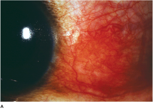
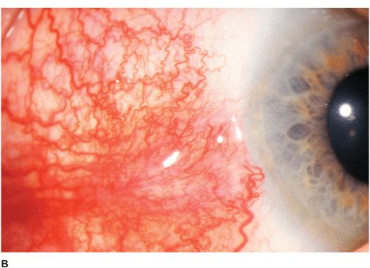
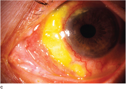
Figure 9-2 Scleritis A.
Stay updated, free articles. Join our Telegram channel

Full access? Get Clinical Tree


