Anatomy of the Ciliary Body and Outflow Pathways
Thomas F. Freddo
Haiyan Gong
An understanding of the glaucomas begins with an understanding of the anatomy of the structures related to the production and drainage of aqueous humor, particularly the ciliary body and the trabecular meshwork. The ciliary body is the sole source of aqueous production. The trabecular meshwork is part of the principal, pressure-dependent pathway for aqueous outflow and harbors the requisite resistance to this flow needed to maintain normal intraocular pressure. Regulation of this flow is dependent, in part, on the anatomic connections between the smooth muscle of the ciliary body and the trabecular meshwork into which its tendons extend.
THE CILIARY BODY AND AQUEOUS PRODUCTION
THE CILIARY BODY
The ciliary body performs three principal functions: (1) It produces and secretes aqueous humor into the posterior chamber of the eye; (2) it contains the smooth muscle that acts on the crystalline lens, via the zonular fibers, to shift the focus of the eye from far to near (accommodation); and (3) by extension of certain smooth muscle fibers and their tendons into the adjacent trabecular meshwork, it can assist in the drainage of aqueous humor from the eye. There is also some evidence supporting a role for the ciliary body in the production and slow turnover of certain molecular components of the vitreous humor.3
The ciliary body extends posteriorly from the root of the iris to the ora serrata. In the adult eye, the distance from the surgical limbus to the ora serrata is approximately 7 mm on the temporal side of the globe and 6 mm on the nasal side. The ciliary body is grossly subdivided into two portions, the pars plicata and the pars plana.
The pars plicata exhibits 70 to 80 corrugated, fin-like ridges on its inner surface that are arranged around the circumference of the crystalline lens (Fig. 1). Each individual ridge is referred to as a ciliary process, and these are divided into categories of major and minor processes based on their relative height. Major processes predominate, and they extend into the posterior chamber of the eye approximately 1 mm. Minor processes are about one third as high. In the scanning electron micrograph shown in Figure 2 these structures and their relationships are clearly discernible. It is important to appreciate, however, that the zonular fibers supporting the crystalline lens have been removed from the surface of this specimen. These fibers form a continuous carpet over the inner surface of the pars plana and are then channeled into the valleys between adjacent processes in the pars plicata on their way to the lens capsule. As such, in vivo, the valleys between adjacent ciliary processes are largely filled with zonules leaving only the crest of each process exposed to the posterior chamber (Fig. 3).
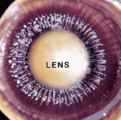 Fig. 1. Macrophotograph of the crystalline lens and surrounding ciliary body. The fin-like ridges representing the ciliary processes of the pars plana region are evident, surrounded by the smooth-surfaced pars plana. |
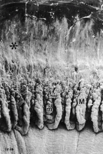 Fig. 2. Scanning electron micrograph of the inner surface of the iris and ciliary body with the lens and zonules removed. The pars plicata region is distinguished from the more posterior pars plana region (asterisk) by the presence of major (M) and minor (m) ciliary processes. (From Morrison JC, Van Buskirk EM, Freddo T. Anatomy, microcirculation and ultrastructure of the ciliary body. In: Ritch R, Shields MB, Krupin T (eds). The Glaucomas. St Louis: CV Mosby, 1989.) |
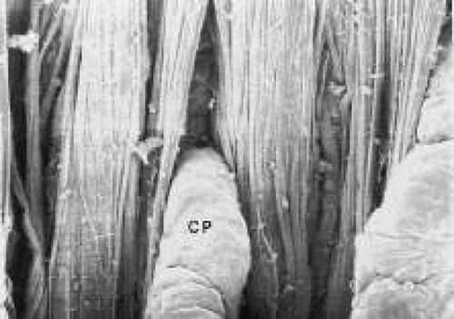 Fig. 3. High magnification scanning electron micrograph showing the confluence of zonular fibers leaving the pars plana (top) channeling into the valleys between adjacent ciliary processes. CP, Ciliary processes. |
The pars plana, as its name implies, exhibits a flat inner surface. This portion of the ciliary body is much thinner and less vascularized than the pars plicata, making it the safest point of entry into the eye for vitreoretinal surgical procedures.
In meridional sections, the ciliary body appears as shown in Figure 4. The entire surface of the ciliary body, facing the inside of the eye, is lined by a bilayered epithelium. These two layers are named for their relative content of melanin pigment. The layer closest to the posterior chamber is devoid of pigment and is called the nonpigmented ciliary epithelium. The layer closest to the ciliary body stroma is called the pigmented ciliary epithelium for its melanin content. The nonpigmented ciliary epithelium is continuous with the posterior pigmented epithelium of the iris anteriorly and with the neurosensory retina posteriorly at the ora serrata. The pigmented ciliary epithelium continues anteriorly as the anterior myoepithelium of the iris (which includes the iris sphincter muscle) and posteriorly as the pigmented epithelium of the retina. The double layer of epithelium covering the ciliary body actually represents two simple epithelia that are joined at their apical surfaces following the invagination of the optic cup during embryogenesis. As a result, the basement membrane of the nonpigmented ciliary epithelium faces the posterior chamber and that of the pigmented ciliary epithelium joins both epithelial layers to the ciliary body stroma.
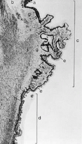 Fig. 4. Light micrograph of ciliary body of a 50-year-old man. The iris root (a) and the anterior chamber angle (b) are seen at the top. The pars plicata (c) and a portion of the pars plana (d) are included within brackets; they are lined by the nonpigmented (e) and pigmented (f) ciliary epithelium. The basement membrane of the pigmented ciliary epithelium increases (h) in thickness with age. The ciliary body stroma (g) and the three portions of the ciliary muscle (i, longitudinal; j, radial; k, circular bundle) are also shown. (From Hogan M, Alvarado J, Wedell J: Ciliary body and posterior chamber. In: Histology of the Human Eye. Philadelphia: WB Saunders, 1971, p 270.) |
The ciliary body stroma is composed of a loose connective matrix that supports the nerves and blood vessels that course within it. This connective tissue matrix extends into the core of each ciliary process. The ciliary body stroma is directly continuous with the stroma of the iris anteriorly and with the choroidal stroma posteriorly. Because the anterior surface of the iris has no epithelial covering, substances released into the ciliary body stroma have access to the anterior chamber by diffusing from the ciliary body stroma to the iris surface.4,5 Similarly, fluid entering the ciliary body stroma from the anterior chamber can reach the choroid by traveling along the uveoscleral outflow pathway described later. Between the loose connective tissue stroma and the inner surface of the sclera is a complex smooth muscle called the ciliary muscle, also described later.
PRODUCTION OF AQUEOUS HUMOR
To provide anatomic correlations for the physiology of aqueous humor formation, it is most convenient to describe the process in two steps: (1) elaboration of a plasma filtrate from which aqueous humor is derived, and (2) formation of aqueous humor from this filtrate. Although these steps are not independent, the first is related primarily to the ciliary body microvasculature and the second to the ciliary epithelium.
Ciliary Body Microvasculature
When viewed in cross section, each ciliary process contains a fibrovascular core that is continuous with the ciliary body stroma and covered by the bilayered ciliary epithelium (Fig. 5). The arterioles that serve the ciliary body stroma, like those serving the iris, arise from the discontinuous major circle of the iris.6 Each major process is served by a set of anterior and posterior arterioles7 (Figs. 6 and 7). The anterior arterioles supply the large diameter capillaries, near the crests of the processes, whereas the posterior arterioles supply the smaller caliber capillaries deep within each process. The direction of blood flow in these systems is from anterior to posterior, toward a network of choroidal veins. The blood from the entire ciliary body ultimately leaves the eye via the vortex veins.
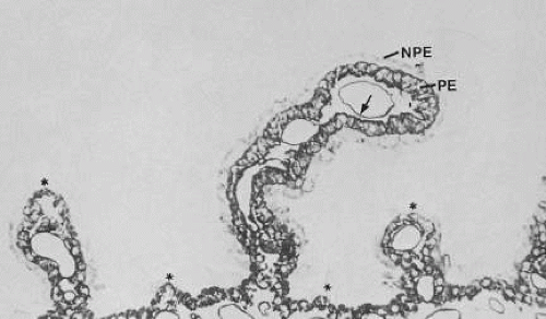 Fig. 5. Light micrograph of ciliary processes (asterisks) shows each process contains a fibrovascular core with large diameter capillaries (arrow). NPE, Nonpigmented ciliary epithelium; PE, pigmented ciliary epithelium. (Lütjen-Drecoll E: Functional morphology of the ciliary epithelium. In: Lütjen-Drecoll E (ed): Basic Aspects of Glaucoma Research. Stuttgart, Germany: FK Schattauer, 1982, p 79.) |
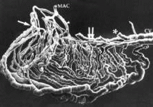 Fig. 6. A cast of the vasculature within a single ciliary process. The major circle of the iris (MAC) gives rise to an anterior (arrow) and posterior (arrowhead) arteriole. Double arrow denotes choroidal vein (CV). Drainage from ciliary muscle to choroidal veins has been removed at asterisk. (From Morrison JC, Van Buskirk EM, Freddo T. Anatomy, microcirculation and ultrastructure of the ciliary body. In: Ritch R, Shields MB, Krupin T (eds). The Glaucomas. St Louis: CV Mosby, 1989.) |
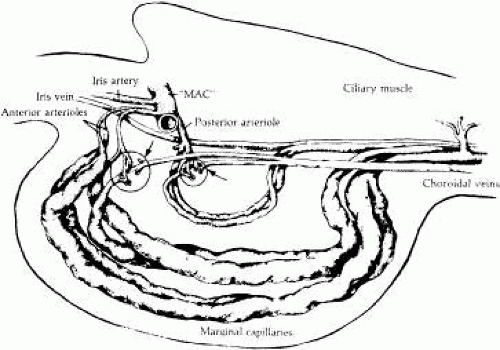 Fig. 7. Diagrammatic representation of Fig. 6 demonstrating the system of anterior and posterior arterioles that enter each ciliary process, ultimately draining into a set of choroidal veins. (From Morrison JC, Van Buskirk EM, Freddo T. Anatomy, microcirculation and ultrastructure of the ciliary body. In: Ritch R, Shields MB, Krupin T (eds). The Glaucomas. St Louis: CV Mosby, 1989.) |
Blood flow in the ciliary body is most likely regionalized and under autonomic control. Scanning electron microscopic studies have revealed the presence of localized constrictions in casts of afferent arterioles in the ciliary body microvasculature that may reflect the presence of a sphincter-like system for controlling blood flow.7,8,9,10 Through these mechanisms, alterations in blood flow can influence aqueous humor production by increasing or decreasing the amount of filtrate made available to the ciliary epithelium.
The capillaries derived from the posterior arterioles that serve the ciliary muscle are nonfenestrated and do not leak plasma proteins under normal conditions (Fig. 8). By contrast, the capillaries derived from the anterior arteriole, which pass within the stromal core of each ciliary process, lack tight junctions and are lined by fenestrated endothelial cells (Fig. 9). Using tracers for plasma protein leakage such as horseradish peroxidase (HRP), the capillaries of the ciliary body stroma are seen to be very permeable to macromolecules, as well as to ions and fluid (Figs. 8 and 10). As such, these vessels are limited in their capacity to serve as a selective permeability barrier. That function is one of several reserved for the ciliary epithelium.
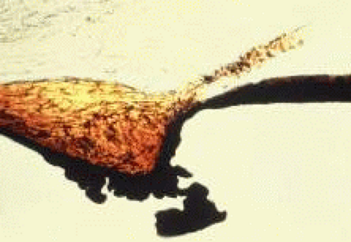 Fig. 8. A 200-μm thick section of the anterior uvea of a monkey eye, reacted for the demonstration of intravenously injected horseradish peroxidase (HRP). The ciliary body stroma is filled with black, HRP reaction product that has leaked from its fenestrated vessels. The ciliary muscle shows relatively little staining resulting from the nonfenestrated vessels that serve it. The dark coloration of the iris is not HRP but the normal dark pigmentation imparted by the pigmented epithelial layers on its posterior surface. |
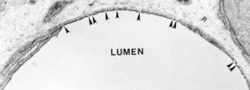 Fig. 9. Transmission electron micrograph demonstrates the appearance of capillaries in the ciliary body stroma. Numerous fenestrations (arrowheads) are evident along the circumference of the vessel wall. (From Morrison JC, Van Buskirk EM, Freddo T. Anatomy, microcirculation and ultrastructure of the ciliary body. In: Ritch R, Shields MB, Krupin T (eds). The Glaucomas. St Louis: CV Mosby, 1989.) |
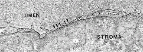 Fig. 10. Transmission electron micrograph demonstrates that granular horseradish peroxidase (HRP) reaction product leaks through fenestrations (arrowheads) into the surrounding ciliary body stroma. (From Morrison JC, Van Buskirk EM, Freddo T. Anatomy, microcirculation and ultrastructure of the ciliary body. In: Ritch R, Shields MB, Krupin T (eds). The Glaucomas. St Louis: CV Mosby, 1989.) |
The Ciliary Epithelium
As noted earlier, the two cell layers that constitute the ciliary epithelium are named for their relative content of melanin pigment. The layer closest to the ciliary body stroma is the pigmented ciliary epithelium and that closest to the posterior chamber of the eye is the nonpigmented ciliary epithelium (Fig. 11). The morphology of the ciliary epithelium varies along the surface of the ciliary body, in accord with the different demands placed on it in various locations.1 From this analysis, it appears that it is primarily the epithelia at the tips of the ciliary processes that are involved in the production of aqueous humor. In these areas, immunoelectron microscopic studies have clearly documented both Na-K-ATPase activity (Fig. 12) and carbonic anhydrase activity11,12 (Fig. 13). Both are known to be central to the production of aqueous humor and one of them, carbonic anhydrase, is regularly targeted for pharmacologic inhibition to reduce intraocular pressure in glaucoma.13
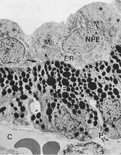 Fig. 11. Electron micrograph of the ciliary epithelium (cynomolgus monkey, pars plicata). The posterior chamber is at the top and the ciliary body stroma, with its fenestrated capillaries (C), is at the bottom. Extensive basolateral infoldings of the nonpigmented ciliary epithelial cells (NPE) are evident (stars). The apical surfaces of both layers face one another (arrow). PE, Pigmented ciliary epithelium; M, mitochondria; ER, endoplasmic reticulum; P, paracellular space. (Lütjen-Drecoll E: Functional morphology of the ciliary epithelium. In: Lütjen-Drecoll E (ed): Basic Aspects of Glaucoma Research. Stuttgart, Germany: FK Schattauer, 1982, p. 79.) |
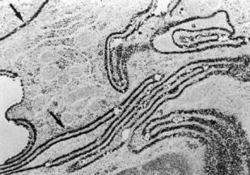 Fig. 12. Ultrahistochemistry of the ciliary epithelium stained for the enzyme Na-K-ATPase. Note basolateral infoldings of the nonpigmented ciliary epithelium show intense reaction product for the enzyme (arrows). (From Flügel C, Lütjen-Drecoll E: Presence and distribution of Na+/K+-ATPase in the ciliary epithelium of the rabbit. Histochemistry 88:613, 1988; Tamm ER, Lütjen-Drecoll E: Ciliary body. Microsc Res Tec 33:390, 1996. Reprinted by permission of Wiley-Liss, a subsidiary of John Wiley and Sons, Inc.) |
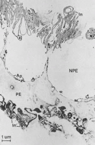 Fig. 13. Electron micrograph of the ciliary epithelium of the albino rabbit after staining for carbonic anhydrase. The reaction product is seen predominantly at the membrane infoldings (in) of the nonpigmented epithelium (NPE) and pigmented epithelium (PE), as well as on the apical cell membranes (Ap). Cytoplasmic staining is found only in the PE. (Lütjen-Drecoll E, Loennerholm G: Carbonic anhydrase distribution in the rabbit eye by light and electron microscopy. Invest Ophthalmol Vis Sci 21:782, 1981. Copyright held by The Association for Research in Vision and Ophthalmology.) |
THE BLOOD-AQUEOUS BARRIER IN THE CILIARY BODY
Aqueous humor is a clear nutrient fluid, derived from plasma, that provides for the metabolic needs of the avascular tissues of the eye. Although free amino acid levels in aqueous humor are nearly equivalent to those found in plasma,14 the protein content of aqueous humor in the anterior chamber is less than 1% of that found in plasma.15 Proteins create turbidity and turbidity scatters light, which degrades the optical efficiency of the eye. To prevent this and to ensure that potential antigens in the bloodstream are prevented from reaching the anterior and posterior chambers of the eye and the lens, vitreous, and retina, a selective barrier must be positioned between the bloodstream and the aqueous humor. As noted earlier, the microvasculature of the ciliary body stroma is composed of fenestrated capillaries that readily leak fluids, ions, and plasma proteins to provide the reservoir from which the ciliary epithelium secretes aqueous humor. To ensure that the protein-laden fluid in the ciliary body stroma and the composition of the aqueous humor remain different, a barrier to macromolecular diffusion is interposed between the ciliary body stroma and the posterior chamber by the ciliary epithelium.16 As noted earlier (see Figs. 8, 9, and 10), following injection of HRP in monkeys, this protein readily leaves the lumen of the fenestrated capillaries of the ciliary body stroma.17 The protein diffuses to the ciliary epithelium and easily moves between adjacent pigmented ciliary epithelial cells, filling the intercellular cleft18 (Fig. 14A). On reaching the interface between the juxtaposed apical surfaces of the pigmented and nonpigmented layers, the HRP is again able to freely permeate the intercellular cleft between these cell layers. But when the HRP attempts to diffuse along the intercellular cleft between adjacent nonpigmented ciliary epithelial cells, its progress toward the aqueous humor in the posterior chamber is blocked by an apicolateral junctional complex composed of a zonula occludens (tight junction), a zonula adherens, and desmosome19,20 (see Fig. 14A). In sections, the tight junction is seen as a series of apparent fusion points between the membranes of adjacent nonpigmented ciliary epithelial cells (see Fig. 14,Inset). Using the technique of freeze-fracture, however, allows an en face view of the tight junctions, revealing that these fusion points are actually cross sections of a complex system of branching and anastomosing protein strands that surround the entire circumference of each cell.19 This web of strands spans the intercellular cleft and is mirrored on the adjacent cell, which blocks the intercellular cleft around the entire circumference of each cell, to limit diffusion of macromolecules (see Fig. 14B). Disruption of this junctional complex has been shown to occur in experimental anterior uveitis, resulting in increased amounts of protein in the aqueous humor.21 This extra protein scatters the light of the slit lamp and is described clinically as flare.
Stay updated, free articles. Join our Telegram channel

Full access? Get Clinical Tree


