Adenovirus
Jerrod S. Kent
Sanjoy K. Gupta
William G. Hodge
OVERVIEW
In the 1950s a novel group of viral agents that were associated with respiratory ailments in humans were discovered. They were named adenoviruses after the adenoids, the original tissue from which the prototypical virus was isolated.1,2 Adenoviruses are a group of nonenveloped double-stranded deoxyribonucleic acid (DNA) viruses that belong to the family Adenoviridae. These viruses are currently classified to include four genera, Aviadenovirus, Mastadenovirus, Atadenovirus, and Siadenovirus.3,4 Mastadenovirus are responsible to infecting humans.
There are at present 51 distinct serotypes of adenovirus that cause self-limiting disease in immunocompetent individuals. These can commonly include acute respiratory infections (including acute respiratory disease),5 hemorrhagic cystitis,6 and diarrhea.7 However, in immunosuppressed patients, such as neonates or those with acquired immunodeficiency syndrome (AIDS), this virus can lead to fatal infections, such as hepatitis, pneumonia, and encephalitis.8,9
Two classic presentations of adenoviral ocular disease are epidemic keratoconjunctivitis (EKC) and pharyngoconjunctival fever (PCF). EKC is highly contagious and is most commonly caused by adenoviral serotypes 8, 11, 19, and 37.10 The clinical manifestations of EKC11,12 include ipsilateral preauricular lymphadenopathy, hyperemia, chemosis, lacrimation, foreign body sensation, photophobia, and acute follicular conjunctivitis, which may be hemorrhagic. The most concerning manifestations include pseudomembranes and subepithelial corneal infiltrates, which can occur in 50% of cases and may account for prolonged visual loss. The incubation period is 5 to 12 days, and patients may be infectious for up to 2 weeks. Many cases are preceded by flulike symptoms such as fever, malaise, upper respiratory tract illness, and myalgia, although systemic manifestations are not common. Transmission occurs through direct personal contact, but the virus may also be transmitted through fomites.13 Epidemic outbreaks usually occur in closed institutions such as hospitals, schools, and in the military and can lead to significant financial consequences and strain on resources.
PCF is most commonly caused by adenoviral serotypes 3, 4, 7, and 14.14 Unlike EKC, PCF commonly presents with systemic manifestations including fever, sore throat, and coryza. Although follicles and preauricular lymphadenopathy are common signs, hemorrhages and pseudomembranes are rare. Corneal signs that are seen in EKC are rare in PCF and, if present, are transitory. Transmission is most commonly believed to occur by aerosolized droplets, ocular secretions, or from contaminated swimming pools and ponds. Outbreaks of PCF are common in school-aged children and in camps.
Most cases of adenoviral infection today have a mixed presentation. Along with the common serotypes causing EKC and PCF (8, 11, 19, 37; and 3, 4, 7, 14, respectively), at least ten other serotypes (1, 2, 6, 9, 10, 15–17, 20, and 22) have been isolated from ocular sources, explaining why most cases cannot be easily distinguished as EKC or PCF.12,15
CLASSIFICATION OF ADENOVIRUS
Human adenovirus of the genera Mastadenovirus is presently classified into six species (A, B, C, D, E, F) based on their ability to agglutinate red blood cells. These species were previously named subgroups or subgenera. Within the 6 species, 51 distinct serotypes of the virus have been isolated16 including 2 new serotypes (50 and 51) in the past 10 years.17 Serotype classification is based on neutralization with specific antisera. Genomic variations within the various serotypes may occur from slight differences in the DNA sequence, which can be identified by restriction fragment length polymorphisms.12
All human adenoviral serotypes share a common antigen (alpha) located on the hexon capsomere.18 The type-specific antigens are the (e) antigen also located on the hexon and (gamma) reactivity located on the capsid fiber.19 Table 96-1 summarizes the known ocular adenoviral serotypes, their original source of isolation, and clinical diagnosis.12
TABLE 96-1. Ocular Adenoviruses | ||||||||||||||||||||||||||||||||||||||||||||||||||||||
|---|---|---|---|---|---|---|---|---|---|---|---|---|---|---|---|---|---|---|---|---|---|---|---|---|---|---|---|---|---|---|---|---|---|---|---|---|---|---|---|---|---|---|---|---|---|---|---|---|---|---|---|---|---|---|
| ||||||||||||||||||||||||||||||||||||||||||||||||||||||
Subclassification of the virus is based on several properties. The first subclassification was based on hemagglutination with monkey and rat red blood cells. This subdivided the virus into four main groups.20 The oncogenic potential of the virus in rodents is another subclassification, which mimics closely the hemagglutination groupings.21 The most confusing subgrouping is composed of the tumor (T) antigens shared within a group. The definition of these antigens has undergone considerable evolution and still has not achieved a consensus.22 DNA restriction endonucleases have allowed for a subclassification scheme based on DNA sequence heterogeneity.23 In 1999 the final subclassification, presented in Table 96-2, categorized six species of human adenovirus4,16
TABLE 96-2. A Summary Classification System for Human Adenoviruses | ||||||||||||||||||||||||||||||||||||||||||||||||
|---|---|---|---|---|---|---|---|---|---|---|---|---|---|---|---|---|---|---|---|---|---|---|---|---|---|---|---|---|---|---|---|---|---|---|---|---|---|---|---|---|---|---|---|---|---|---|---|---|
| ||||||||||||||||||||||||||||||||||||||||||||||||
MOLECULAR BIOLOGY OF ADENOVIRUS
GENOME STRUCTURE AND GENE EXPRESSION
The 51 serotypes of human adenovirus are divided into six species (A–F).12 The human adenovirus serotypes 2 and 5 (Ad2 and Ad5, respectively), both from species C, have been most extensively characterized and have been used recently for construction of viral vectors for gene therapy. (See the later section, Adenovirus-Mediated Gene Transfer.) Ad2 and Ad5 genomes have been completely sequenced and are 95% identical at the nucleotide level, with a similar organization of transcriptional units (reviewed in Berk16). The Ad5 genome is a linear double-stranded DNA of approximately 36 kilobases (kb), with at least 30 different messenger ribonucleic acids (mRNAs). The mRNAs may be grouped into early and late transcripts, according to whether they are expressed before or after viral DNA replication (Fig. 96-1). Early transcripts (E1A, E1B, E3, and E4) encode proteins that modulate the host cell cycle, mediate viral DNA replication, and control the host immune response to viral invasion.24 Three delayed early transcription units (IX, IVa2, and E2 late) and five late transcripts (termed L1 through L5) transcribe RNA polymerase II and encode viral structural proteins such as hexon, penton base, and fiber. The L1 to L5 transcripts are derived from this common transcript through alternative splicing. Other small transcripts that have important functions in the viral life cycle encode pIX (a structural protein), IVa2 (a protein that activates the MLP), and VA RNA I and II (RNAs that block activation of the host interferon response).
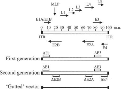 Figure 96-1. Transcription map and genome organization of adenovirus and adenovirus-derived gene transfer vectors. A schematic representation of the adenovirus genome is shown at the top of the figure. The length of the linear double-stranded adenovirus genome is approximately 36 kb and is arbitrarily divided into 100 map units. Black boxes at the terminal ends of the genome represent inverted terminal repeats (ITR). Arrows indicate the direction of transcription for the major messenger ribonucleic acids (mRNAs). E1–E4 indicate early transcripts, L1–L5 represent late transcripts, and MLP is the position of the major late promoter. Below are represented the first-, second-, and third-generation (or gutted) adenovirus vectors. The transcription regions that are deleted are indicated in each vector. (Adapted with permission from Wu Q, Moyana T, Xiang J: Cancer gene therapy by adenovirus-mediated gene transfer. Curr Gene Ther 1:101, 2001.) |
STRUCTURE OF THE VIRUS
Adenoviruses are nonenveloped viruses. The viral structural proteins are DNA associated or involved in formation of the complex capsid encapsulating the viral DNA. The DNA-associated proteins include the terminal protein (TP) that binds to the 5′ termini of the viral genome containing the inverted terminal repeats (ITRs).25 Other core proteins include polypeptides V and VII and protein X. Like cellular histones, these proteins are rich in the basic amino acid arginine, but unlike histones they contain appreciable amounts of tryptophan.26,27 The precise function of each of the core proteins has yet to be elucidated, but protein V may play a role in interfering with host cellular function by redistributing nucleolar antigens.28
The icosahedral capsid structure of adenovirus is made up primarily of three proteins: hexon, penton base, and fiber as shown in Figure 96-2. The capsid is formed from 252 unit structures, known as capsomeres, arranged in an icosahedral structure with 20 triangular sides. Hexagonal capsomeres or hexons constitute 240 of the structural subunits.29 Each hexon is composed of three different polypeptides: VI, VII, and IX. There are also 12 pentagonal capsomeres or pentons. Each penton is composed of an 85-kilodalton (kDa) protein, polypeptide III. Polypeptide III is the largest protein of the entire viral capsid. Projecting from each penton is a rodlike structure called a fiber made up of three molecules of protein IV, a 62-kDa polypeptide.30 The fiber and penton base not only contribute to the capsid structure but also are intimately involved in the attachment of the viral particle to the host cell surface.
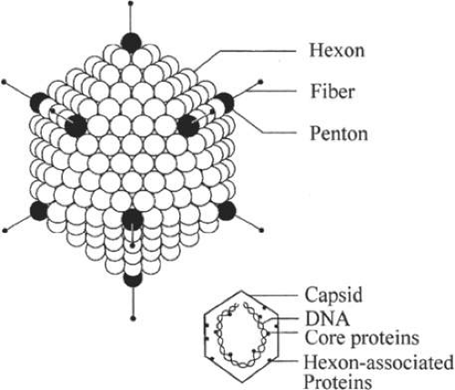 Figure 96-2. Structure of adenovirus. The icosahedral capsid structure of adenovirus is made up of three proteins, hexon, penton base, and fiber. A cross-sectional view of the viral capsid. Some proteins are associated with viral DNA, whereas others are associated with hexon and are involved in the formation of the capsid. |
VIRAL LIFE CYCLE
The replication cycle of adenovirus may be divided into three stages: host infection, DNA replication, and construction of new viral particles. The fiber protein of the viral capsid attaches to a primary receptor on the host cell surface, much like an anchor. Three types of host cell-surface molecules can act as the primary receptor: the coxsackievirus group B and adenovirus receptor (CAR),31 the major histocompatibility complex (MHC)-I α2 subunit,32 and sialic acid residues on glycoprotein.33 Once the viral particle is tethered through the primary interaction, a second interaction between the penton base and a secondary cell-surface receptor, the integrins αvβ3 and αvβ5, takes place. The integrins, in turn, promote internalization of the viral particle through receptor-mediated endocytosis.34 Once inside the cell, the outer capsid protein is partially disassembled because of acidification of the contents of the endosome. This allows the virus to escape the endosome before the formation of the lysosome35,36 and enter the cytoplasm. The process of viral internalization and release to the cytosol takes about 15 minutes.37 Virus particles are then translocated to the nucleus along the microtubule network.38 The adenoviral particle undergoes further disassembly, and, as a final step, the Ad hexon proteins remain at the nuclear membrane while the DNA is released into the nucleus.37
Stay updated, free articles. Join our Telegram channel

Full access? Get Clinical Tree


