Actinomyces Organisms in Ocular Disease
Carolyn Y. Shih
Michael P. Vrabec
George J. Florakis
Taxonomy
Actinomycetes are facultatively anaerobic or strictly anaerobic gram-positive rods that tend to grow slowly as branching filaments. Because the filamentous growth leads to mycelial colonies and some cause chronic subcutaneous granulomas such as those caused by fungi, the actinomycetes have been regarded as fungi. However, they are true bacteria in that they are prokaryotic, they have cell walls with muramic acid and diaminopimelic acid, their cell membranes do not contain sterols and are therefore insensitive to fungicidal polyenes, and their growth is inhibited by antibacterial drugs such as penicillins, tetracyclines, and sulfonamides, which are innocuous for fungi. Within the order Actinomycetales are the following genera: Mycobacterium, Actinomyces, Dermatophilus, Nocardia, and Streptomyces. There are numerous species of actinomyces that have been described, including Actinomyces israelii, Actinomyces meyeri, Actinomyces naeslundii, Actinomyces odontolyticus, and Actinomyces viscosus. However, A. israelii is the species that has been implicated most frequently in ocular infections.1
Detection and Isolation
Direct microscopic examination of clinical specimens for gram-positive rods may permit a diagnosis of actinomyces (Figs. 60.1,60.2,60.3). Typically, extensive granulation tissue containing polymorphonuclear leukocytes, lymphocytes, plasma cells, and fibroblasts is noted. Specimens stained with the Brown-Brenn stain show sulfur granules, which are diagnostic for actinomycosis.2 With hematoxylin-and-eosin stain, the core of the granule is basophilic, and the periphery is eosinophilic. Peripheral club-shaped filaments surround the central mass of filaments in a radial arrangement.3,4,5 The acid-fast Putt modification of the Ziehl-Neelsen stain (using a weaker decolorizer) makes Actinomyces species appear partially acid fast (i.e., speckled red).
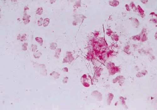 Figure 60.1 Gram stain of actinomyces; gram-positive, branching organisms are noted. Source: Courtesy of David Meisler, MD, Division of Ophthalmology, Cleveland Clinic, Cleveland, OH |
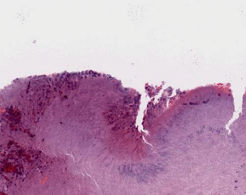 Figure 60.2 Hematoxylin-and-eosin stain of a dacryolith. Source: Courtesy of Robert Folberg, MD, and Marsha Apushkin, MD, Department of Pathology, University of Illinois at Chicago, Chicago |
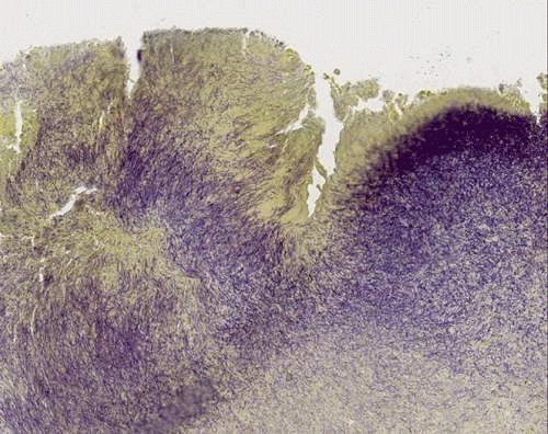 Figure 60.3 Gram stain of dacryolith showing actinomyces. |
Actinomyces are mobile or nonmotile chemo-organotrophs (requiring preformed organic compounds as a source of carbon and oxidizing organic compounds as a source of energy) and can be differentiated by their ability (saccharoclastic) or inability (nonsaccharoclastic) to cleave complex sugars. Some species are catalase positive, whereas others are catalase negative. Energy is provided by fermentative glucose metabolism. The metabolic products include succinic acid, lactic acid, and small amounts of acetic acid. There is no indole production. Nitrate reduction is variable among species. Other traits that differentiate species of actinomyces include gelatin hydrolysis and H2S production on triple iron agar. It is, however, difficult to tell some of the species apart.
Actinomyces species can be divided into strict anaerobes (Actinomyces bovis, A. israelii, and A. meyeri) and facultative anaerobes (all others).2 Most strains of A. Israelii form rough colonies on agar and grow at the bottom of broth tubes as discrete aggregated clumps—often called “bread crumbs.” These organisms are anaerobic or microaerophilic and require rich media (e.g., containing blood or brain-heart infusion); growth is stimulated by CO2 and is poor at temperatures <37°C.
Transport and Storage of Specimens
Samples obtained from sites of infection should be treated as anaerobic, although some species are aerotolerant. The most important parameter in the successful isolation of Actinomyces species is minimizing the time between specimen collection and the incubation of the inoculated media.
Usually, the specimen is collected from the punctum or canaliculus because this is the most common site of infection involving the eye. If the material can be expressed from the punctum to a cotton-tipped applicator, it is best directly placed in an anaerobic transfer media for plating in the laboratory. Sometimes a concurrent canalostomy, along with curettage, must be performed to obtain an adequate sample.
Clinical Significance
Disease caused by actinomyces is termed actinomycoses. Although it has been long assumed that actinomycosis arose from exogenous infection by organisms in the soil, most actinomycoses are of endogenous origin. Actinomyces organisms colonize the upper respiratory, gastrointestinal, and female genital tracts. The organisms have a low virulence potential and cause disease only when the normal mucosal barriers are disrupted by trauma, surgery, or infection. The most common etiologic agent in humans is A. israelii, although most infections in humans are polymicrobic.2,6 A. viscosus and A. naeslundii play a significant role in dental caries and periodontal disease.7
Stay updated, free articles. Join our Telegram channel

Full access? Get Clinical Tree


