8
Intermediate Uveitis
John J. Huang and Vicente Diaz
INTRODUCTION
Intermediate uveitis (IU) is the term for a subset of uveitis in which the vitreous is the major site of inflammation, including those cases in which there is inflammation of the pars plana and peripheral retina. Macular edema and vascular sheathing occasionally occur in IU.
IU may be associated with systemic inflammatory diseases such as multiple sclerosis (MS) or sarcoidosis as well as infectious conditions such as syphilis, Lyme disease, cat scratch disease, and viral infections. IU with pars plana exudation in the absence of an underlying systemic disease is termed “pars planitis.”
The first part of this chapter will cover the management steps for IU, and then specific diseases causing IU will be covered.
EPIDEMIOLOGY
IU accounts for between 4% and 25% of all uveitis cases and somewhere between 16% and 33% of uveitis in children. The overall prevalence of IU is estimated to be 5.9/100,000. Patients as young as 5 years old are well recognized, and it has been reported that there is a bimodal age distribution with one peak occurring in the second decade and another peak in the third/fourth decade of life. There does not appear to be an association with regard to gender or race.
CLINICAL FEATURES
The most common presenting symptoms are blurry vision and floaters. Pain and photophobia are unusual. The disease is noted to be bilateral in nearly 80% of cases, and it appears that one third of unilateral cases will eventually become bilateral. Visual loss in IU most commonly occurs due to macular edema, while uveitic glaucoma, cataracts, and vitreous hemorrhage are less common causes. Cyclitic membrane formation is a complication of severe prolonged disease.
Vitritis is a defining examination; its absence precludes the diagnosis. Notable anterior inflammation is uncommon, is usually only in young patients, and is very early in the disease process. Aggregates of inflammatory cells may appear in the lower vitreous and are widely referred to as “snowballs.” A “snowbank” is the term for grayish yellow exudation found along the inferior ora serrata, and in severe cases, this exudation may encircle the entire ora serrata over 360 degrees.
CLINICAL MANAGEMENT
We present below a schema for the practical management of patients with IU.
As with all uveitis, the steps for management are (i) classify the process thoroughly, (ii) attempt to determine an etiology, (iii) establish immediate control, and (iv) maintain long-term control.
Management Step 1: Classify The Process
We classify uveitis so as to enable subsequent diagnostic efforts.
As delineated elsewhere, uveitis is classified as
- Acute, recurrent, or chronic in duration
- Unilateral or bilateral
- Granulomatous or nongranulomatous in character
- Anterior, intermediate, or posterior in location
It is essential that practitioners who treat uveitis are familiar with the classification scheme described in Chapter 6.
The recommendations in this chapter apply to patients with IU, that is, centered in the vitreous cavity with a less significant iritis and without evidence of retinitis or choroiditis.
Determining Whether Intermediate Uveitis Is Acute, Recurrent, or Chronic
For all patients with uveitis, it can be difficult to ascertain how long his or her disease has been active, or whether he or she had a prior episode in the remote past, since the disease onset can be quite insidious and patients may be unaware of their disease for a long time. Nevertheless, certain findings generally indicate that a patient with IU has probably had it for more than 3 months (i.e., chronic disease):
- In a young patient whose vitreous should not normally be liquefied, a history of visual floaters more than 3 months before (In older patients with vitreous syneresis, this symptom is much less specific.)
- Macular edema severe enough to cause visual loss
- Fibrotic appearing and hyalinized exudation at the pars plana
These findings indicate chronic disease. We rely on a patient’s history to differentiate chronic from recurrent disease.
Determining Whether Intermediate Uveitis Is Bilateral or Unilateral
One may occasionally find a patient with symptomatic IU in one eye but no symptoms in the other. Fortunately, almost any episode of IU leaves behind a very obvious footprint, in the form of residual vitreous cells, making it easy to determine if the asymptomatic eye is or has ever been affected. If the fellow eye has even a few vitreous cells, active or not, then the process is bilateral.
Determining Whether Intermediate Uveitis Is Granulomatous
Most IU is nongranulomatous. Examination findings indicating a granulomatous process are found in the anterior chamber, and IU by definition has relatively little anterior chamber inflammation. One may, however, see slight keratic precipitates or iris nodules or residua of keratic precipitates (which may look like hyalinized and glassy or like pigmented flecks or as endothelial “footprints” notable as a slightly hazy dot on the endothelium).
Management Step 2: Try to Ascertain The Etiology
The list of systemic illnesses that cause IU is short, and most of these entities are suggested from the examination and the patient’s history and review of systems. While there are no ancillary tests that are “routine” in our workup for IU, it is important to consider the disease listed below, which are intended as a working guide to assist practitioners in the evaluation and management of most cases of IU that one is likely to encounter. Most of these diseases are described further in Chapter 15.
Sarcoidosis
This disease can present with virtually any form of ocular inflammation, including purely IU, which may be acute or chronic, and is usually bilateral, granulomatous or nongranulomatous. If patients have skin lesions, pulmonary complaints, or some other reason to suggest sarcoidosis, we check an angiotensin converting enzyme (ACE) level and usually a chest X-ray. If the presentation is granulomatous or if pulmonary symptoms are present, we prefer a chest CT scan with contrast. Sarcoidosis is discussed in more detail in Chapter 15.
Multiple Sclerosis
Most patients with MS-related uveitis already have established MS by the time their uveitis develops; it is in fact quite rare that uveitis is the presenting problem for which patients with undiagnosed MS seek medical care. MS-related uveitis can present as either IU or diffuse uveitis, which is generally chronic or recurrent, bilateral, and may be granulomatous. In patients with IU who note any unexplained neurologic deficits such as extremity tingling, numbness or weakness, especially when these symptoms are made worse by heat, the test of choice is a magnetic resonance imaging (MRI) scan of the brain and spinal cord, with gadolinium contrast. We do not pursue—or really entertain—the diagnosis of MS in patients who do not have neurologic symptoms.
Lyme Disease
IU due to Lyme disease is generally chronic, bilateral, and may be granulomatous. If patients give a history of a rash or joint pain within months prior to presentation, we order Lyme serologies. We do not order Lyme serologies without suggestive findings on review of systems, specifically arthritis. Lyme disease is discussed in Chapter 15.
Syphilis
Most IU presents during the latent stage of syphilis infection and is generally chronic, bilateral, and may be granulomatous. We check a fluorescent treponemal antibody absorption (FTA-ABS) or other treponemal syphilis test in patients whose history suggests they are at risks for the disease or in those who have any element of retinal or choroidal infiltration, or in patients whose IU has not responded to corticosteroid therapy. We do not routinely order syphilis serologies. Syphilis is discussed in Chapter 15.
Tuberculosis
Uveitis due to tuberculosis (TB) is rare as a cause of uveitis in developed countries and would most commonly be chronic, unilateral, and may be granulomatous. We suspect TB in patients who have immigrated from areas where TB is endemic, or who live in contact with persons who have the disease, and we test for it with a purified protein derivative (PPD) test, which we confirm with a quantiferon gold assay. TB is discussed in Chapter 15.
Herpes Zoster Ophthalmicus
IU due to herpes zoster is generally chronic, always unilateral, and usually nongranulomatous. The history will almost always indicate this diagnosis, which does not require ancillary testing. Herpes zoster is discussed in Chapter 15.
Lymphoma of the Central Nervous System
We suspect lymphoma in patients whose vitreitis is chronic, unilateral or bilateral, nongranulomatous, presenting in older patients, and associated with infiltrates in the retina or choroid. If patients with IU develop such infiltrates, neurologic symptoms, we order a central nervous system (CNS) magnetic resonance imaging (MRI), refer the patient for lumbar puncture, and strongly consider vitreous biopsy if syphilis testing is negative. Lymphoma is discussed in Chapter 15.
Summary
In the absence of history or review of systems findings suggesting one of these entities, we do not feel compelled to order ancillary tests. IU that is not attributable to a systemic illness “defaults” to the diagnosis of pars planitis if pars plana exudation is noted (which is typically the case). This diagnosis accounts for the majority of IU in our practice, and a more complete discussion of pars planitis is presented at the end of this chapter.
These disease entities and other more unusual causes of IU are explained in greater detail below.
Management Step 3: Treat for Immediate Control
We consider “immediate” to mean the first 6 months after presentation in patients with IU, during which time we feel control of the process should be achieved. In most cases of IU, no etiology is forthcoming, and thus treatment is empiric.
General Treatment Considerations
- Corticosteroids, systemically or by periocular injection, are the mainstay of initial treatment for most patients—including those cases due to infection.
- Patients whose disease is known to be due to infection should also receive antimicrobial therapy concomitantly.
- Topical corticosteroids are not suitable for treating IU, since eyedrops generally do not penetrate much deeper than the lens/iris diaphragm.
Perhaps the most challenging aspect of managing IU is assessing the efficacy of therapy. The reason for this is that, unlike aqueous humor, vitreous gel traps cells such that the cells one sees on examination could have been there for many months or years, making it very difficult to detect all but very large changes in a patient’s vitritis. Our preference is therefore to measure and follow macular thickness with optical coherence tomography (OCT) as a surrogate indicator inflammation in patients with IU.
Treatment Steps for Intermediate Uveitis on Initial Presentation
The following guidelines should enable the management of most patients who present with IU:
1. Determine whether treatment is indicated. Not all cases of IU require treatment, especially if the extent of inflammation and secondary involvement are not severe. A substantial minority of patients with IU will have disease that waxes and wanes of its own accord without affecting visual function. We treat if any of the following are true or present:
- Macular edema detectable by OCT regardless of visual acuity. Most patients whom we treat with IU are in this category.
- Secondary retinal vaculitis detectable on fluorescein angiography (FA).
- Any known infectious etiology. The great majority of IU is not infectious, and so in the absence of convincing evidence to the contrary, we assume IU to be a primary immune-mediated (i.e., not infectious) process.
- The patient wants treatment and understands the risks of intraocular pressure (IOP) elevation and cataract formation. We find that patients with good vision and no macular edema frequently ask for treatment because they are bothered by visual floaters, although for many patients the main issue is fear of eventual visual loss. We do not discourage this, because we believe that a lot of symptomatic IU will eventually cause macular edema and/or epimacular fibrosis if left untreated, and thus the benefits of treatment generally justify the associated risks.
2. If treatment is indicated, start with oral corticosteroids, which should achieve control of inflammation that is immune mediated. A 1- to 2-week course of prednisone 1 mg/kg/day (or equivalent other corticosteroid) in a single dose after breakfast or lunch is satisfactory; nearly all immune-mediated uveitis will improve substantially with this treatment course. If the disease does not improve convincingly, we suspect infection or (in an older patient) malignancy.
3. Treat with topical corticosteroids concomitant with oral corticosteroids as a trial therapy to “tease out” a propensity for very early and marked steroid-induced IOP elevation. Prednisolone 1% or dexamethasone 0.1% for at least 2 weeks is an appropriate agent. These drops will not, by themselves, treat the patient’s IU. IOP elevation above 30 mm Hg by this point is a contraindication to local ocular corticosteroids (we skip to step 6 below).
Note: IOP elevation can occur after corticosteroid use of any duration, and this brief trial will detect only patients with an extraordinarily severe propensity for steroid-induced IOP elevation. Ideally, one would use topical corticosteroids for 6 weeks to detect a much larger percentage of the “steroid responders,” and while we occasionally do this in very mild cases of IU in which a patient nevertheless requests treatment, we tend to reason that since most steroid-induced IOP elevation is easily manageable, it is not in the patient’s best interest—especially if macular edema is present—to use topical corticosteroids for 6 weeks without actually treating his or her disease.
4. Give a periocular corticosteroid injection if the IOP has not risen above 30 after 2 weeks of topical corticosteroid treatment. Our preferred agent is triamcinolone acetonide 40 mg/mL, of which we typically inject 20 to 40 mg superotemporally along the equator of the globe in the subtenon space or inferotemporally through the eyelid along the orbital floor.
5. Reevaluate 3 to 6 weeks later with OCT and/or FA. If there is still more than mild macular edema or secondary retinal vasculitis, repeat the injection, and reevaluate again after 3 to 6 weeks, anticipating near total control of the process. Note that if patients have severe macular edema on presentation, such that we believe that one periocular injection will not likely achieve sufficient control, we may opt to treat initially with intravitreous corticosteroid injections (see special case d, below);
6. Special cases
- Patients are not good candidates for oral corticosteroid therapy (usually due to diabetes or a history of steroid-induced psychosis). There is no way to confirm that these patients’ IU is immune- mediated before proceeding to periocular injection. We use the standard 2 or more weeks of trial therapy with topical corticosteroids to assess for severe and early IOP elevation, and then go directly to injections (it is important that the patient understands the slight risk that this approach entails, since uveitis that is infectious will not improve and may even worsen after periocular corticosteroid injection).
- Patients cannot be treated with periocular corticosteroid injections or they decline this approach. Use oral corticosteroids, anticipating at least 6 weeks of therapy. Start with prednisone 1 mg/kg/day and taper by 10 mg weekly down to 20 mg per day. Calcium and vitamin D supplementations are important in these cases, and we use bisphosphonate therapy in patients who are not women of child bearing age or younger. We schedule follow-up weekly, and patients generally notice improvement after 2 weeks of therapy.
- Patients show no improvement in inflammation after 2 weeks of oral corticosteroids or a periocular injection. Suspect infection or malignancy, and order tests for syphilis, Lyme disease, and, possibly, magnetic resonance imaging of the CNS.
- There is very severe macular edema on initial presentation. This scenario merits treatment with intravitreous corticosteroid injections. We use triamcinolone 2 mg in 0.05 ml injected through the pars plana for these patients. While this approach is effective in controlling inflammation and macular edema, the secondary cataract and IOP elevation can be severe, and so we try to avoid repeat injections and do not consider this therapy suitable for long-term management.
- Severe IOP elevation occurs following periocular injection. This phenomenon can occur at any point following the injection and is usually treatable with IOP-lowering eyedrops. If the IOP does not come down in response to eyedrop therapy, we remove the drug deposit under local anesthesia in our office (this is enabled by placing the triamcinolone injection along the equator of the globe, where it is accessible fairly easily). Obviously, these patients are not candidates for repeat injections.
In summary, immediate control of IU centers around corticosteroids, either by periocular injection or orally. Infectious causes are treated concomitantly. We use OCT to measure small changes in the degree of intraocular inflammation. Having achieved immediate control, we look toward maintaining control over the long term.
Management Step 4: Maintain Long-Term Control
Once we have achieved immediate control of a patient’s IU, we generally encounter one of three scenarios:
1. The disease resolves and no further anti-inflammatory treatment is necessary. This is certainly true of uveitis due to syphilis and other bacterial infections, as well as herpetic infections that are treated with chronic antiviral drugs. Some cases of noninfectious IU may also resolve after initial treatment.
2. The disease becomes quiescent for many months or years before recurring.
3. The disease recurs soon after the initial period of immediate control.
It is patients in this last group who require long-term treatment either with serial periocular corticosteroid injections or with immunosuppression or surgical therapy. Decision making at this point depends greatly on the individual circumstances of each patient, and each approach requires modification to fit the circumstances. Acknowledging the need for nuanced consideration, we observe the following:
- Periocular corticosteroid injections are appropriate for some patients as the sole means of treating their disease. We opt for this approach in patients (i) who do not require systemic corticosteroid therapy for the management of a systemic autoimmune disease (such as sarcoidosis), (ii) who do not have a substantial degree of corticosteroid-induced IOP elevation, and (iii) who understand that they are likely to develop posterior subcapsular cataracts that may require cataract surgery. A substantial percentage of our patients meet these criteria, and this approach is common in our practice. How frequently can periocular injections be administered? Practitioners’ views vary greatly along these lines (as do their injection techniques), and patients’ tolerance is also variable. We generally give injections as often as needed until the first signs of toxicity become apparent. In our experience, it is unusual that patients require more than one periocular injection of triamcinolone 20 mg (given in the superotemporal or inferotemporal subtenon space) every 4 months.
- Immunomodulatory therapy (also termed “immunosuppressive therapy” or “immunosuppression” or “immunomodulation”) is appropriate for patients who cannot tolerate as many periocular corticosteroid injections as they need or who have a systemic autoimmune disease underlying their uveitis. We discuss the use of these agents in Chapter 19. We typically start with methotrexate. Since this drug takes approximately 8 weeks to show a clinical effect, we achieve immediate control with periocular triamcinolone injections or oral corticosteroids in the meantime, anticipating that the immunosuppressive drug will take effect as the triamcinolone effect begins to wear off. If methotrexate does not work, we face the choice of recommending stepped-up immunosuppression, usually with mycophenolate and possibly cyclosporine, or surgical therapy.
- Surgical therapy for IU includes therapeutic vitrectomy, pars plana cryopexy, pars plana photocoagulation, and the use of an intraocular corticosteroid implant. We consider surgical treatment in patients who are not candidates for immunosuppression or in whom this approach is not well tolerated or has not been effective.
- Pars plana vitrectomy is occasionally helpful in decreasing the severity of patients’ IU, and we opt for this approach when attempts at immunosuppression have either failed or are contraindicated. This approach does not work in all patients, and there is unfortunately no way to identify likely beneficiaries in advance. At the very least, however, vitrectomy makes it easy to follow pars planitis, since by making the vitreous cavity liquid, one can more easily assess for changes in the severity of vitreitis simply by counting cells. When vitrectomy works well, we find that patients can be treated with periocular triamcinolone injections at less infrequent intervals than was previously necessary. Thus, vitrectomy is useful for enabling treatment with corticosteroids rather than being a “one-off” treatment by itself.
- Pars plana cryopexy or laser photocoagulation. It was observed in the 1960s that, in patients with pars planitis, destruction of the retina with cryopexy just posterior to areas of pars plana exudation often had the effect of diminishing the severity of patients’ IU. Indirect laser photocoagulation was then found to have a similar result. The mechanism of this therapeutic effect is not known, and the benefit is often short-lived, but it is a useful adjunct for many patients. We opt for it as second- or third-line therapy in patients with marked pars plana exudation, and we prefer to perform it in conjunction with pars plana vitrectomy.
- An intraocular fluocinolone acetonide intravitreal implant (Retisert, Bausch & Lomb, Rochester, NY) is very effective for treating severe uveitis affecting the posterior segment. We consider this a therapy of last resort for patients in whom other approaches, including surgical approaches, have not effected lasting disease control. Most of these patients have already had cataract extraction, many have required at least some IOP-lowering eyedrops, and most have also been vitrectomized. The field of intraocular implant devices is evolving, and we believe it will have an increasing role in the management of patients with eye-limited inflammation.
- Pars plana cryopexy or laser photocoagulation. It was observed in the 1960s that, in patients with pars planitis, destruction of the retina with cryopexy just posterior to areas of pars plana exudation often had the effect of diminishing the severity of patients’ IU. Indirect laser photocoagulation was then found to have a similar result. The mechanism of this therapeutic effect is not known, and the benefit is often short-lived, but it is a useful adjunct for many patients. We opt for it as second- or third-line therapy in patients with marked pars plana exudation, and we prefer to perform it in conjunction with pars plana vitrectomy.
- Pars plana vitrectomy is occasionally helpful in decreasing the severity of patients’ IU, and we opt for this approach when attempts at immunosuppression have either failed or are contraindicated. This approach does not work in all patients, and there is unfortunately no way to identify likely beneficiaries in advance. At the very least, however, vitrectomy makes it easy to follow pars planitis, since by making the vitreous cavity liquid, one can more easily assess for changes in the severity of vitreitis simply by counting cells. When vitrectomy works well, we find that patients can be treated with periocular triamcinolone injections at less infrequent intervals than was previously necessary. Thus, vitrectomy is useful for enabling treatment with corticosteroids rather than being a “one-off” treatment by itself.
In summary, not all patients with IU require long-term therapy, but for those who do, the options are serial periocular injections, immunosuppression, or surgical therapy with vitrectomy, pars plana ablation, or corticosteroid implants.
PARS PLANITIS/IDIOPATHIC INTERMEDIATE UVEITIS
Pars planitis is idiopathic IU that usually affects persons in the second to third decades of life. It accounts for well over 50% of patients with IU. The course of this disease is variable and ranges from a self-limited disease to a severe and vision-threatening illness. The most frequent complaint is decreased visual acuity. Histopathologic and clinical findings suggest an autoimmune etiology.
Clinical Findings
Systemic Findings
- None, by definition.
Ocular Findings
- Vitritis, bilateral 80% of the time.
- Vitreous snowballs (Fig. 8.1).
- Anterior infl ammation, with as much as ten cells/hpf on initial presentation in young patients, otherwise less.
- Peripheral retinal vasculitis, usually in areas of obvious vitreous inflammation.
- Papillitis is common on initial presentation, and if severe and prolonged, it may induce lasting optic neuropathy.
- Pars plana exudation, which generally requires scleral depression to be seen clearly.
- Macular edema is a common complication of a prolonged disease.
- Cyclitic membranes form adherent to the ciliary processes in very severe and prolonged disease and often cause hypotony.
- Optic nerve neovascularization indicates a severe, prolonged disease.
- Macular edema is a common complication of a prolonged disease.
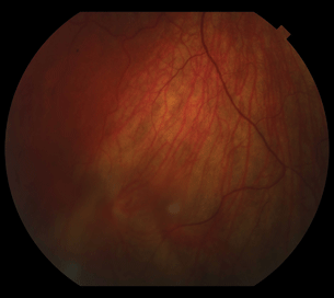
Figure 8.1 Snowball formation in the inferior retina of a patient with idiopathic pars planitis.
Stay updated, free articles. Join our Telegram channel

Full access? Get Clinical Tree


