Nonvalved Implants
Molteno: The original drainage device, introduced in 1969. A third-generation model is available as a 175-mm2 or 230-mm2 single plate, made of rigid polypropylene (FIG. 5.1.1C). Older models are still available, including a double plate implant (FIG. 5.1.1D).
Baerveldt: A pliable silicone reservoir with lateral “wings” that must be tucked beneath the lateral and superior rectus muscles. It is available in 250-mm2 and 350-mm2 sizes (FIG. 5.1.1B).
Tube Placement
Tubes are most commonly placed in the anterior chamber, but can also be introduced through the ciliary sulcus or pars plana in aphakic or pseudophakic eyes.
Pars plana placement requires vitrectomy because strands of vitreous would otherwise obstruct the tube and prevent drainage.
ALTERNATIVES
Medical/Laser Therapy
Maximum topical IOP-lowering therapy is typically pursued prior to considering incisional surgery. In some cases, topical therapy may be ineffective in lowering IOP, may cause adverse side effects, or the patient may be unable to afford medications or comply with their administration.
Laser trabeculoplasty (argon laser trabeculoplasty [ALT] or selective laser trabeculoplasty [SLT]): Application of laser spots to the pigmented trabecular meshwork stimulates trabecular meshwork function and increases aqueous drainage, lowering the IOP. This procedure should be used with caution in uveitic eyes and may not lower pressures sufficiently in glaucoma with low or average IOP.
Timely intraocular anti-VEGF (vascular endothelial growth factor) injections (bevacizumab or ranibizumab) or panretinal photocoagulation (see Chapter 8.3 Retinal Laser Photocoagulation) can inhibit or reverse the development of anterior chamber neovascularization and scarring. This may prevent the onset of NVG.
Surgery
- Trabeculectomy: A guarded fistula is created between the anterior chamber and sub-Tenon space, reducing IOP by permitting the aqueous to drain beneath the scleral flap, filter into a conjunctival bleb, and reabsorb into the blood, lymph, and tear film or evaporate after transudation.
- Canaloplasty: A “nonpenetrating” glaucoma surgery that reduces IOP by dilating Schlemm’s canal to increase outflow through the trabecular meshwork. After injection of viscoelastic, a Prolene suture is threaded through the canal and tightened to stent open the meshwork and increase its porosity. This procedure is generally not effective in neovascular, inflammatory, or chronic angle closure glaucomas.
- Trabectome (i.e., ab interno trabeculotomy): A specialized bipolar electro-ablation device is introduced into the anterior chamber through a clear corneal incision to remove a segment of the trabecular meshwork and “unroof” the underlying Schlemm’s canal. This lowers pressure by permitting more direct outflow through the collector channels and episcleral veins.
- Ex-PRESS Mini Glaucoma Shunt: A 3-mm stainless steel drainage tube is inserted in the anterior chamber angle to bypass the trabecular meshwork. The implant is placed beneath a partial-thickness scleral flap to regulate outflow and create a trabeculectomy-like filtration bleb.
- iStent: Using a clear corneal incision, a 1-mm heparin-coated titanium drainage tube is inserted through the trabecular meshwork and into Schlemm’s canal, reducing the resistance to aqueous outflow.
- Cyclodestruction: Diode laser cyclophotocoagulation or cryotherapy (which has fallen out of favor as too destructive and inflammatory) can reduce aqueous production by ablating a portion of the ciliary body, either transsclerally or via an endoscopic approach. Ciliary body destruction is generally irreversible, although the aqueous humor producing nonpigmented epithelium may partially regenerate (Note: This is more common in children, but can occur in adults as well). The IOP response is not immediate and repeat administration may be required to achieve satisfactory results.
NOTE: Low IOPs can be irreversible and may lead to phthisis, so the procedure is generally reserved for patients with poor preoperative vision or for those who are not good surgical candidates.
RELEVANT ANATOMY AND PATHOPHYSIOLOGY
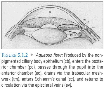
Anatomy of Aqueous Drainage (FIG. 5.1.2)
Neovascular Glaucoma (NVG)
Ischemic retinal disorders (e.g., diabetic retinopathy or central retinal vein occlusion) can cause neovascularization of the iris and anterior chamber angle due to VEGF release (also known as rubeosis iridis).
This initially causes secondary open angle glaucoma due to obstruction of the trabecular meshwork by a fibrovascular membrane. In later stages, it leads to secondary angle closure as formation of anterior synechiae pulls the peripheral iris toward the cornea.
Anti-VEGF agents (bevacizumab and ranibizumab) and PRP are the most effective initial therapy because they reduce the stimulus for neovascularization. Once NVG has developed, drainage implants become the therapy of choice.
Uveitic Glaucoma
Protein and inflammatory cells accumulate due to the breakdown of the blood-ocular barrier.
Inflammation within the anterior chamber leads to formation of synechiae between the peripheral iris and cornea.
As with NVG, this causes angle closure and increased intraocular pressure.
Iridocorneal Endothelial (ICE) Syndrome
A group of disorders characterized by abnormal corneal endothelium.
These disorders cause iris atrophy, corneal edema, and secondary angle closure due to the formation of peripheral anterior synechiae.
PREOPERATIVE SCREENING
Establish whether current glaucoma therapy is adequate using serial IOP measurements. Determine the rate of disease progression and the target IOP with serial evaluation of optic disc morphology and visual fields.
Conduct a careful external exam to determine whether there is conjunctival scarring from prior surgery or inflammation.
Perform a full slit-lamp exam with gonioscopy to examine the angle morphology and to check for signs of anterior chamber neovascularization or inflammation (e.g., cell and flare).
ANESTHESIA
Retrobulbar, peribulbar or topical/subconjunctival anesthesia is sufficient for most patients, and it can be supplemented by local infiltration of the tissues that are dissected during plate placement.
General anesthesia may be necessary for pediatric and uncooperative patients.
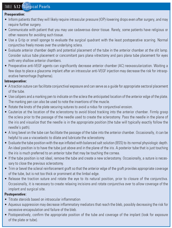
PROCEDURE
 SEE TABLES 5.1.1 Rapid Review of Steps and 5.1.2 Surgical Pearls, and WEB TABLES 5.1.1 to 5.1.4 for equipment and medication lists.
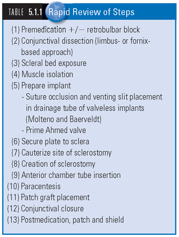
- Preoperative Medication: If IOP is greater than 35 mm Hg, the patient can be pretreated with IV mannitol or other pressure-lowering medications.
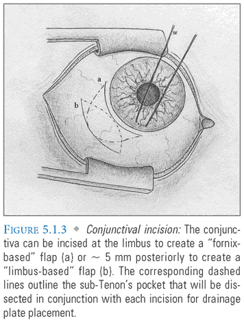
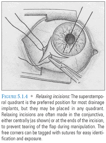
- Conjunctival Incision: The conjunctiva is generally incised at the limbus to create a fornix-based flap. If there is extensive conjunctival scarring or a history of prior filtration surgery, a limbus-based flap may be substituted (FIG. 5.1.3). It is important to avoid prior trabeculectomy sites as disruption may result in overfiltration and hypotony. Single-plate implants require dissection in only one quadrant and can be placed through a shorter conjunctival incision. The superotemporal quadrant is preferred, but any quadrant can be used if necessary. Double plate implants require dissection of two quadrants (most often the superotemporal and supranasal), and therefore require a longer incision. Relaxing incisions are often made in the conjunctiva to prevent tearing of the flap during manipulation. The free corners can be tagged with sutures for easy identification and to aid in exposure (FIG. 5.1.4).
- Scleral Bed Exposure: Conjunctiva and Tenon’s capsule are elevated from the underlying sclera with blunt dissection (FIG. 5.1.5). This creates a pocket for plate placement.

- Muscle Isolation: The scleral insertions of the adjacent rectus muscles are identified and captured with muscle hooks. The muscle bodies are dissected free of surrounding fascial tissue. Thick sutures can then be passed beneath the muscles to facilitate their manipulation during plate placement.
- Plate Placement: Implants are generally secured to the sclera about 8 mm from the limbus by passing partial-thickness sutures through anterior holes in the plate(s). This position offers protection while still allowing implant visualization through the conjunctiva on downgaze. Each device also has specific placement guidelines, depending on the number and shape of drainage plates.
Ahmed: This valved device requires priming prior to implantation; forced irrigation through the drainage tube separates the valve leaflets and ensures their subsequent function. Because of its longer anterior–posterior dimension and narrow horizontal dimension, the plate does not overlap with the adjacent rectus muscles.
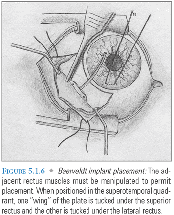
Baerveldt: This valveless implant requires temporary suture occlusion to prevent hypotony (see Suture Occlusion). It is significantly wider than the Ahmed, and the adjacent rectus muscles must be manipulated to permit placement. When positioned in the superotemporal quadrant, one “wing” of the plate is tucked under the superior rectus and the other is tucked under the lateral rectus (FIG. 5.1.6).
Double Plate Molteno: Like the Baerveldt, this valveless device requires temporary suture occlusion to prevent hypotony (see Suture Occlusion). Usually, one plate is positioned in each superior quadrant. The plates are joined with a connecting tube that is routed above the superior rectus.
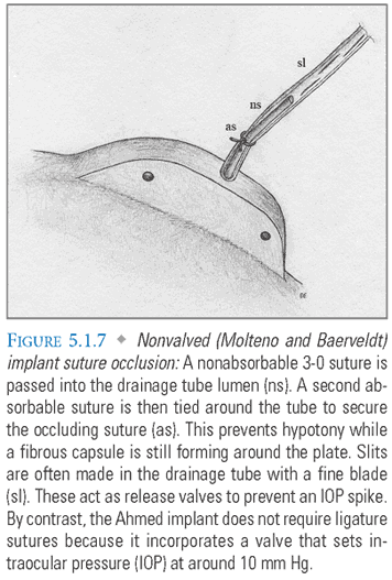
- Suture Occlusion (Molteno and Baerveldt Only): A nonabsorbable 3-0 suture is passed into the drainage tube lumen. A second absorbable suture is then tied around the tube to secure the occluding suture. This prevents hypotony while a fibrous capsule is still forming around the plate. The surgeon can then remove the nonabsorbable suture, or the absorbable suture will generally dissolve after 4 to 6 weeks, restoring flow into the capsule. Slits are often made in the drainage tube with a fine blade. These act as release valves to prevent an IOP spike (FIG. 5.1.7). The Ahmed does not require an occluding suture as the valve mechanism sets the IOP at approximately 10 mm Hg.
- Anterior Chamber Tube Insertion: The drainage implant tube is cut obliquely at a length that permits it to approach the center of the anterior chamber but not to obstruct the visual axis. This cut is oriented so that the bevel faces upward to avoid occlusion by iris tissue. The tube is then inserted through a narrow sclerostomy tunnel made with a 23-gauge needle (FIG. 5.1.8).

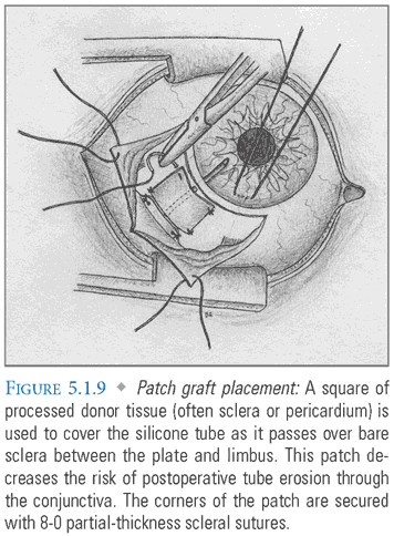
- Patch Graft: A square of processed donor tissue (e.g., pericardium or sclera) is used to cover the silicone tube as it passes over bare sclera between the plate and corneal limbus. This patch decreases the likelihood that the tube will erode through the conjunctiva postoperatively. Its corners are secured with partial-thickness 8-0 scleral sutures (FIG. 5.1.9).
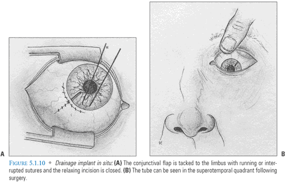
- Conjunctival Closure: The conjunctival flap is then tacked to the limbus with running or interrupted sutures (FIG. 5.1.10A,B). With valveless implants, the free end of the occluding suture is brought forward beneath the conjunctiva for easy postoperative access.
COMPLICATIONS
Intraoperative
- Bleeding: This occurs frequently with NVG due to disruption of fibrovascular membranes in the anterior chamber. Significant hyphema should be evacuated to prevent occlusion of the drainage tube.
- Tube misdirection: If the tube is directed anteriorly, it may abrade the corneal endothelium, causing edema. If the tube is directed posteriorly, it may occlude with iris tissue, induce traumatic cataract formation, or inadvertently enter the posterior chamber.
- Anterior chamber loss: This may occur due to aqueous leakage during sclerostomy creation, inadequate suture ligation of a nonvalved implant, or a faulty Ahmed valve mechanism. It can be reversed with viscoelastic injection.
- Suture perforation of the globe: This can occur when the drainage plate is anchored to the sclera. The risk is increased in patients with thin sclera. Perforations often close without significant repercussions, but they occasionally cause retinal detachment or vitreous hemorrhage.
Early Postoperative
- Hypotony: Low IOP due to rapid aqueous drainage, which is usually seen prior to maturation of the plate capsule. It is treated by injecting viscoelastic or temporarily reoccluding the tube with a suture ligature.
- Increased IOP: There are many potential causes of increased intraocular pressure in the early postoperative period. These include tube blockage by iris tissue, vitreous, blood clots, fibrin or retained viscoelastic, and Ahmed valve failure.
- Choroidal effusion: Fluid accumulates in the choroid, often as a complication of hypotony. Small effusions often resolve spontaneously, but larger effusions may require evacuation.
Late Postoperative
- Increased IOP: The “hypertensive phase” occurs 4 to 6 weeks after tube placement due to excess deposition of fibrous tissue in the drainage plate capsule. Fibrous tissue growth can occasionally be controlled with IOP lowering agents, capsule aspiration, antimetabolites, or corticosteroids. Other late causes of increased pressure include occlusion of the tube lumen, which is sometimes treated with Nd:YAG (neodymium-doped yttrium aluminum garnet) laser application or tube replacement.
- IOP decrease: Sudden drops in intraocular pressure are generally due to wound dehiscence or drainage plate extrusion.
- Corneal decompensation: Close proximity of, or contact between, the drainage tube and endothelial cells increases the risk of corneal edema.
- Diplopia: This is most common with larger, high-profile implants, but is often self-limiting.
- Plate migration: This can occur if scleral fixation is disrupted prior to complete encapsulation of the drainage plate.
- Tube erosion: The tube can wear through the underlying sclera or overlying patch and conjunctiva, potentially leading to endophthalmitis and/or implant extrusion. If the eroded site is not infected, the patch can be replaced. If it is infected, the tube may require removal.
POSTOPERATIVE CARE
Administer topical corticosteroids for up to 2 months.
Stop antibiotics after 1 week if there is no evidence of wound leakage and the corneal epithelium is intact on slit lamp examination.
With valveless implants, remove the occluding suture 4 to 6 weeks after the operation. At this point, a fibrous capsule has started to form over the drainage plate and the IOP should begin to rise gradually. A small conjunctival incision is made over the tail of the occluding suture, and it is removed with gentle traction. Absorbable sutures will spontaneously dissolve at a relatively predictable rate (about 6 weeks post-op for a 7-0 polygalactin suture). Ligation release prior to capsule formation can result in severe hypotony and must be avoided.
Further Reading
Text
Freedman J, Trope GE. Drainage implants. In: Yanoff M, Duker J, eds. Ophthalmology. 3rd ed. Philadelphia, PA: Mosby; 2009:1277–1283.
Kahook MY. Neovascular glaucoma. In: Yanoff M, Duker J, eds. Ophthalmology. 3rd ed. Philadelphia, PA: Mosby; 2009:1178–1182.
Lang GK. Glaucoma. In: Ophthalmology: A Pocket Textbook Atlas. 2nd ed. New York, NY: Georg Thieme Verlag; 2007:chap 10.
Minckler DS, Francis BA, Hodapp EA, et al. Aqueous shunts in glaucoma: a report by the American Academy of Ophthalmology. Ophthalmology. 2008;115(6):1089–1098.
Sarkisian SR Jr. Tube shunt complications and their prevention. Curr Opin Ophthalmol. 2009;20(2):126–130.
Schwartz KS, Lee RK, Gedde SJ. Glaucoma drainage implants: a critical comparison of types. Curr Opin Ophthalmol. 2006;17(2):181–189.
Primary Sources
Gedde SJ, Schiffman JC, Feuer WJ, et al. Treatment outcomes in the tube versus trabeculectomy study after one year of follow-up. Am J Ophthalmol. 2007;143(1):9–22.
Nguyen QH. Primary surgical management refractory glaucoma: tubes as initial surgery. Curr Opin Ophthalmol. 2009;20(2):122–125.
Tsai JC, Johnson CC, Kammer JA, et al. The Ahmed shunt versus the Baerveldt shunt for refractory glaucoma II: longer-term outcomes from a single surgeon. Ophthalmology. 2006;113(6):913–917.
- Creation of a full-thickness defect in the peripheral iris with a Nd:YAG (neodymium-doped yttrium aluminum garnet) or argon laser equalizes the anterior and posterior chamber pressures, eliminating pupillary block and opening the angle to promote aqueous outflow.
INDICATIONS
Stay updated, free articles. Join our Telegram channel

Full access? Get Clinical Tree


