- Muscle origins:
- All four rectus muscles originate from the Annulus of Zinn, a fibrous ring that encloses the optic canal and nerve at the orbital apex.
- The superior oblique and levator palpebrae superioris arise from the sphenoid bone, just outside of the annulus.
- The inferior oblique arises just deep to the medial orbital rim.
- All four rectus muscles originate from the Annulus of Zinn, a fibrous ring that encloses the optic canal and nerve at the orbital apex.
- Muscle insertions:
- The distance between the corneal limbus and scleral insertions of the rectus muscles is described by the Spiral of Tillaux:
- Medial rectus: 5.5 mm
- Inferior rectus: 6.5 mm
- Lateral rectus: 6.9 mm
- Superior rectus: 7.7 mm
- Medial rectus: 5.5 mm
- After passing through the trochlea, the superior oblique fans out to insert beneath the superior rectus, posterior to the axis of rotation.
- The inferior oblique passes beneath the inferior rectus and inserts in the inferior temporal quadrant of the posterior globe.
- The distance between the corneal limbus and scleral insertions of the rectus muscles is described by the Spiral of Tillaux:
- Vascular supply:
- Each rectus muscle is accompanied by two anterior ciliary arteries, except for the lateral rectus, which usually has just one.
- These muscular branches arise from the ophthalmic artery and contribute to an anastomotic circle at the root of the iris.
- Disruption of multiple arteries can produce anterior segment ischemia. Surgery on extraocular muscles, therefore, is usually limited to two rectus muscles per eye and session.
- Each rectus muscle is accompanied by two anterior ciliary arteries, except for the lateral rectus, which usually has just one.
- Key anatomic associations:
- The sclera is thinnest just posterior to the rectus muscle insertions (~300 microns vs. ~700 microns elsewhere).
- Near their insertions, the oblique muscles travel below the corresponding rectus muscles.
- The inferior oblique inserts over the macula.
- The sclera is thinnest just posterior to the rectus muscle insertions (~300 microns vs. ~700 microns elsewhere).
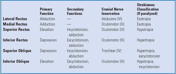
Extraocular Muscle Function
See table above.
PREOPERATIVE ASSESSMENT
- Stability of ocular deviation is usually confirmed by at least two sets of measurements prior to surgical intervention, commonly performed 4 to 6 weeks apart.
- Ocular alignment is assessed for distance and near vision in the nine diagnostic gaze positions (straight, left, up/left, up, up/right, right, down/right, down, and down/left; these are measured in 30 degrees excentric gaze).
- Forced ductions are used to rule out external impingements on ocular motility. They are tested by moving the eye with forceps, either in the operating room under general anesthesia or in clinic under topical anesthesia (if the patient is able to cooperate). A cotton-tip applicator can also be used to move the eye but is less accurate.
ANESTHESIA
- General anesthesia produces the best globe akinesia and is typically required in younger patients.
- Retrobulbar anesthesia is occasionally used in older, cooperative patients.
PROCEDURE
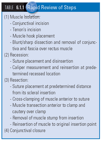
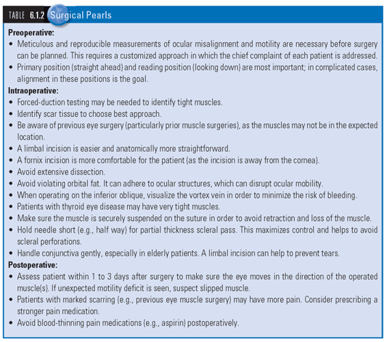
 SEE TABLES 6.1.1 Rapid Review of Steps and 6.1.2 Surgical Pearls, and WEB TABLES 6.1.1 to 6.1.4 for equipment and medication lists.
Stay updated, free articles. Join our Telegram channel

Full access? Get Clinical Tree


