4
Laboratory Tests Commonly Used in Evaluating Ocular Inflammation
Paul A. Gaudio
This chapter serves as a reference guide for understanding commonly ordered laboratory tests in ocular inflammatory diseases, including how to order and interpret them. See Table 4.1 for further details.
Table 4.1 Laboratory tests commonly used in ocular inflammatory diseases
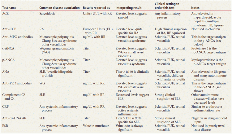
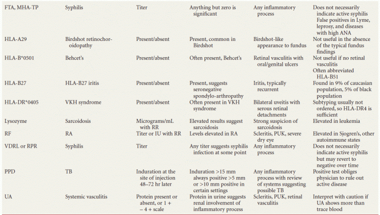
PUK, peripheral ulcerative keratics; TB, tuberculosis.
BLOOD TESTS
Angiotensin-Converting Enzyme
Angiotensin-Converting Enzyme (ACE) testing is sometimes abnormal in patients with sarcoidosis, and we test it when we suspect this disease. ACE is the enzyme that converts Angiotensin I to vasopressor Angiotensin II. It is normally found in high concentrations in the kidney and in low concentrations in peripheral tissues and blood. Epithelioid macrophages (the histopathologically defining feature of granulomatous inflammation) produce ACE pathologically, so increased serum ACE levels suggest systemic granulomatous disease. The result is reported as a level (usually in Units per liter, or U/L) with a laboratory reference range (RR). Elevated levels suggest sarcoidosis, but false positives are seen in hyperthyroidism, acute hepatitis, multiple myeloma, diabetes, tuberculosis (TB), and leprosy, among others. Being just over 60% sensitive, the test is not useful as a screening tool, but since it is over 90% specific, a positive result in the appropriate clinical setting strongly supports the diagnosis of sarcoidosis.
This blood test is easily obtained in most laboratories, although we have never seen it on preprinted laboratory “menu” forms, and we are careful to write out the complete test name, since “ACE” has occasionally been interpreted as “acetone” by laboratory staff. The RR changes depending on the method used, and some laboratories perform the test on plasma (recall that plasma is whole blood minus red blood cells, whereas serum is plasma minus the clotting factors).
Anti–Cyclic Citrullinated Polypeptide Antibodies
This test is used adjunctively in the diagnosis of adult rheumatoid arthritis (RA), being over 90% specific. The result is reported in “ELISA Units/ml,” or EU, with a RR. We generally leave this blood test to the rheumatologists, but if we do order it (usually to clinch a diagnosis), we spell out the test name and keep in mind that not all laboratories can perform it. The utility of this test is not established in children.
Antineutrophil Cytoplasmic Antibodies
We order this test in patients with necrotizing scleritis, peripheral ulcerative keratitis, and retinal vasculitis. Antineutrophil cytoplasmic antibodies (ANCA) are autoantibodies directed against the cytoplasmic portion of neutrophils. Classically, the ANCA was an immunofluorescence assay using fluorescein labeled antibodies against the patient’s antibodies in the cytoplasm of the white blood cells (hence the term “c-ANCA”) or around the nucleus (perinuclear, “p-ANCA”). Subsequently, the target antibodies for both assays were identified as anti-proteinase 3 (anti-PR3) in the c-ANCA and anti-myeloperoxidase (anti-MPO) in the p-ANCA. Laboratories may use the immunofluorescence technique, antigen-specific antibody assays, or a combination of both. Immunofluorescence assay results are usually reported as a titer, while direct antibody assays are reported as serum levels. Nobody should really have a detectable ANCA, so we consider a positive test to indicate a potentially severe immunologic disease. Elevated c-ANCA or PR3 levels suggest small vessel vasculitis, most commonly Wegener’s. Elevated p-ANCA or MPO levels are less disease specific, being found in microscopic polyangitis, crescentic glomerulonephritis, and occasionally ulcerative colitis and ankylosing spondylitis. Frequently, the term “ANCA positive vasculitis” is used to describe patients whose inflammatory disease does not fit any classic clinical description. Note that the ANCA can remain elevated for a prolonged period, so this test is not useful for short-term follow-up of disease activity. Neither of these tests is useful for screening.
This test is easily ordered in any clinical immunology laboratory. The terms “p- and c-ANCA” are standard terminology, and the laboratory will perform the test according to its routine method.
Antinuclear Antibodies
We order this test in the setting of scleritis or peripheral ulcerative keratitis, in young people with anterior uveitis in whom we suspect juvenile idiopathic (rheumatoid) arthritis, and in retinal or choroidal vasculitis. The Antinuclear antibodies (ANA) test looks for any antibodies against nuclear proteins such as DNA, RNA, histones, and others. We generally should not have ANA present, although with increasing age many people develop them at a low level. The test is performed by layering the patient’s serum over epithelial cells, such that any antinuclear antibodies in the serum will bind to the epithelial cell nuclei and can be detected with fluorescein-labeled antibodies directed against all human antibodies (the nuclei should fluoresce if ANA are present).
The result is reported as a titer, this being the greatest dilution at which fluorescence was still observed. Titers above 1:160 are significant and are commonly seen in systemic lupus erythematosus (SLE) and juvenile idiopathic arthritis.
Other immune diseases, notably RA, scleroderma, and mixed connective tissue diseases can also cause elevated ANA. It is worth noting that a pattern of nuclear fluorescence is sometimes reported (homogenous, nucleolar, speckled, etc.) and that only the “speckled” pattern is informative, being specific for SLE. A negative test more or less excludes SLE, while a high titer suggests a more severe disease. This test is easily obtained in most laboratories and is usually on most preprinted laboratory forms.
The ANA titer fluctuates with disease activity and is useful in following patients during treatment.
Complements C3 and C4 (C3, C4)
Diseases with activation of the complement pathways will generally show decreased levels of C3 and C4. C4 is part of the classic pathway, and C3 is at the convergence of the classic and alternate complement pathways. Complement activation thus consumes C3 and C4, causing decreased serum levels. We order C3 and C4 levels when we suspect SLE, usually in patients with scleritis, peripheral ulcerative keratitis (PUK), or retinal vasculitis.
Most laboratories can perform these tests, and they are ordered by writing “complement C3 and C4” on the laboratory slip. Results are reported in mg/dL with a RR.
C-Reactive Protein (CRP)
We order serum C-reactive protein (CRP) levels when we suspect a systemic inflammatory process in patients with scleritis, PUK, or retinal vasculitis. CRP is an acute phase reactant protein which is produced by the liver in response to inflammatory serum cytokines. Levels are normally low, although they increase with age. The test is performed on serum, and results are reported in mg/dL with a laboratory RR. Women and men have the same reference values. CRP levels are elevated in the setting of systemic infection, inflammation, or malignancy. Levels increase slightly in old age. This test’s utility is similar to that of the erythrocyte sedimentation rate (ESR), except that CRP levels normalize more quickly when the inflammatory process comes under control.
This blood test is standard in any hematology laboratory and is generally on preprinted laboratory forms. Note: related test called the “high sensitivity C-reactive protein” or hs-CRP is used in evaluating risk factors for heart disease. It is sometimes simply called “cardiac CRP.” Many laboratories will include both types of CRP on preprinted test order forms.
Double-Stranded DNA Antibody (ds-DNA Ab)
This serum test is useful in identifying and following patients with SLE. This antibody correlates fairly well with SLE disease activity and moderately well with glomerulonephritis. We do not routinely order this test but find it helpful in patients with scleritis, PUK, and retinal vasculitis in which the ANA is elevated and we suspect SLE. Results should be negative in drug-induced lupus, so this test is also helpful in evaluating patients in whom drug-related elevated ANA is suspected, as occurs with infliximab (Remicade) therapy.
This test is easily obtained in clinical laboratories by writing “ds-DNA” antibodies on the lab slip. Results are reported as a titer, and greater than 1:10 is 95% specific for SLE.
Erythrocyte Sedimentation Rate
ESR is helpful in determining the presence of a potentially severe systemic inflammatory process in patients with scleritis, PUK, and retinal vasculitis. The test measures the distance which the patient’s erythrocytes (RBCs) fall in a column in 1 hour, and the result is thus reported in units of mm/hour. RBCs normally fall slowly, but they fall much faster if the patient’s blood contains increased levels of blood proteins like fibrinogen and other acute phase reactants which cause RBC aggregation. Acute phase reactants are increased in inflammatory diseases. A markedly elevated ESR (indicating rapidly aggregating, fast-falling RBCs), generally over 80 mm/hour, suggests a potentially severe systemic inflammatory or infectious process or malignancy. Low or intermediate levels are not helpful. We do not order this test in the workup of uveitis, but we routinely order it for scleritis, PUK, and retinal vasculitis.
This test is routine in nearly all hematology laboratories and is usually listed on preprinted laboratory forms as “ESR” or occasionally “Sed rate.”
Fluorescent Treponemal Antibody—Absorption
This blood test looks for antibodies to Treponema pallidum. A patient’s serum is placed over a slide containing T. pallidum, and fluorescein-labeled antibodies are used to detect any patient antibodies that have bound there. The result is reported as a titer, and any titer is considered significant and usually indicative of syphilis. Lyme, leprosy, and ANA elevation can cause false positives. This test generally remains positive after treatment and so is not useful in assessing disease activity. It is rarely used on cerebrospinal fluid.
This test is easily obtained, although many laboratories treat this as a confirmatory test for Rapid Plasma Reagin (RPR) or Venereal Disease Research Laboratories (VDRL). It is thus important to indicate that either of these tests is not needed before performing the Fluorescent treponemal antibody (FTA) or that the FTA is necessary regardless of what the RPR or VDRL shows. Many laboratories may substitute another treponemal assay, frequently the microhemagglutination-Treponema pallidum, or MHA-TP, in place of FTA. This is fine, since all treponemal assays work quite nearly the same way as the FTA and are interpreted the same way. The MHA-TP may be falsely elevated in mononucleosis.
Human Leukocyte Antigen (HLA), or Major Histocompatibility Complex
This is a set of genes that encode the cell surface markers by which our immune system recognizes self from nonself. There are two broad classes of Human leukocyte antigen (HLA) genes, denoted I and II. The HLA class I allele has three major loci, designated A, B, and C. Humans thus have six major HLA I loci in total: two As, two Bs, and two Cs (one allele inherited from each parent). The gene products of each locus are molecules found on the surface of nucleated cells and play a critical role in antigen presentation to T lymphocytes. A number of serotypes for each locus are found in the human population, and serotypes are designated with numbers, for example: A*01, A*02…, B*01, B*02… etc. Multiple subtypes are recognized for each serotype, and subtype numbers follow the serotype number, for example, A*0102 indicates subtype 2 of serotype I at the A locus of the HLA class I gene. In the entire human population, the total number of subtypes at each locus (A, B, or C) numbers in the hundreds. It is possible, but quite rare, to have two copies of the same subtype; most humans are heterozygous at each HLA allele.
Class II HLA designation works similarly. The major classes are DR, DQ, and DP, and these are expressed only on antigen-presenting cells. Humans have two copies of the DP and DR molecules and four copies of the DQ, for a total of eight major Class II HLA molecules. Serotypes and subtypes are indicated with numbers as with class I antigens.
Some HLA molecules are linked with certain diseases. Common HLA types involved in ocular inflammation are
- HLA-B27, which is associated with recurrent anterior uveitis
- HLA-A29, associated with Birdshot retinochoroidopathy
- HLA-B*0501, associated with Behcet disease
- HLA-DR*0405, associated with Vogt-Koyanagi-Harada (VKH) syndrome
In evaluating patients with ocular inflammation, one usually orders a test for the single HLA type in question, by simply writing, for example, “HLA-B27” on a laboratory slip. Testing is usually performed with polymerase chain reaction. The result is returned as “present” or “absent” or some similar terminology. Rarely, one will want to know all of a patient’s HLA serotypes, or all those in either class, in which case an entire panel can be ordered.
Lysozyme (Muramidase)
Lysozymes are proteins that assist in damaging bacterial cell walls. These compounds are found in the granules of neutrophils and in many bodily secretions (and, incidentally, in very high levels in egg whites). The serum lysozyme level can be used in establishing the diagnosis of sarcoidosis. It is reported as a concentration in μg/mL. We generally only use it to shore up the diagnosis in strongly suspected cases; it is not sufficiently specific to be useful routinely. Lysozyme levels are elevated in several forms of leukemia and can be elevated in TB as well.
Rheumatoid Factor
This test is useful in patients with scleritis or peripheral ulcerative keratitis, because it suggests the presence of a systemic immune process, most notably RA. Rheumatoid Factors (RF) are heterogeneous antibodies of the IgM class that are directed at the Fc region of human IgG class immunoglobulins. We generally have very low levels of RF antibodies, although this increases with age and even some normal individuals have them. Elevated RF nevertheless suggests immune activation in some form. The result is reported as a titer or as international units (IU) with a laboratory RR. Elevated RF is seen most commonly in RA, although this test is far from perfect, and many RA patients do not have RF, while Sjogren syndrome, SLE, and scleroderma patients often do. Methyldopa can increase RF titers, as can viral infections and chronic bacterial diseases.
RF is a standard blood test in most laboratories and is generally found on preprinted laboratory “menu” forms, occasionally listed as “RA,” usually for “rheumatoid antibodies.”
Venereal Disease Research Laboratories or Rapid Plasma Reagin Tests
These are blood tests to determine the presence of antibodies that are produced when T. pallidum interacts with host tissues. Since they do not detect antitreponemal antibodies, these tests are considered “nontreponemal.” The results are reported as a titer, and titers over 1:32 are generally considered positive for active syphilis. The test should be positive 1 to 3 weeks after the appearance of a genital chancre. This test will often revert to negative when syphilis is treated or becomes latent, and for this reason, they are less sensitive than the treponemal tests. Mononucleosis, Lyme disease, pregnancy, collagen vascular diseases, leprosy, and malaria may cause false positives, which are unfortunately common.
We generally do not use nontreponemal tests, since uveitis tends to occur in the later stages of syphilis at which patients are often seroreverted and hence the test becomes nonreacative. Almost any laboratory can perform a VDRL or RPR, which are essentially interchangeable.
OTHER TESTS
Purified Protein Derivative
The purified protein derivative (PPD) is a skin test used to determine if an individual has been exposed to the tubercle bacillus, which causes TB. In this test, a small amount of biologically standardized reagent is injected subcutaneously, generally in the skin of the dorsal forearm. The reagent is commercially available, and most uveitis clinics stock it. The standard test involves injecting 0.l mL of reagent, containing 5 tuberculin units (TU—an internationally established measure of biologic activity), although other TU concentrations are available for special situations. The arm is examined 48 to 72 hours later, and induration at the injection site indicates prior exposure to the tubercle bacillus.
The size of induration at the injection site is determined. For most individuals, a PPD is considered positive if the induration is 15 mm or greater. In patients with impaired immunity or in whom the pretest likelihood of disease is high, 5 mm induration is considered significant. This group includes patients taking immunosuppressive medications including corticosteroids at a dose equivalent to prednisone 15 mg per day for a month, patients with active human immunodeficiency virus (HIV) infection, or patients with radiographic evidence of healed TB, as well as close contacts of patients with known TB.
There is an intermediate group in whom 10 mm of induration is considered positive. These are patients whose medical history puts them at risk for progression to active TB, such as those with diabetes, silicosis, or carcinoma of the head, neck, or lung. In addition, this group includes patients with a somewhat elevated pretest likelihood of TB, including immigrants from TB-endemic countries or intravenous drug users. Children under 4 years of age are also included in this group.
A positive PPD test obliges the physician to rule out active TB. By itself, a PPD does not indicate active disease.
Quantiferon Gold (QFT-g)
This is a whole blood test used to diagnose infection with Mycobacterium tuberculosis. It was approved by the U.S. Food and Drug Administration in 2005, being a modification of an earlier test called Quantiferon. The test is based on the fact that an infected individual’s white blood cells will produce interferon gamma (IFN-γ) if exposed to certain M. tuberculosis antigens. In the test, the patient’s whole blood is incubated with two such antigens for 16 to 24 hours, and the level of INF-γ released is quantified. This test may eventually take the place of PPD, but its use in the setting of diseases that affect immune system function (HIV infection, malignancy, diabetes mellitus) is not clearly delineated.
At the time of this writing, laboratory experience with this test is quite variable, and one should contact the receiving laboratory for its collection protocol.
Urinalysis
We evaluate patients for proteinuria when we suspect a systemic vasculitis, since renal involvement of the disease generally informs the prognosis and treatment. A simple Urinalysis (UA) achieves this.
Stay updated, free articles. Join our Telegram channel

Full access? Get Clinical Tree


