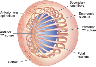
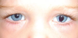
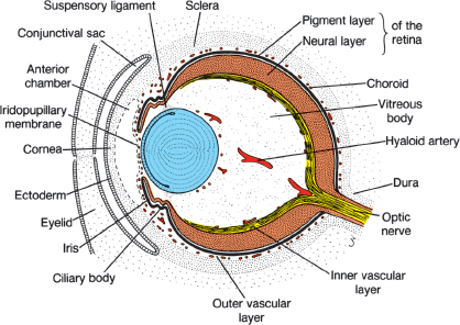
LENS ANATOMY AND PHYSIOLOGY Normal Lens Anatomy See Figure 2.1. Biconvex shape, more convex posteriorly. Infant: 6.4-mm wide and 3.5-mm long. Weighs approximately 90 mg. Adult: 9.0-mm wide and 5.0-mm long. Weighs approximately 255 mg. Type IV collagen, which is the basement membrane for the lens epithelial cells. Thickness: 2 to 23 μm. Thickest near periequator, thinnest central posteriorly. Single row of nucleated lens epithelial cells beneath anterior capsule only. Mitotically active lens epithelial cells found at equator of lens (lens bow). FIGURE 2.1. Anatomy of the fetal lens. Note the location of nucleated lens epithelial cells anteriorly and along the lens equator. The ends of the elongating lens fibers form the anterior and the posterior “Y” sutures. Lens fibers at equator elongate and wrap around to join other epithelial cells forming anterior “Y” suture and posterior inverted “Y” suture. Fibers are 5 to 30 μm in diameter and provide support for the lens. Originate from the nonpigmented epithelium of the pars plana and pars plicata of the ciliary body. Insert on the lens capsule in the periequatorial region, 1.5 mm anteriorly and 1.25 mm posteriorly. Lens Embryology and Development Time after gestation and lens development: Day 25: formation of optic vesicles from the forebrain (diencephalons). Day 27: elongation of surface ectodermal cells to form lens plate (placode). Day 29: invagination of surface ectoderm to form lens pit (fovea lentis). Day 33: formation of lens vesicle complete; optic vesicles form two-layered optic cup. Day 40: Primary lens fibers fill lens cavity forming embryonic nucleus. Week 7: Secondary lens fibers with nuclei at equator elongate and form Y sutures (upright Y suture anteriorly, inverted Y suture posteriorly) (Figure 2.2). Vasculature surrounding and nourishing the lens during development. Usually regresses by birth (remnants include Mittendorf dot and persistent pupillary membranes). Lens Biochemistry Lens proteins (one third of lens mass): Alpha crystallin: 32% of lens proteins, largest of the crystallins. Beta crystallin: 55% of lens proteins. Gamma crystallin: 1.5% of lens proteins. Urea-insoluble fraction: mostly plasma membrane proteins. Urea-soluble fraction. Lens Physiology Within the lens: Na+ = 20 mmol, K+ = 120 mmol. Aqueous humor: Na+ = 150 mmol, K+ = 5 mmol. Accommodation Helmholtz: Most widely held theory Schachar FIGURE 2.3. Cascade of accommodation. Lens Coloboma Summary A notch in the peripheral lens caused by focal absence of the ciliary body and the zonules. Signs and Symptoms Irregular pupil. May be asymptomatic. Ophthalmic Findings FIGURE 2.4. Photograph of a typical iris coloboma with a keyhole pupil located inferiorly and slightly nasally at approximately 7 o’clock position. The eye is slightly smaller than normal. Systemic Findings None. Disease Course Typically stationary. Treatment and Management Lens extraction if associated with visually significant cataract. Mittendorf Dot Etiology Remnant of the posterior tunica vasculosa lentis. Signs and Symptoms Usually asymptomatic. Ophthalmic Findings Systemic Findings None. Disease Course Stationary. Treatment and Management None required. Ectopia Lentis Ectopia lentis describes the condition of the natural lens being either partially or fully displaced from its normal anatomic position. There are many causes; the most common is trauma. Signs and Symptoms Monocular diplopia. Change in refraction (marked astigmatism). Ophthalmic Findings Phacodonesis (tremulous lens on eye movement). Iridodonesis (tremulous iris). Cataract. Pupil block with angle-closure glaucoma if lens displaced into anterior chamber. Marfan Syndrome Summary Marfan syndrome is an autosomal dominant disorder of collagen synthesis characterized by skeletal, cardiac, and ocular findings. Etiology An abnormality of fibrillin, a structural protein. Demographics Autosomal dominant inheritance. Ophthalmic Findings Systemic Findings Tall, thin stature. Mitral valve prolapse. Dilated aortic root and aortic dissection. Arachnodactyly. Hyperextensible joints. Disease Course Variable findings may present in adulthood. Treatment and Management Homocystinuria Summary Homocystinuria is an autosomal recessive systemic disorder of amino acid metabolism resulting in lenticular zonular fragility, seizures, and an increased risk for thromboembolic events. Etiology Abnormality of methionine metabolism. Demographics Autosomal recessive. Ophthalmic Findings Ectopia lentis (characteristically inferonasal subluxation). Systemic Findings Tall with light colored hair. Seizures. Osteoporosis. Mental retardation. Increased incidence of thromboembolism. Disease Course Eighty percent of patients have subluxed lenses by age 15 years. Treatment and Management A diet low in methionine, with supplemental cysteine and vitamin B6 may reduce the incidence of ectopia lentis in these patients. Miscellaneous Causes Hyperlysinemia Inborn error of metabolism associated with ectopia lentis, muscular hypotony, and mental retardation. Sulfite Oxidase Deficiency Microspherophakia Summary Microspherophakia is a congenital abnormality of lens development resulting in a small, spherical shaped lens that can dislocate anteriorly and cause pupillary block with resulting angle-closure glaucoma. Etiology Abnormal formation of secondary lens fibers during embryogenesis. Ophthalmic Findings Systemic Findings Usually associated with Weill-Marchesani syndrome. May be an isolated abnormality or found in: Lowe syndrome. Alport syndrome. Congenital rubella. Disease Course May develop angle-closure glaucoma. Treatment and Management Weill-Marchesani Syndrome Summary Weill-Marchesani syndrome is an autosomal recessive condition characterized by short stature, small hands and feet, and microspherophakia. Etiology Autosomal recessive inheritance. Signs and Symptoms Myopia Angle closure that is due to pupillary block. Ophthalmic Findings Microspherophakia. Systemic Findings Short stature. Disease Course May develop angle-closure glaucoma. Treatment and Management Traumatic Cataract Summary Blunt, nonperforating injury to the eye may cause lens opacification either as an acute event or as a late sequela. Etiology Traumatic lens damage caused by blunt, nonperforating mechanical injury. Signs and Symptoms Blurry vision. Fluctuating vision. Ophthalmic Findings Pigment from pupillary ruff imprinted on anterior lens capsule (Vossius ring). Axially located stellate or rosette-shaped posterior lens capsule opacification that may progress to opacification of the entire lens. Phacodonesis Iridodonesis. Retinal dialysis. Rhegmatogenous retinal detachment. Systemic Findings Related to trauma. Periorbital trauma (eyelid ecchymosis, bone fractures). Disease Course Treatment and Management Traumatic Subluxation and Dislocation of Lens Summary Blunt ocular trauma can cause stretching of the zonular ring, stretching and rupture of the lens zonules, and lens subluxation. Etiology Concussive force transmitted to the lens with zonular disruption. Signs and Symptoms Fluctuating vision. Impaired accommodation High astigmatism Monocular diplopia. Ophthalmic Findings Phacodonesis. Iridodonesis.
 Lens dimensions:
 Lens capsule:
 Lens epithelium:
 Zonules:
 Tunica vasculosa lentis:
 Water soluble (crystallins):
 Water insoluble
 Proportion of water-insoluble proteins is increased in cataractous lenses.
 Ionic gradient:
 Lens epithelial cells supply metabolic demands for the lens.
 Anaerobic glycolysis responsible for bulk of adenosine triphosphate (ATP) production, since oxygen tension low.
 Hexose monophosphate shunt produces about 5% of ATP.
 Glutathione, vitamins C and E are antioxidants counteracting the damage of free radicals.
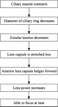
 Accommodation is a change in lens shape to allow focusing from distance to near (Figure 2.3).
 Theories of mechanism:
 Circular muscle fibers of ciliary body contract decreasing equatorial circumlenticular space.
 This reduces zonular tension, which allows the lens to become more spherical and increases optical power.
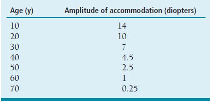
 Amplitude of accommodation decreases with age as the lens substance becomes less flexible resulting in presbyopia (Table 2.1).
 Circular muscle fibers of ciliary body contract increasing equatorial zonular tension while simultaneously decreasing zonular tension of anterior and posterior zonules.
 This causes the central surface of the lens to steepen, the anterior-posterior diameter to increase, and the peripheral surface to flatten increasing optical power.
 Amplitude of accommodation decreases with age as the equatorial diameter of the lens increases causing diminished baseline zonular tension due to decreasing distance between the lens and the ciliary body.
 Usually located inferonasally.
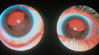
 May be associated with other colobomata (iris, optic nerve, choroid) (Figure 2.4).
 Lens opacification may be noted in the region of the coloboma.
 Whitish opacity generally connected inferonasal to the posterior capsule of the lens.
 May have remnant of hyaloid artery attached.
 Bilateral ectopia lentis (characteristically superotemporal) (Figure 2.5).
 Myopia.
 Increased incidence of retinal detachment.
 Most patients can be treated with spectacle correction.
 Pupillary dilation may be helpful.
 Lens extraction may be difficult.
 A very rare autosomal recessive deficiency in sulfur metabolism.
 Associated with ectopia lentis, seizures, and mental retardation.
 Myopia.
 Angle closure due to pupillary block.
 Lens may dislocate into anterior chamber.
 Abnormally small lens may be entirely visible through well-dilated pupil.
 Iridotomy for pupillary block.
 Avoid miotics as they aggravate pupillary block.
 Cycloplegics may be helpful by moving the lens posteriorly and decreasing its anteroposterior diameter.
 Iridotomy for pupillary block.
 Avoid miotics as they aggravate pupillary block.
 Cycloplegics may be helpful by moving the lens posteriorly and decreasing its anteroposterior diameter.
 Progression of cataract.
 May have progression to subluxation of lens.
 Surgical removal of cataract (intracapsular or pars plana approach if inadequate zonular support).
 Thorough examination of peripheral retina.
Stay updated, free articles. Join our Telegram channel

Full access? Get Clinical Tree


