16
Endophthalmitis
Ron A. Adelman and Rajeev K. Seth
DEFINITION, CLASSIFICATION, CLINICAL SIGNS, AND TREATMENT
Infectious endophthalmitis is defined as severe, diffuse intraocular inflammation caused by a microbial agent. Fortunately, it is a rare event, but because of its potentially rapid and devastating course, it remains an important entity to quickly recognize and appropriately treat.
Endophthalmitis can be classified on the basis of time course, by the route of entry of microorganisms into the eye, and by the type of offending organism (bacterial or fungal). Endogenous endophthalmitis is when there is hematogenous spread of organisms to the eye, whereas exogenous endophthalmitis occurs when organisms gain entry into the otherwise sterile eye from an external source, most commonly after ocular surgery or trauma.
Patients typically present with rapidly progressive pain, redness, discharge, decreased vision, and possibly with complaints of floaters. Common clinical signs include purulent discharge, conjunctival injection, significant anterior chamber cell and flare with fibrin and hypopyon, vitritis, and retinitis. Treatment depends mainly on the particular type of endophthalmitis (endogenous vs. exogenous) and presenting signs. Endophthalmitis treatment was evaluated by the Endophthalmitis Vitrectomy Study (EVS), which was a large, randomized, multicenter clinical trial evaluating the treatment of bacterial endophthalmitis. The EVS included only those patients with endophthalmitis following cataract surgery or secondary intraocular lens (IOL) implantation.
This chapter reviews the major forms of endophthalmitis, as well as their presentation, diagnosis, and treatment.
POSTOPERATIVE ENDOPHTHALMITIS
Postoperative endophthalmitis represents the most common cause of endophthalmitis, accounting for approximately 70% of all cases endophthalmitis. Because of the high frequency of cataract surgery, endophthalmitis associated with cataract surgery remains to be the most common cause of postoperative endophthalmitis, accounting for approximately 70% to 90% of all postoperative endophthalmitis. However, endophthalmitis can occur after any intraocular surgery, including vitrectomy, glaucoma filtering surgery or glaucoma drainage implant surgery, corneal transplantation, and even strabismus surgery with inadvertent penetration of the sclera.
Factors that predispose to the development of postoperative endophthalmitis include blepharitis, acute conjunctivitis, posterior capsular rupture, vitreous loss, prolonged surgical time, inadequate wound closure, wound leak, vitreous incarceration into the wound, chronic immunosuppressive therapy, and the presence of diabetes mellitus.
Studies have shown that in many cases of postoperative endophthalmitis, the infecting organism is genetically identical to organisms found as part of the normal skin flora of the patient’s periocular tissue. This emphasizes the importance of methods that reduce the risk of developing postoperative endophthalmitis. The wide-spread method of either irrigating the fornices with or placing a few drops on the conjunctiva of half-strength (5%) povidone-iodine has been shown to effectively decrease the number of bacterial colonies on the conjunctiva.
ACUTE POSTOPERATIVE ENDOPHTHALMITIS
Acute postoperative endophthalmitis refers to endophthalmitis within 6 weeks following surgery, with most cases presenting within the first 1 to 2 weeks.
A 10-year retrospective study at the Bascom Palmer Eye Institute revealed an incidence of acute endophthalmitis following intraocular surgery to be 0.093%.
The overall incidence of endophthalmitis following cataract surgery has been reported to be 0.04% to 0.30%. The rate of post–cataract surgery endophthalmitis evaluated at the Bascom Palmer Eye Institute was 0.05% from 1995 to 2001 and 0.04% from 2000 to 2004. There is some concern that there are increasing rates of endophthalmitis associated with clear corneal incisions for cataract surgery, with an approximate fourfold increase in the risk of endophthalmitis with clear corneal temporal incision over superior scleral tunneled incisions. Taban et al. performed the largest metaanalysis of post–cataract surgery endophthalmitis that revealed an overall incidence of endophthalmitis following cataract surgery of 0.13%. There was a significant increase in the rate of endophthalmitis in the 2000s (0.265%) when compared to the 1990s (0.087%), a time that temporally coincides with the development of clear corneal incisions.
The incidence of acute postoperative endophthalmitis is 0.03% to 0.046% following pars plana vitrectomy (PPV), 0.08% to 0.178% following penetrating keratoplasty, 0.2% to 0.366% following secondary IOL implantation, and 0.124% to 0.2% following glaucoma surgeries.
Analysis of the EVS revealed that 94% of culture-confirmed cases involve Gram-positive bacteria and that approximately 70% of cases were culture positive. Diagnosis is based on clinical findings with a high index of suspicion and can be confirmed with Gram stain and culture of either aqueous or vitreous.
Treatment has been modified based on the results of EVS, and visual acuity plays a large role in the decision-making process. The EVS was a randomized, multicenter trial that evaluated the role of 3-port PPV and systemic antibiotics in the management of acute postoperative endophthalmitis. All patients received intravitreal antibiotics (vancomycin 1 mg and amikacin 0.4 mg), topical, and systemic steroids. Those patients with presenting visual acuity of light-perception or worse had a threefold increase in the frequency of achieving 20/40 or better acuity if they underwent immediate PPV versus tap/injection. No difference in visual acuity outcomes between PPV and tap/injection were seen in the group with hand-motions vision or better (hand-motions vision was defined as seeing movement of the hand at a distance of 1 m).
It is important to note that patients included in the EVS were post–cataract surgery or secondary IOL placement, and therefore these results do not pertain to endogenous endophthalmitis or postoperative endophthalmitis following trauma or other types of ocular surgery.
Clinical Findings
- Ocular pain
- Photophobia
- Eyelid swelling
- Conjunctival injection
- Purulent discharge
- Decreased vision
- Floaters
- Wound leak
- Corneal edema
- Significant anterior chamber cell and flare
- Hypopyon (Fig. 16.1)
- Anterior chamber fibrin
- Vitritis
- Retinitis
- Retinal phlebitis
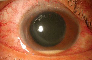
Figure 16.1 Acute bacterial endophthalmitis after temporal incision cataract extraction. Patient with dense vitritis, hypopyon, and ciliary injection.
Differential Diagnosis
- Retained lens material
- Toxic anterior segment syndrome secondary to noninfectious agents that gained entry into the eye intraoperatively (possible preservatives, medications, cleaning compounds, detergents, or intraocular irrigating solutions with improper pH or osmolality)
- Severe postoperative inflammation
- Preexisting uveitis
Microbiology
- Coagulase-negative staphylococci
- Staphylococcus aureus
- Streptococcus species
- Gram-negative organisms
- Fungus (rare)
Diagnostic Testing
- Ultrasound, if no view of posterior segment
- Needle vitreous aspiration or PPV vitreous biopsy for Gram stain and culture (aerobic, anaerobic, and fungal)
- Aqueous aspiration for Gram stain and culture (aerobic, anaerobic, and fungal)
- Note that anaerobic or fungal cultures may not become positive for several days or weeks
Treatment
- If visual acuity is hand motions or better in patients who are not diabetic and who would have fit the EVS criteria, the technique of vitreous tap or plana vitrectomy should be used to obtain diagnostic specimen. All patients require intravitreal injection of vancomycin (1 mg), along with ceftazidime (2.25 mg) or amikacin (0.4 mg).
- Visual acuity of light-perception: immediate PPV and intravitreal antibiotics as above.
- Topical broad-spectrum antibiotic, for example, fourth-generation fluoroquinolone every 1 hour or fortified vancomycin (25 to 50 mg/mL) and fortified tobramycin (14 mg/mL) alternating every 30 to 60 minutes.
- We often use oral corticosteroids (prednisone 1 mg/kg or equivalent) after the first antibiotic injection or after some clinical improvement has been noted.
- Topical cycloplegic (atropine 1% or cyclopentolate 1%).
- If there is minimal or no improvement 48 hours after intravitreal antibiotic injection, then perform reinjection of antibiotics or if not done initially, PPV with intravitreal antibiotic injection.
- If fungal endophthalmitis is suspected, then vitreous tap is NOT sufficient, and PPV should be performed, with intravitreal injection of amphotericin B 5 to 15 µg (hospital pharmacies often have their own standard formulas for preparing a dose in this range, and we go along with them) in 0.1 mL.
- Diabetics with hand-motions or better visual acuity in the EVS trended towards better visual acuity with PPV with antibiotics injection.
- EVS showed no benefit in the final visual outcomes with the use of systemic antibiotics. However, the systemic antibiotics used in the EVS did not have good vitreous penetration, as opposed to the good vitreous penetration of the newer fourth generation fluoroquinolones. Oral fluoroquinolones can be considered as adjuvant therapy to intravitreal injection.
CHRONIC POSTOPERATIVE ENDOPHTHALMITIS
Chronic postoperative endophthalmitis presents 6 weeks following surgery, and as opposed to acute postoperative endophthalmitis, has a much more indolent course with less severe inflammation and little to no pain. The presence of hypopyon and fibrin in the anterior chamber is rare, and vitritis, if present, is usually mild. Patients often present with prolonged inflammation not responsive to corticosteroid therapy, and a high index of suspicion is needed to make the diagnosis.
Bacterial microorganisms are most commonly the offending agent, with Propionibacterium species accounting for nearly 63% of cases and Staphylococcus epidermidis accounting for 16%. Candida species account for another 16% of cases. Most cases of Propionibacterium acnes infection is associated with a whitish intracapsular plaque involving the lens capsule or IOL. This has been shown to be composed of the bacterial organisms with some retained lens cortical material. Cases of chronic fungal postoperative endophthalmitis may have whitish/pale yellowish fluffy lesions in the vitreous. Diagnosis is based on Gram stain and culture of aqueous or vitreous aspirates, but it should be noted that many of the organisms that cause chronic postoperative endophthalmitis are slow-growing organisms, and growth may not be seen in culture until 1 to 2 weeks following inoculation.
Treatment generally involves surgery, but the prognosis is overall better than seen with acute postoperative endophthalmitis, with more than 50% attaining 20/40 visual acuity or better.
Clinical Findings
- Little to no pain.
- Photophobia.
- Minimal conjunctival injection.
- Keratic precipitates on corneal endothelial surface (usually granulomatous).
- Anterior chamber cell, usually without fibrin or hypopyon.
- Whitish/gray intracapsular plaque involving lens capsule or IOL.
- Mild vitritis.
- In cases caused by fungus, whitish/pale yellowish fluffy lesions in the vitreous or adjacent to the posterior lens capsule may be seen.
Differential Diagnosis
- Retained lens material
- Prolonged inflammation
- Uveitis-glaucoma-hyphema (UGH) syndrome secondary to IOL contact with iris or ciliary body
- Preexisting uveitis
- Retained foreign body (e.g., cotton fiber)
- Vitreous incarcerated into wound— Masquerade syndromes
Microbiology
- P. acnes
- S. epidermidis
- Candida species
- Corynebacterium species
Diagnostic Testing
- Aqueous aspirate for Gram stain and culture (aerobic, anaerobic, and fungal).
- Vitreous tap or biopsy for Gram stain and culture (aerobic, anaerobic, and fungal).
- Polymerase chain reaction may be useful in culture-negative cases.
- Note that anaerobic or fungal cultures may not become positive for several days or weeks.
Treatment
- PPV with removal of plaque or fluffy lesions, and we favor either total capsulectomy with IOL removal, although some patients may get away with less extensive capsulectomy and without removing the IOL initially.
- Injection of vancomycin 1 mg in the capsular bag and/or intravitreal amphotericin B 5 to 15 µg (if fungal endophthalmitis is suspected).
- If unresponsive to above treatment, IOL removal with total capsulectomy and repeat intravitreal antimicrobial injection may be needed.
BLEB-RELATED ENDOPHTHALMITIS
The term blebitis refers to an infection of the bleb without vitreous involvement that may be accompanied by mild to moderate anterior chamber reaction. These infections can often times be treated with topical antibiotics alone. However, bleb-related endophthalmitis is a more severe infection, in which the vitreous and the posterior segment are involved as well, with more intensive therapy necessary.
Endophthalmitis following trabeculectomy surgery presents similarly to acute postoperative endophthalmitis. However, bleb-related endophthalmitis can present days to years following trabeculectomy surgery, as the overlying conjunctiva is susceptible to developing leaks and/or microbial invasion at anytime following initial surgery. The mean time between surgery and development of endophthalmitis has been reported to be around 19 months.
Risk factors for the development of bleb-associated endophthalmitis include an inferior filtering bleb, use of contact lenses, conjunctivitis, blepharitis, manipulation of the bleb (e.g needling), bleb leaks, and possibly the use of antifibrotic agents (e.g., mitomycin-C or 5-fluorouracil).
No prospective, randomized clinical trial has addressed the management of bleb-related endophthalmitis. In cases of blebitis alone, topical fortified or broad-spectrum antibiotics are often used successfully. In cases of early-onset bleb-related endophthalmitis, the infecting microorganism most likely gained entry into the eye in the perioperative period and therefore could be treated similarly to acute postoperative endophthalmitis. However, there are no guidelines based on a randomized, prospective study for the treatment of late-onset bleb-related endophthalmitis. Late-onset bleb-related endophthalmitis is associated with more virulent organisms and therefore more aggressive treatment may be warranted. Retrospective studies have shown that immediate PPV with intravitreal antibiotics leads to better visual outcomes than vitreous tap/inject.
Overall, the prognosis of bleb-related endophthalmitis is poor, with final visual acuity reported as worse than 20/200 in 94% of cases in one series and count fingers or worse in 66% in another series.
Clinical Findings
- Ocular pain
- Photophobia
- Eyelid swelling
- Conjunctival injection
- Purulent discharge
- Decreased vision
- Floaters
- Bleb leak
- Purulent bleb (Fig. 16.2)
- Corneal edema
- Anterior chamber cell and flare
- Hypopyon
- Anterior chamber fibrin (Fig. 16.3)
- Vitritis
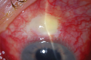
Figure 16.2 Patient with history of glaucoma filtering surgery with infected bleb.
Stay updated, free articles. Join our Telegram channel

Full access? Get Clinical Tree


