(1)
University of Sydney, Sydney, Australia
Overview
Visual electrophysiology is the recording of electrical signals produced by the visual system.
It allows assessment of the entire visual pathway, from RPE cells to the visual cortex.
Most electrophysiology tests are evoked potentials.
An abnormality at a proximal location along the visual pathway usually gives abnormal signal proximally as well as distally.
Hence, tests should be evaluated in the context of a full clinical history, examination, and other structural and visual electrophysiology investigations, and not interpreted in isolation.
Common Visual Electrophysiology Tests
Different testing strategies are used to assess separate parts of the visual pathway (Fig. 10.1).
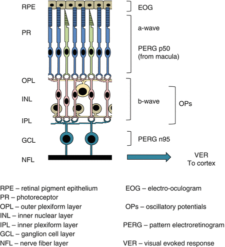
Fig. 10.1
Origin of the electroretinogram components and other electrophysiology studies
These include:
(i)
Electrooculogram
(ii)
Electroretinogram (ERG)
(a)
Full field
(b)
Focal
(c)
Pattern
(d)
Multifocal
(iii)
Visual evoked potentials
(a)
Pattern
(b)
Flash
(c)
Multifocal
The Electrooculogram
The electrooculogram (EOG) is a recording of the slow change in resting potential (voltage) of the retinal pigment epithelium (RPE) from a dark-adapted to a light-adapted state.
The resting potential is measured as the difference in voltage between the front (positive) and the back (negative) of the eye.
It is used as a measure of RPE function.
1.
Origin of the retinal pigment epithelium resting potential
RPE cells have a barrier layer of adjoining tight junctions near their apical surface (see Chap. 9, The Retinal Pigment Epithelium).
This separates the cell apical and basal membranes, causing a standing potential difference (voltage) across the RPE layer that is positive apically.
This results in the back of the eye (basal RPE layer) being negatively charged relative to the front of eye (cornea) (Fig. 10.2a).
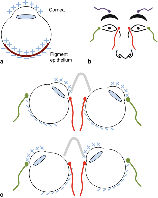
Fig. 10.2
(a) Origin of the ocular resting potential. (b, c) The electrooculogram test procedure
Dark adaptation results in a slow reduction in the RPE resting potential.
2.
Electrooculogram test procedure [5]
The test is performed binocularly, with the pupils dilated.
The electrode placement is outlined in Table 10.1 and Fig. 10.2b.
Table 10.1
Electrode placement for the electrooculogram
Electrode
Site
Active (+)
Inner canthi
Reference (−)
Outer canthi
Earth
Forehead, above the respective eyebrows
The patient shifts gaze repeatedly from left to right (Fig. 10.2c).
3.
Electrooculogram recording
The shifts in voltage on alternating gaze are recorded (Fig. 10.3).
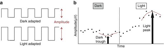
Fig. 10.3
Recording the electrooculogram. (a) Shifts in voltage on alternating gaze change in dark and light conditions. (b) This change can be plotted over time
The test is performed under light and dark adaptation states.
The dark trough is the minimal voltage amplitude in the dark.
The light peak is the maximal voltage amplitude in the light.
The Arden ratio is calculated:
Arden ratio = dark trough to light peak
The Arden ratio is normally >1.85
Values <1.85 are subnormal, and <1.3 are severely subnormal or extinguished.
4.
Electrooculogram: uses and limitations
The EOG depends on the function of both the RPE and the overlying photoreceptors [6].
Hence, it is most specific for the RPE when other tests of retinal function are normal.
The Full-Field Electroretinogram
The full-field ERG (ffERG) is a mass retinal potential evoked by a brief flash of light.
It is important in diagnosing retinal dystrophies or degenerations and can be used to evaluate global retinal dysfunction due to trauma or drug toxicity.
1.
Electroretinogram test procedure [7]
The test is performed monocularly with the subject’s pupils dilated.
Brief light flashes are presented full field to the retina, dispersed using a Ganzfeld bowl.
The bowl surface allows light to diffuse uniformly, stimulating the whole retina (Fig. 10.4).
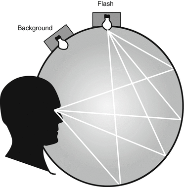
Fig. 10.4
The Ganzfeld bowl
A fixation point is incorporated into the bowl.
The electrodes placed on the face are outlined in Table 10.2.
Electrode
Site
Function
Active (+)
Contact lens (provides the most accurate and reproducible results) OR
Measurement of retinal electrical activity
Lower fornix, OR
Skin over eyelid (suboptimal trace)
Reference (–)
Incorporated into speculum (contacting the conjunctiva), OR
A baseline for measurement
Close to lateral canthus
Earth
Distant to the eyes
Elimination of electrical interference
2.
Recording the electroretinogram [7]
Recordings are usually made in two retinal adaptation states:
(a)
Scotopic trace: after 20 min of dark adaptation.
(b)
Photopic trace: after 10 min of light adaptation.
Recording the ERG in these two states helps separate the rod and cone system responses.
3.
Components of the electroretinogram (Fig. 10.5)
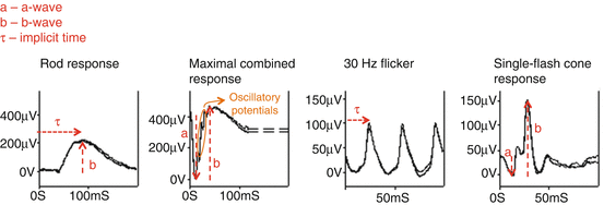
Fig. 10.5
Waveforms of standardized electroretinogram responses
In general, the ERG is characterized by:
(a)
An a-wave (negative waveform), followed by
(b)
A b-wave (positive waveform), followed by
(c)
A c-wave (positive waveform)
(i)
The a-wave
The a-wave is generated by light-induced photoreceptor hyperpolarization, with some postreceptoral contributions from OFF bipolar cells [8–11].
In the dark, cationic nucleotide-gated (CNG) channels are open resulting in the dark current (see Chap. 8, The Retina).
Light stimulation halts this cationic transfer, resulting in photoreceptor hyperpolarization.
Cones respond more quickly than rods, so cone hyperpolarization gives earlier negative deflection.
(ii)
(iii)
(iv)
Electroretinogram duration
The duration of the response is usually <150 ms.
The implicit time (τ) is from stimulus onset to the trough of the a-wave or peak of the b-wave.
4.
Standardized electroretinogram responses
Recording the ERG along international standards allows consistent and reproducible results [15].
5 standard ERG responses are typically recorded (Table 10.3 and Fig. 10.5).
Table 10.3
Standardized electroretinogram tests (flash strength in candela seconds per square meter) [7]
Test
Former name
Response
Dark-adapted 0.01 ERG
Stay updated, free articles. Join our Telegram channel

Full access? Get Clinical Tree

 Get Clinical Tree app for offline access
