(1)
University of Sydney, Sydney, Australia
Overview
2.
Listing’s plane and axes of Fick (Fig. 17.1)
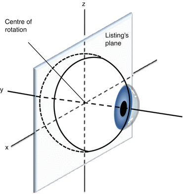
Fig. 17.1
Listing’s plane and the axes of Fick
All eye movements are composed of rotations of the anterior pole (central cornea) around one of three geometric axes of Fick: horizontal (x), anteroposterior (y), or vertical (z) [2].
The y-axis is a sagittal axis passing through the pupil; it is perpendicular to Listing’s plane.
The x-, y-, and z-axes meet at the center of rotation.
Vertical rotations occur about the x-axis, horizontal about the z-axis, and torsional about the y-axis.
3.
Positions of gaze
Primary position of gaze for each eye is directed straight ahead of the face.
Secondary positions are up, down, right, and left gaze.
These are achieved by pure rotations about the horizontal (x) or vertical (z) axes.
Tertiary positions are oblique positions: up and right, up and left, down and right, and down and left [4].
4.
Donders’ law, Listing’s law, and false torsion
Donders’ law: For any gaze direction, the eye assumes a specific three-dimensional orientation [1].
The orientation is always the same irrespective of where the eye came from.
Listing’s law (an extension of Donders’ law): All gaze positions can be reached by rotation around a single axis that lies on Listing’s plane [5, 6].
Each tertiary gaze position has a vertical and horizontal component that causes a degree of torsion: this is false torsion [4].
5.
Point of tangency and arc of contact
The extraocular muscles (EOMs) arise from the bony orbit and travel anteriorly to reach the globe.
The point of tangency is the point where the muscle first contacts the globe: this is the point of effective insertion that determines the vector of force exerted by the muscle.
The arc of contact is the area where EOM lies in contact with the globe, between the point of tangency and the anatomical insertion [7].
Actions of the Extraocular Muscles (Table 17.1) [8, 9]
Muscle | Action | ||
|---|---|---|---|
Primary | Secondary | Tertiary | |
Medial rectus | Adduction | ||
Lateral rectus | Abduction | ||
Superior rectus | Elevation | Incyclotorsion | Adduction |
Inferior rectus | Depression | Excyclotorsion | Adduction |
Superior oblique | Incyclotorsion | Depression | Abduction |
Inferior oblique | Excyclotorsion | Elevation | Abduction |
Each muscle changes tone with every ocular rotation.
All other EOMs have primary, secondary, and tertiary movements.
The relative amounts of each vary with gaze position, determined by the direction, origin, and insertion of the muscle in the particular direction of gaze [12].
1.
Superior rectus (SR)
The SR is a pure globe elevator on 23° abduction (along the geometric axis of the orbit).
In other gaze positions, it also incyclotorts (principally in adduction) and adducts (principally in abduction beyond 23°).
2.
Inferior rectus (IR)
The IR is a pure globe depressor on 23° abduction (along the geometric axis of the orbit).
In other positions it also excyclotorts (principally in adduction) and adducts (principally in abduction beyond 23°).
3.
The superior oblique (SO) and inferior oblique (IO)
The SO primarily incyclotorts, while the IO primarily excyclotorts the eye.
These actions are maximal in abduction.
In adduction their role in depression (SO) and elevation (IO) becomes more prominent.
4.
Field of action and field of activation
The field of action is the gaze direction for a muscle where its effect is most evident (Fig. 17.2).
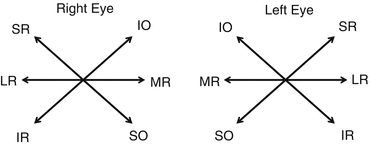
Fig. 17.2
Fields of action and yolk pairs
The field of activation is the direction of rotation from primary position if the muscle was the only one to contract.
For the LR and MR, these directions are the same (abduction and adduction, respectively).
They are not the same for the vertical recti or oblique muscles; e.g., the inferior oblique, acting alone, is an elevator and abductor; however, the elevatory function is best observed in adduction.
5.
Yoke muscles and Hering’s law
Yoke muscles describe muscle pairs (1 from each eye) that work together, sharing common fields of action (Fig. 17.2) [13].
Stay updated, free articles. Join our Telegram channel

Full access? Get Clinical Tree


