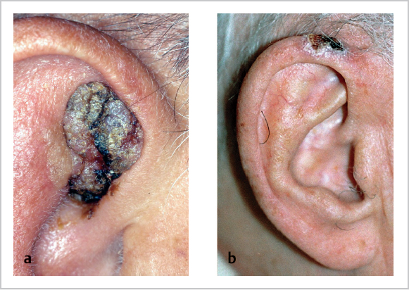
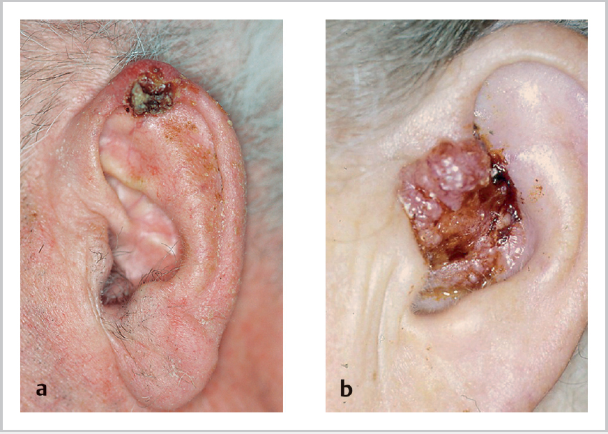
11 Tumours of the Ear and Skin • Tumours of the ear are rare: ~70% pinna, 20% external auditory canal (EAC), 10% middle ear (ME) • Squamous cell carcinoma (SCC) and basal cell carcinoma (BCC) most common (respective incidences vary country to country) (Figs. 11.1 and 11.2a) • Rx: primary resection with appropriate margins; minor superficial lesions with no cartilage invasion or fixation may have radiotherapy (RT) alone • Benign include nevus, epithelioma adenoides cysticum, molluscum contagiosum • SCC (Fig. 11.2b) • Adenocarcinoma: very aggressive (usually fatal within 4 years), often presenting with VII n palsy • Adenoid cystic: most common “ceruminous” tumour; long natural history, painful • Also adenoma, mucoepidermoid carcinoma; malignant melanoma very rare, as is BCC of EAC • Choristoma = normal tissue in abnormal site (e.g., salivary gland in ME): rare; may be attached to VII n • Adenoma (benign): cause unilateral progressive conductive hearing loss (HL), TM intact in 75% cases; 2nd most common after glomus tumours, but still rare • Adenocarcinoma: very rare; cause deafness, pain, VII n palsy • SCC: • Other benign tumours: Fig. 11.1a, b a Senile keratosis of the auricle. b Basal cell carcinoma. • Pathology: • Epidemiology:
11.1 Incidence and Site
11.2 Pinna
11.3 EAC
 90% of malignant tumours, slow-growing, spread via cartilage portion and later into ME, parotid, sternocleidomastoid muscle
 20% lymph node (LN) spread (posteriorly to mastoid nodes, inferiorly to level III, anteriorly to preauricular)
 Associated with chronic inflammation, RT
 Rx: consider lateral temporal bone resection, ± neck dissection; post-operative RT
 Prognosis very poor if node positive
11.4 Middle Ear
 Most common epithelial ME malignancy, but rare
 Association with chronic otitis media in 85%, irradiation
 Extensive bony destruction, most easily through tegmen to middle cranial fossa (dura is a relatively strong barrier); intracranial invasion along VII n and VIII n in internal auditory canal; IX n and XII n involvement shows skull base involved; can also extend to temporomandibular joint, parotid, via eustachian tube to nasopharynx
 LN involvement in 10 to 15% at presentation, rare distant metastasis; death by cachexia secondary to pain because cranial nerve (CN) involvement, though sometimes invasion of meninges leads to intracranial complications and erosion of jugular bulb or carotid is possible
 Rx: surgery (petrosectomy) + RT; palliative
 5-year survival ~35% (better for EAC than ME); prognosis very poor if node positive
 Osteoma
 VII n schwannoma (usually at geniculate ganglion)
 Hemangioma, neurogenic
 Sarcoma
 Multiple myeloma
 Malignant xanthoma
 Rhabdomyosarcoma most common in children; may require surgery ± chemoradiotherapy
 Also direct extension (e.g., from nasopharynx, external ear, parotid, meningioma) and distant metastases (kidney, lung, prostate, thyroid, breast)
11.5 Glomus Tumours
 Correct (WHO) term is paragangliomas; aka chemodectomas; arise from paraganglionic tissue (chemoreceptor cells)
 Common embryological origin (neural crest) and association with neural structures
 90% adrenal (phaeochromocytomas); 10% nonadrenal (85% of which are abdominal, only 3% H&N); subdivided into abdominal and extra-abdominal
 H&N paragangliomas associated with parasympathetic system
 Relative incidence: carotid > jugulotympanic > vagal
 5th decade, F>M, may be multiple (show multicentricity); ~10%, autosomal dominant; usually benign and slow-growing but ~5% malignant; rarely (<1%) secrete neuropeptides (e.g., serotonin, vasoactive intestinal peptide)
![]()
Stay updated, free articles. Join our Telegram channel

Full access? Get Clinical Tree


