Topography, Bones, Vessels, Sensory Nerves


Topography, Bones, Vessels, Sensory Nerves


1 The ocular globes reside in two symmetrical bony cavities called the orbits.

2 Nerves and vessels enter the orbit, and their course and distribution must be understood. The aerated sinuses as well as the anterior and middle cranial fossae are intimately related to the orbital walls. The first edition, Surgical Anatomy of the Orbit, focused on this orbit and its contents, but very little on the neighboring areas. This edition expands widely, leaving the orbital confines to expose structures around it. Some basics are still required, so this initial chapter was rewritten to present an updated compendium of the essential data.

3 In this portion, you will learn certain orbital distances and relationships that you, the surgeon, will need to commit to memory. For that reason, some key numbers may be repeated.

Notes to the reader: Throughout this book, all dissections use the right orbit. Labels are kept to a minimum so as not to distract the reader; perusing the legends will allow the pictures to really tell the story. This approximates “truth” where labels do not exist.


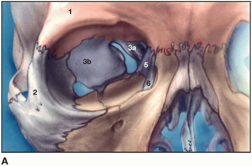
Figure 1.1A Bony components of the right orbit

1. Frontal bone (orange)

2. Zygoma (light gray)

3. Sphenoid bone (purple)

3a. Lesser wing

3b. Greater wing

Maxilla (tan)

Ethmoid bone (purple – 5)

Lacrimal bone (6)

Palatine bone (no. 4 below)


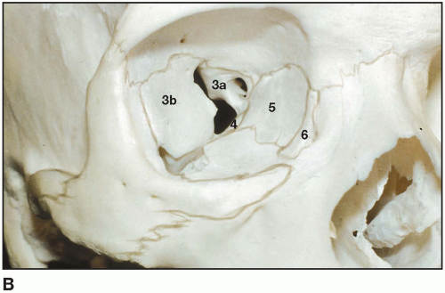
Figure 1.1B Dried skull with bony borders accented

3a. Lesser wing of sphenoid

3b. Greater wing sphenoid

4. Palatine bone

5. Ethmoid bone

6. Lacrimal bone


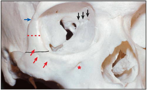
Figure 1.2 Anatomic points

1. The infraorbital foramen(*) opens downward and medially.

2. Red arrows point to exits of zygomaticofacial sensory nerve branches which supply the malar eminence and below.

3. The width of the lateral orbital rim inferior line is 1.3 to 1.5 cm.

4. The width of the lateral orbital rim at canthal level is 1.0 to 1.1 cm (dotted), and thinner as it approaches zygomatico-frontal suture (blue arrow).

5. Intraorbital arrows:

Anterior and posterior ethmoidal foramina separate frontal bone above from ethmoid bone below it at the frontoethmoidal suture.

The anterior foramen is 20 mm behind the anterior orbital margin, and the posterior is 12 mm behind this. The ophthalmic artery gives off the posterior ethmoidal arteries for the posterior ethmoidal air cells, anterior cranial fossa dura, and upper nasal mucosa. The anterior ethmoidal artery enters the anterior cranial fossa, and then via the cribriform plate into the nose.

The anterior ethmoidal nerve, a sensory branch of the nasociliary, supplies anterior ethmoidal air cells, mucosa of the upper nose, and then exists as the dorsal nasal nerve (responsible for herpes zoster of the nasal tip).


Figure 1.3 The walls

The roof. This wall is composed mainly of the orbital plate of the frontal bone. Posteriorly, it receives a minor contribution from the lesser wing of the sphenoid bone.

Located within the anterolateral portion of the roof, note a smooth, wide depression for the lacrimal gland, the lacrimal fossa (LGF). Approximately 5 mm posterior to the medial aspect of the rim, the trochlear fossa (TF) denotes the attachment of the cartilaginous pulley for the tendon of the superior oblique muscle. The triangular roof narrows as it proceeds toward the apex, where a vertically oval opening, the optic foramen (OF), marks the orbital end of the optic canal.

The lateral wall. The lateral wall is formed primarily by the orbital surface of the zygomatic bone (Z) and the greater wing of the sphenoid bone (S).

A small, bony promontory may be noted just within the orbital rim. This important landmark, Whitnall’s tubercle (WT), is the point of attachment for several structures 11 mm below the zygomaticofrontal suture. At the anterior end of the inferior orbital fissure, a small groove may be noted for the passage of the zygomatic nerve and vessels. The groove often develops into a canal that divides within bone to conduct the zygomaticofacial vessels and nerves onto the face and the zygomaticotemporal vessels and nerves into the temporal fossa. The zygomaticofacial and zygomaticotemporal complexes may pass through separate foramina.

The floor. The inferior orbital fissure (IOF) separates the greater sphenoid wing portion of the lateral wall from the floor. The fissure communicates with the pterygopalatine fossa as well as the infratemporal fossa. Through this fissure pass (1) the maxillary division of the trigeminal nerve, V2, and its branches; (2) the infraorbital artery; (3) branches of the sphenopalatine ganglion; and (4) branches of the inferior ophthalmic vein to the pterygoid plexus.
The thin orbital floor is composed of the orbital plate of the maxilla, the zygomatic bone anterolaterally, and the orbital process of the palatine bone posteriorly. A shallow rough area at the anteromedial angle (OBL) marks the origin of the inferior oblique muscle. The infraorbital groove runs forward from the inferior orbital fissure. Anteriorly, the groove becomes a canal within the maxilla, finally forming the infraorbital foramen on the face of the maxilla. The groove and canal transmit the infraorbital nerve and artery. From the lower aspect of this nerve, middle superior alveolar nerves (occasionally) emanate to supply the bicuspid teeth. More anteriorly and 5 to 20 mm prior to the infraorbital nerve exit from its foramen, the anterior superior alveolar nerves descend medially along the inner face of the maxilla or within a canal to supply sensation to the anterior three teeth and gingiva.

The medial wall. The medial wall is quadrangular in shape and is composed of four bones: (1) the ethmoid bone centrally; (2) the frontal bone superoanteriorly; (3) the lacrimal bone inferoanteriorly; and (4) the sphenoid bone posteriorly. The inferior orbital margin continues upward into the anterior lacrimal crest (ALC), part of the frontal process of the maxilla. The superomedial margin continues downward into the posterior lacrimal crest (PLC) part of the lacrimal bone. Between these rests the fossa for the lacrimal sac (LF). Usually, the fossa is approximately 14 mm in height.

The medial wall is quite thin, and the ethmoidal portion has been termed lamina papyracea. The anterior and posterior ethmoidal foramina (AE, PE) are noted at the frontoethmoidal suture and denote the level of the cribriform plate. The anterior ethmoidal foramen transmits the anterior ethmoidal artery and the anterior ethmoidal nerve branches of the nasociliary nerve. The posterior ethmoidal foramen provides a passage for the posterior ethmoidal artery and, occasionally, for a sphenoethmoidal nerve branch from the nasociliary nerve.


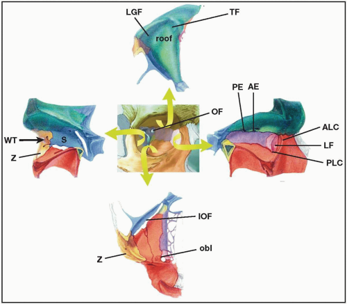
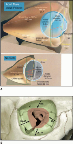
Figures 1.4(A-B)

Volumes and distances: Males/females and babies

In theory, everything outside the annulus (Fig. 1.4A) and superior orbital fissure is safe. As the surgeon realizes the distance to the optic foramen is approximately 50 mm (men) and 45 mm (women) and gives at least 1 cm margin, the surgeon may dissect 35 to 40 mm back on the floor and roof region. Lateral dissection may even go a bit farther back.

Only gold members can continue reading. Log In or Register to continue

Stay updated, free articles. Join our Telegram channel

Sep 23, 2016 | Posted by in OTOLARYNGOLOGY | Comments Off on Topography, Bones, Vessels, Sensory Nerves

Full access? Get Clinical Tree

Get Clinical Tree app for offline access