Fig. 8.1
Ultrasound presentation of a subacute de Quervain thyroiditis. Two hypoechoic areas with undefined margins and vascular spots are present at the superior and inferior thirds of the right thyroid lobe (a, longitudinal scan; b, transversal scan). Mild-moderate pain is detectable at physical examination and ultrasound evaluation
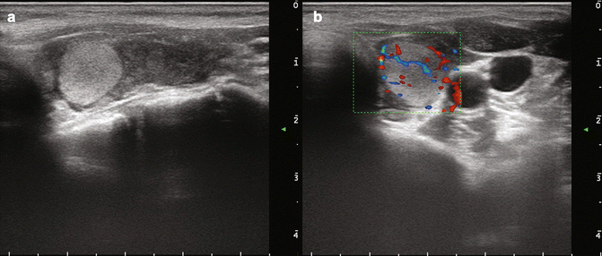
Fig. 8.2
Ultrasound presentation of a case of Hashimoto thyroiditis with a well defined hyperechoic area. The gland has an extended hypoechogenicity as a sign of tissue destruction by Hashimoto disease. At the base of left lobe there is a normoechoic (hyperechoic with respect to the surrounding tissue) nodular area (a) with normal vascularization (b) of about 1 cm. This area is not a “true” nodule but represents a part of the gland not involved by Hashimoto autoimmune pathways
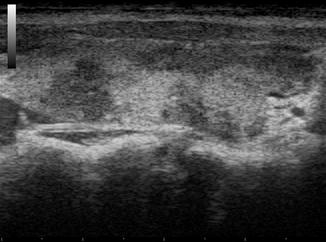
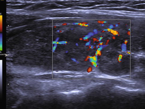
Fig. 8.3
Longitudinal ultrasonography scan of right thyroid lobe with Hashimoto’s thyroiditis. The structure is inhomogeneously damaged, the majority of the tissue appears hypoechoic due to the damage by autoimmune pathway. Normoechoic areas represent the normal (normofunctioning) tissue
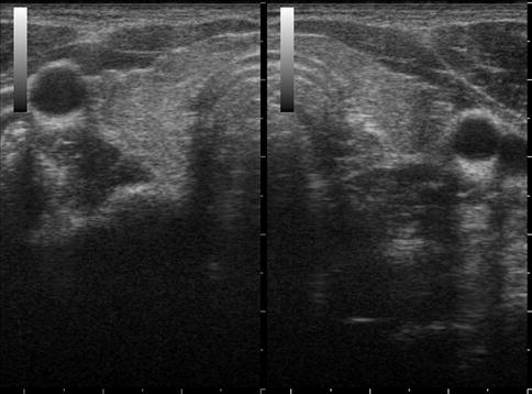
Fig. 8.4
Transverse ultrasonography scan of thyroid gland with thyroiditis. Thyroid volume is reduced, margins are irregular, echostructure is slightly inhomogeneous, echogenicity is poor. This thyroid US aspect frequently correlates with hypofunction (hypothyroidism)
Only gold members can continue reading. Log In or Register to continue
Stay updated, free articles. Join our Telegram channel

Full access? Get Clinical Tree


