Fig. 16.1
Well-differentiated neuroendocrine tumor of the right middle ear detected by fused somatostatin receptor PET/CT. PET with somatostatin analogs labeled with gallium-68 (upper row) detected a focal area of increased radiopharmaceutical uptake (arrows) corresponding to a subcentimeter nodule in the right middle ear at CT (mid row) and fused PET/CT images (lower row)
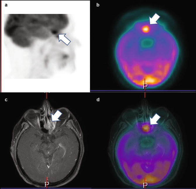
Fig. 16.2
Sinonasal small cell carcinoma of the left ethmoidal sinus (arrows) imaged by fluorine-18 fluorodeoxyglucose positron emission tomography (18F-FDG PET) in sagittal (a) and axial projection (b), magnetic resonance imaging (MRI) in axial projection (c), and fused PET/MRI (d). This tumor was well-characterized by MRI which showed invasion of the adjacent structures. PET demonstrated increased glucose metabolism of this tumor without metastatic spread
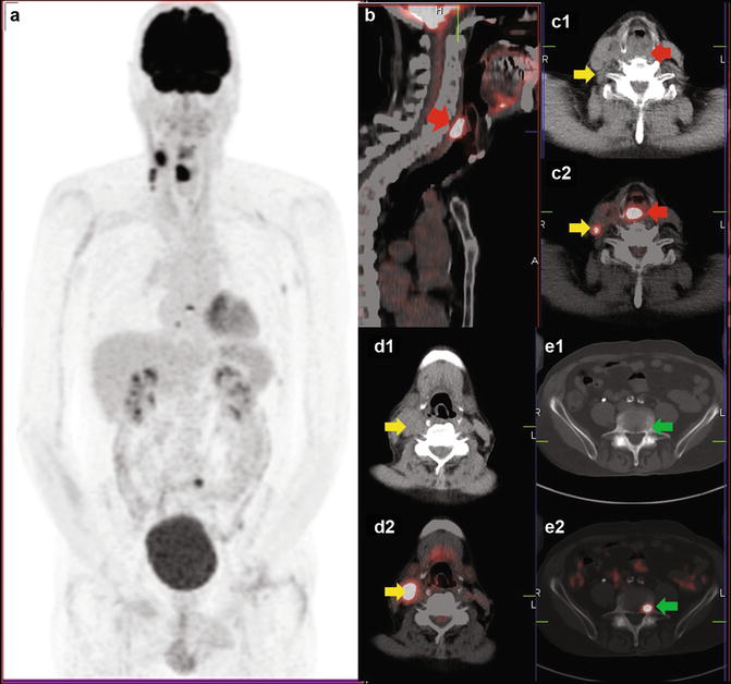
Fig. 16.3
Metastatic laryngeal small cell carcinoma imaged by fluorine-18 fluorodeoxyglucose positron emission tomography/computed tomography (18F-FDG PET/CT). 18F-FDG PET (a) showed an area of increased tracer uptake corresponding to a laryngeal tumor (red arrow) at CT (c1) and fused PET/CT images in sagittal (b) and axial projection (c2). Furthermore several right cervical lymph nodal metastases (yellow arrows) were detected by PET (a), CT (c1, d1), and fused PET/CT images (c2, d2). Interestingly, 18F-FDG PET detected bone metastases (green arrows) without significant morphological abnormalities at co-registered CT (e1, e2)
< div class='tao-gold-member'>
Only gold members can continue reading. Log In or Register to continue
Stay updated, free articles. Join our Telegram channel

Full access? Get Clinical Tree


