Purpose
To report the use of a sterile, γ-irradiated corneal tissue without viable endothelium (VisionGraft Sterile Cornea; Tissue Banks International) in lieu of fresh donor cornea in Boston type 1 keratoprosthesis (KPro) implantation.
Design
Retrospective, interventional small case series.
Methods
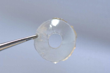
Eleven eyes of 11 patients underwent Boston type 1 keratoprosthesis implantation using VisionGraft Sterile Cornea between April 2009 and October 2010. Precut donut-shaped corneal lenticules, using femtosecond laser, measuring 8.5 mm in diameter with a 3-mm central hole were used.
Results
Surgical procedures were uneventful. Complete corneal re-epithelization was noted within 10 days in all cases. No complications related to the donor cornea (eg, stromal necrosis, wound leak, or device extrusion) occurred over an average follow-up of 16.5 months.
Conclusions
VisionGraft Sterile Cornea eliminates the need for use of fresh donor corneal tissue for Boston type 1 keratoprosthesis procedures, which makes this procedure a viable sight-restoring option when donor corneal tissues are not readily available.
Boston type I keratoprosthesis (KPro) implantation is a viable alternative to penetrating keratoplasty in cases where donor corneal transplantation has a high risk of failure. Although the device was granted 510(k) clearance by the United States Food and Drug Administration in 1992, its use has increased significantly within the last decade, with approximately 6000 devices implanted worldwide. Recent design modifications, advances in surgical technique, and improvements in postoperative management have reduced the frequency of postoperative complications such as corneal necrosis with device extrusion and endophthalmitis, which were associated more commonly with KPro implantation in the past. Although retroprosthetic membrane formation and glaucoma continue to be problematic, multiple published reports consistently have demonstrated favorable outcomes, with a retention rate between 80% and 100% over an average follow-up of 8.5 to 33.6 months. Rapid rehabilitation of vision with excellent uncorrected acuity are added benefits with KPro, even in aphakic patients. Despite these encouraging results, however, the use of KPro has remained limited outside the United States. Only approximately one third of the 1200 devices used in 2010 were implanted outside the United States. Although this is the result of multiple factors, one significant issue is the unavailability of fresh donor cornea to serve as a carrier for the KPro.
We herein present our experience in a small group of patients using sterile, γ-irradiated corneal tissue without viable endothelium (VisionGraft Sterile Cornea; Tissue Banks International [TBI], Baltimore, Maryland, USA) in lieu of fresh donor cornea as a carrier tissue in KPro implantation.
Methods
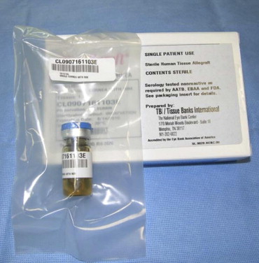
The VisionGraft Sterile Corneas were supplied by TBI in sterile glass bottles stored in albumin solution ( Figure 1 ). The tissue consists of a full-thickness, clear, corneal lenticule that is precut using a femtosecond laser (IntraLase; Abbott Medical Optics, Abbott Park, Illinois, USA) with an 8.5-mm outer diameter and a 3-mm inner diameter ( Figure 2 ). The step of preparation and trephination of the donor tissue thus is fully eliminated with this technique. The device is assembled on a side table by sandwiching the donor tissue between the front plate and the back plate of the KPro (supplied by Massachusetts Eye and Ear Infirmary, Boston, Massachusetts, USA). The assembly then is locked together with a titanium C-locking ring that is snapped into a groove on the stem of the optic, securing the back plate into position. The assembly is demonstrated in the Supplemental Video (available at AJO.com ). The entire unit then is sutured to the host corneal bed as in standard penetrating keratoplasty.
At the end of each operation, a large-diameter soft contact lens (16-mm diameter and 9.8-mm base curve, plano; Kontur Kontact Lens Co., Hercules, California, USA) that comes with the KPro device in the same packaging is placed in the operative eye. Subconjunctival injections of cefazolin and dexamethasone are administered. A combination antibiotic and steroid ointment is used to dress the eye before the sterile eye patch and a shield are placed. Daily topical antibiotic prophylaxis with fourth-generation fluoroquinolones and anti-inflammatory treatment with 1% prednisolone are used in all patients indefinitely. A soft bandage contact lens is maintained in most eyes to ensure hydration and to prevent epithelial defect formation overlying the donor cornea. The contact lens is exchanged every 1 to 3 months to decrease the risk of microbial colonization and possible infectious keratitis. The postoperative management after KPro implantation continues to be largely surgeon driven. The regimen mentioned here is the preference of the authors and may not necessarily be consistent with other previous reports.
Results
Since the VisionGraft Sterile Cornea became commercially available in the United States in April 2009, more than 1700 grafts have been used: 50 of them in KPro surgeries as a carrier tissue (Pennington D, TBI, The National Eye Bank Center, person communication, February 28, 2012). We herein report available detailed patient information, surgical technique, and follow-up on 11 of these patients who underwent surgery at 3 tertiary eye care centers (Wilmer Eye Institute, Jules Stein Eye Institute, and Flaum Eye Institute) performed by 3 experienced KPro surgeons (E.K.A., A.J.A., and J.V.A., respectively). The demographic and clinical information as well as outcomes of the patients are summarized in the Table . The surgeries were performed between April 2009 and October 2010. All but 2 patients had failed multiple donor corneal transplants before receiving the KPro. Three of 11 (30%) the patients had associated inflammatory ocular surface diseases and clinically significant tear film abnormalities (graft-versus-host disease, n = 1; mucous membrane pemphigoid, n = 1; and Stevens-Johnson syndrome/toxic epidermal necrolysis, n = 1). In all patients, resolution of the epithelial defect that invariably was present on the donor cornea on postoperative day 1 was achieved by postoperative day 10. The most common postoperative complication was retroprosthetic membrane formation, which developed in 30% (3/11) of patients. None of the recognized postoperative complications possibly attributable to the donor cornea, including persistent corneal epithelial defect formation, infectious keratitis, and sterile stromal necrosis, developed during an average follow-up of 16.5 months (range, 8 to 24 months). Slit-lamp appearance of an eye (Patient 1) 3 months after KPro surgery using VisionGraft Sterile Cornea is depicted in Figure 3 . A small air bubble is apparent underneath the anterior plate, perhaps suggesting a mild thinning of the tissue around the stem. This patient now has more than 3 years of follow-up with no complications.
| Patient No. | Age (y) | Sex | Preoperative VA | Indication for KPro | Initial Corneal Diagnosis | Comorbid Conditions | Postoperative Complications | Follow-up (mos) | CDVA at Last Visit | KPro Status at Last Visit |
|---|---|---|---|---|---|---|---|---|---|---|
| 1 | 59 | M | HM | Failed PK | Bullous keratopathy | Glaucoma | RPM | 25 | 20/20 | Retained |
| 2 | 75 | F | HM | Failed PK | Bullous keratopathy | Glaucoma | None | 24 | CF | Retained |
| 3 | 71 | F | HM | Failed PK | Sterile corneal ulceration and scarring | Glaucoma | Optic nerve atrophy | 13 | 20/80 | Retained |
| 4 | 64 | F | HM | Failed PK | Bullous keratopathy | None | None | 19 | 20/40 | Retained |
| 5 | 62 | M | HM | Mentally Challenged | Keratoconus | Cataract | RPM | 22 | Functional | Retained |
| 6 | 70 | F | 20/100 | Failed PK | Keratoconus | None | None | 20 | 20/20 | Retained |
| 7 | 62 | M | HM | Corneal scarring | Keratoconus | None | RPM | 20 | Functional | Retained |
| 8 | 89 | M | HM | Failed PK | Sterile corneal ulceration and scarring | GvHD | None | 24 | 20/100 | Retained |
| 9 | 55 | F | CF | Failed PK | Sterile corneal ulceration and scarring | MMP, cataract | None | 13 | 20/60 | Retained |
| 10 | 26 | F | 20/400 | Failed PK | Aniridia | Aphakia | None | 9 | 20/70 | Retained |
| 11 | 10 | M | CF | Failed KPro | SJS/TEN | Retinal detachment | None | 8 | 20/60 | Retained |
Stay updated, free articles. Join our Telegram channel

Full access? Get Clinical Tree


