The Use of Fluorescein Angiography in Acquired Macular Diseases
Antonio P. Ciardella
Stephen R. Kaufman
Lawrence A. Yannuzzi
Fluorescein angiography (FA) has been widely used clinically for more than 3 decades, and it has been valuable in the understanding, diagnosis, and treatment of acquired macular diseases. Extensive use of FA, combined with growing knowledge of the range of clinical presentations and natural histories of the acquired macular diseases, has helped clinicians obtain an appreciation of the indications for FA. Our aim in this chapter is to illustrate useful parameters of FA and to provide guidance for the optimal use of this technique. Comprehensive reviews of the interpretation of the fluorescein angiogram may be found elsewhere.1,2
AGE-RELATED MACULAR DEGENERATION
DIAGNOSIS
Age-related macular degeneration (AMD) may be divided into two types. Nonexudative (“dry”) AMD has several morphologic forms, including “hard” discrete drusen, shallow retinal pigment epithelial detachments associated with thickened Bruch’s membrane (“soft” drusen), and geographic atrophy (GA) of the retinal pigment epithelium (RPE).3 On FA the area of GA appears hyperfluorescent for window defect from the early frames of the angiogram, with late staining of the underlying sclera (Fig. 1). However, these pathologic changes can usually be assessed by clinical examination, and FA is generally not necessary to diagnose nonexudative AMD. An exception is cuticular drusen, which may appear clinically as a subtle disturbance of the RPE; FA reveals multitudes of small, discrete drusen described as “stars in the sky” (Fig. 2). The second type of AMD, which is associated with soft drusen, is known as exudative (“wet”) AMD. It is due to a choroidal neovascular membrane that has incompetent vessels resulting in detachments of the RPE and the neurosensory retina. Consequently, in patients with a large RPE and/or serous neurosensory detachment, FA is often necessary to rule out a choroidal neovascularization (CNV). In general, a small pigment epithelium detachment (PED) and a larger neurosensory detachment overlie CNV, while the opposite is generally the case in a nonexudative PED. Additionally, CNV often presents as a “notched” PED (Fig. 3).4 The presence of subretinal blood or pigment at the border of a PED strongly indicates that the detachment is exudative in origin (Fig. 4). Similarly, a rip in the RPE generally reflects subretinal fibrosis from a CNV (Fig. 5 and 6). The diagnosis is more difficult in patients who have a chronic, organized PED. Such a lesion may be due to either nonexudative AMD or to an organized, fibrotic CNV. Clinically and angiographically, it may be impossible to distinguish between these two conditions. In most cases, however, FA does assist in making the diagnosis.
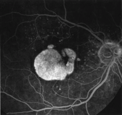 Fig. 1. Late-phase fluorescein angiography of an eye with a central area of geographic atrophy, which appears hyperfluorescent. A few soft drusen are also present. |
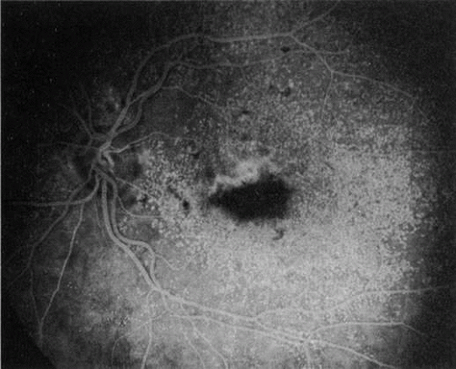 Fig. 2. Multiple cuticular drusen and subretinal neovascularization. This patient had some central soft drusen, which, unlike cuticular drusen, are associated with neovascularization. The inferior aspect of the choroidal neovascular membrane has blocked fluorescence due to hemorrhage. Note the presence of the small, hyperfluorescent cuticular drusen that confer a “stars in the sky” appearance. |
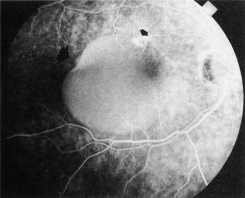 Fig. 3. Notched retinal pigment epithelium (RPE) detachment. There is a notch at the superotemporal border of a large RPE detachment that fills unevenly with fluorescein (large arrowhead). There is also a superior neovascular complex (small arrowhead) that hyperfluoresces. (Courtesy of Dr. Kenneth G. Noble.) |
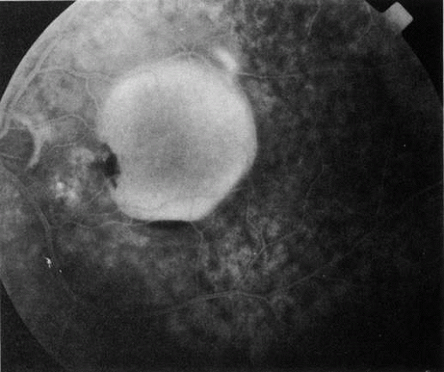 Fig. 4. Subretinal blood from a choroidal neovascular membrane. A small hemorrhage has layered out at the inferior aspect of a large retinal pigment epithelium (RPE) detachment. A shallow overlying neurosensory detachment can be appreciated as the slightly darkened, narrow band that surrounds the RPE detachment. The neurosensory detachment is being filled with fluorescein through a break in the RPE. The subretinal neovascular membrane, not clearly evident in this early-phase fluorescein angiogram, is at the nasal edge of the RPE detachment, near the optic disc. |
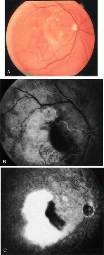 Fig. 5. A. Clinical photograph of a large, crescent-shaped rip of the retinal pigment epithelium (RPE) in the temporal macula. B. Early-phase fluorescein angiography study demonstrates the presence of a window defect corresponding to the RPE rip, which exposes the choroidal vasculature. Where the RPE is redundant in the central macula there is blockage of the normal choriocapillaris fluorescence. C. Late-phase angiogram reveals intense hyperfluorescence seen through the RPE defect. |
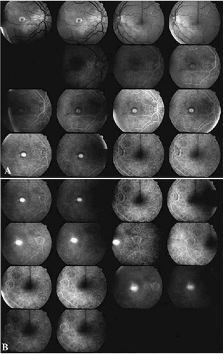 Fig. 6. Classic, submacular choroidal neovascularization (CNV) and retinal pigment epithelium (RPE) rip. Fluorescein angiography reveals the presence of a well-defined subfoveal CNV and hyperfluorescent area in the inferior macula corresponding to the RPE rip (yellow arrow). |
In patients with a shallow neurosensory detachment, the Amsler grid test and visual acuity may be normal. If there is subtle elevation of the neurosensory retina on biomicroscopy examination, FA may demonstrate a CNV before it is symptomatic. It is often easier to evaluate both RPE and neurosensory detachments with good stereoscopic FA pictures than with direct examination.5 Consequently, FA can be helpful in determining the presence and extent of these processes. This is particularly important in patients with CNV due to AMD, because its aggressive course often requires prompt intervention to save central vision.6,7
Furthermore, FA helps in recognizing two types of CNV: classic and occult. Classic CNV consists of a well-defined neovascular membrane, which is apparent in the early phase of the angiogram and shows late leakage of dye beyond its boundaries (Fig. 7 and 8). Occult CNV is seen on by FA as an area of late hyperfluorescence of undefined origin or as a neovascularized PED (Fig. 9 and 10). Mixed-type CNV is predominately classic or minimally classic depending on whether the classic component is more or less than 50% of the entire lesion (Fig. 11).
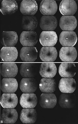 Fig. 7. Composite photograph of fluorescein angiography study in a patient with classic, subfoveal choroidal neovascularization (CNV) in the right eye. A The classic neovascular membrane appears as a well-defined area of hyperfluorescence in the early phases of the angiogram. There is leakage of dye from the classic net in the subretinal space throughout the study. B. In the late phase of the study, the edges of the CNV are fuzzy and indistinguishable. |
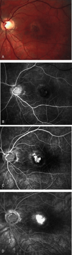 Fig. 8. A. Color photograph of subfoveal classic choroidal neovascularization (CNV). The neovascular membrane appears as a dirty gray, subfoveal lesion surrounded by exudative neurosensory detachment. B–D. Fluorescein angiography demonstrates early hyperfluorescence and late leakage of the CNV. |
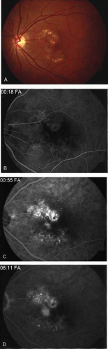 Fig. 9. A. Clinical photograph of the left eye of a patient with exudative neurosensory macular detachment. There were also intraretinal and subretinal hard exudates, subretinal hemorrhage, and retinal pigment epithelium (RPE) changes. B–D. Fluorescein angiography of the same eye demonstrates the presence of stippled hyperfluorescence from the RPE, and late-phase oozing of dye of undefined origin. There was occult choroidal neovascularization. |
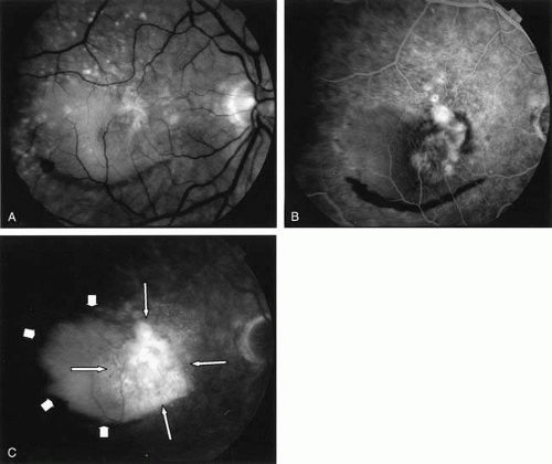 Fig. 10. A. Red-free photograph of the right eye of a patient with wet age-related macular degeneration. There was a large, exudative pigment epithelium detachment (PED), with a narrow band of subretinal hemorrhage at its inferior border. A notch in the PED is present at its nasal edge. There were also soft drusen. B–C. Fluorescein angiography demonstrates pooling of dye into the PED (short arrows). There was also late hyperfluorescence of undefined origin consistent with occult choroidal neovascularization (long arrows). There was blockage of fluorescence at the inferior border of the PED caused by subretinal hemorrhage. |
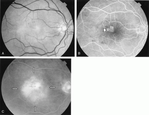 Fig. 11. A. Red-free photograph of the right eye of a patient with wet age-related macular degeneration reveals exudative, neurosensory detachment in the macula and a few subretinal hemorrhages. B. Early-phase fluorescein angiography demonstrates well-defined classic choroidal neovascularization (CNV) (arrowhead). C. Late-phase fluorescein angiography shows leakage of dye from the classic CNV surrounded by an area of late hyperfluorescence consistent with occult CNV (arrows). |
FA is also useful in characterizing two other subgroups of CNV: retinal angiomatous proliferation (RAP)8,9,10,11,12,13,14,15,16 and polypoidal choroidal vasculopathy (PCV).17,18,19,20,21,22,23,24,25,26,27,28,29,30,31,32,33,34,35,36,37,38,39,40,41,42,43,44,45,46,47,48,49,50 RAP begins in the deep retinal complex, forming intraretinal neovascularization (IRN), which may subsequently progress to extend beneath the neurosensory retina, forming subretinal neovascularization (SRN), and a vascularized PED.8 In the later phases of the process there may be a retinal-choroidal anastomosis (RCA). Clinical features of RAP include intraretinal hemorrhages, cystoid macular edema, and associated vascularized PED. FA is useful in revealing the presence of the angiomatous intraretinal vascular complex and the extension of the associated PED (Figs. 12 and 13). However, other diagnostic techniques such as indocyanine green (ICG) angiography, and optical coherence tomography (OCT) may be able to better demonstrate the presence of the RAP lesion.
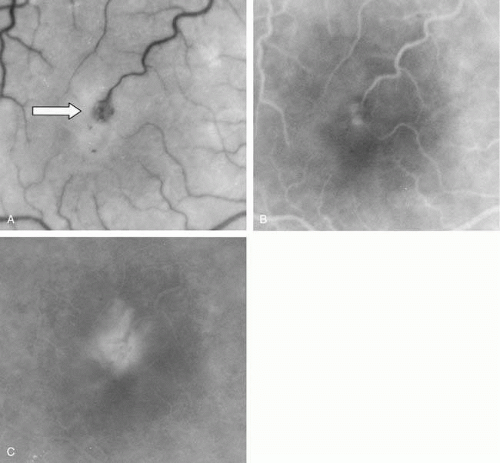 Fig. 12. A. Clinical photograph of a retinal angiomatous proliferation (RAP) lesion (arrow). Note the intraretinal angiomatous proliferation, a feeding retinal arteriole, and a draining retinal venule, as well as the presence of intraretinal hemorrhages. B–C. Fluorescein angiography reveals late leakage from the RAP lesion. |
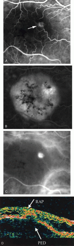 Fig. 13. A. Early-phase Fluorescein angiography demonstrating the presence of an intraretinal angiomatous lesion (arrow). There is an associated pigment epithelium defect (PED), which is still hypofluorescent. B. Late-phase fluorescein angiography shows leakage from the retinal angiomatous proliferation (RAP) lesion and polling of dye into the PED. C. Indocyanine green angiogram of the same eye better demonstrates the presence of a hot spot corresponding to the RAP lesion. The PED remains hypofluorescent. D.Optical clearance tomography image demonstrates the presence of a serous PED and of intraretinal neovascularization. |
PCV is characterized by the presence of dilated, choroidal vascular channels ending in orange bulging polyp-like dilations in the peripapillary and macular area. Associated features are recurrent subretinal hemorrhage and vitreous hemorrhage, relatively minimal fibrous scarring, absence of retinal vascular disease, pathologic myopia, and signs of intraocular inflammation. FA demonstrates the presence of the dilated vascular channel (Fig. 14 and 15). However, the presence of blood and exudation may block the details of the choroidal circulation on the angiogram. In these cases, ICG angiography can better demonstrate the presence of a distinct network of vessels within the choroid because the larger choroidal vessels are filled with dye.
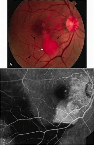 Fig. 14. A. Color photograph of the right eye shows a ramified pattern of choroidal vascular abnormality irradiating from the peripapillary area toward the macula. The dilated vascular channels end with bulging polyp-like structures. A larger, orange, saccular dilation is seen inferior to the macula (white arrow); leakage of fluid from this vascular abnormality results in serosanguineous pigment epithelium detachment (black arrows). B. The corresponding fluorescein angiogram composite highlights the vascular lesion in the peripapillary area and the serosanguineous detachment of the pigment epithelium that extends inferiorly and temporally off the macula. |
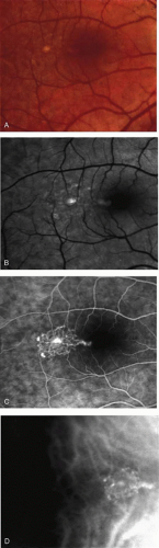 Fig. 15. A 51-year-old Caucasian woman was referred with diagnosis of central serous chorioretinopathy in her right eye. A and B. Color photograph and red-free photograph of the right eye show the presence of atrophic changes in the retinal pigment epithelium consistent with central serous chorioretinopathy. C, Fluorescein angiogram of the right eye reveals the presence of avascular network of small-caliber vessels terminating in polyp-like structures. D. Indocyanine green angiogram of the right eye confirms the presence of the polypoidal lesion. |
The FA can also distinguish CNV from simulating lesions. For example, a dark mound of blood due to hemorrhage from a CNV will block choroidal fluorescence, whereas vascular tumefactions such as choroidal hemangiomas leak fluorescein. Choroidal melanomas frequently block early choroidal fluorescence and then leak fluorescein from their intrinsic vascular network in later phases of FA.
TREATMENT
FA is vital for the management of CNV.51,52,53,54,55 It can define the borders of the membrane and help localize the fovea. The final determination whether the CNV is subfoveal, juxtafoveal, or extrafoveal requires use of both FA to outline the membrane with respect to the retinal vasculature and clinical examination to define the precise location of the fovea (Fig. 16). Some patients, particularly those with high myopia, have an indistinct foveal avascular zone on FA. Other FA clues, such as the location of the macula lutea pigment, can be deceptive, because fixation does not necessarily correspond to the center of the macula lutea. The value of FA is also limited in cases of occult or poorly defined CNV, in which the exact location of the leaking CNV vessels cannot be angiographically determined (Fig. 17), and in patients with subretinal hemorrhage that obscures the membrane. In these patients, ICG angiography may be the most precise means of localization.
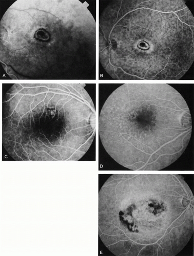 Fig. 16. Subfoveal, juxtafoveal, and extrafoveal choroidal neovascular membranes. A and B. Large subfoveal choroidal neovascularization (CNV) in a 69-year-old man with blood and pigment blocking central fluorescence on both the early-phase (A) and late-phase (B) photographs. The hypofluorescence surrounding the membrane is commonly seen in CNV and may be due to lipofuscin. C. Juxtafoveal CNV in a 37-year-old man with idiopathic CNVM. D. Cuticular drusen in same patient as in C were asymptomatic. E. Years later, this same patient developed a large extrafoveal CNV with central macular pigment abnormalities. A large neurosensory detachment was responsible for the disappearance of the drusen. (Courtesy of Dr. Kenneth G. Noble.) |
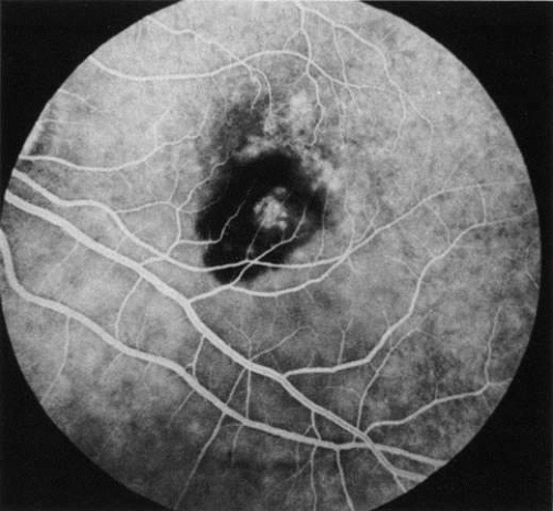 Fig. 17. Poorly defined choroidal neovascularization (CNV). This woman has a CNV that hyperfluoresces in the juxtapapillary area; however, its full extent cannot be determined owing to blockage of fluorescence by blood. (Courtesy of Dr. Kenneth G. Noble.) |
Conventional laser thermophotocoagulation is the treatment of choice for extrafoveal, well-defined, classic CNV. Photodynamic treatment (PDT) is the treatment of choice for subfoveal, predominantly classic CNV. FA is used to localize the lesion in relation to the fovea, classify the subtype, choose the type of procedure, and guide the treatment (Figs. 18, 19, and 20).56,57,58,59,60,61,62,63,64,65,66,67,68,69,70,71,72,73
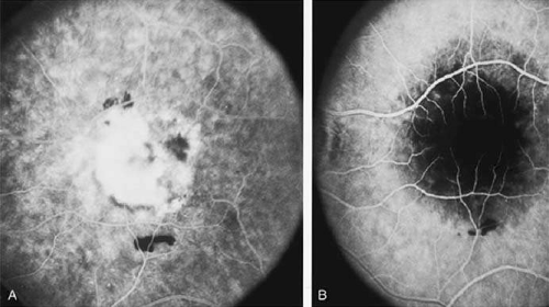 Fig. 18. A. Late-phase fluorescein angiogram demonstrates the presence of actively leaking, subfoveal, classic choroidal neovascularization. B. After photodynamic treatment (PDT) with Visudyne there was complete closure of the neovascular membrane. The round, hypofluorescence corresponds to the area treated with PDT. There was no damage to the retinal vasculature. |
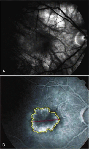 Fig. 19. A. Red-free photograph of the right eye of a patient with exudative angiomatous macular degeneration. B. Fluorescein angiography reveals the presence of subfoveal, classic choroidal neovascularization (CNV). The boundaries of the CNV are digitally traced (yellow), and the greater linear dimension of the lesion is measured (red) to guide the PDT. |
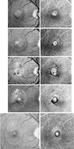 Fig. 20. A. Red-free photograph of a 20-year-old patient with sudden loss of vision to the level of 20/200. There is exudative, neurosensory macular detachment, a few hemorrhages, and lipid exudates. B. Fluorescein angiography reveals the presence of classic choroidal neovascularization (CNV), which appears to be juxtafoveal (<200 μ from fixation). Given the size of the CNV and its proximity to the fovea, it was decided to treat the patient with photodynamic treatment (PDT). C. Red-free photograph of the same eye 2 weeks after PDT; there is increased subretinal exudation D. Fluorescein angiography demonstrates that the CNV is still actively leaking. E. Red-free photograph 4 weeks after PDT demonstrates further increase in the size of the neurosensory macular detachment, subretinal hemorrhages, and lipid exudation. F. Fluorescin angiography reveals that the CNV has extended under the fovea. Given the young age of the patient, an inflammatory component of the neovascular process was suspected. It was decided to give a posterior, subtenon injection of triamcinolone acetonide, 40 mg/1 mL. G. Two weeks after steroid treatment there is partial reabsorption of the subretinal fluid. H. Fluorescein angiography demonstrates contraction of the CNV. I. Four weeks after injection of triamcinolone there is further reduction in the degree of neurosensory detachment; vision had improved to 20/60. J. Fluorescein angiography demonstrates that the CNV is smaller and less active (less leakage). |
FOLLOW-UP
FA is needed to assess response to laser photocoagulation of a CNV and to diagnose recurrent membranes.51,54 The authors generally obtain angiograms 2 weeks, 1 month, 3 months, and 6 months after treatment. The risk of recurrence is greatest during the first 3 months, and the patient, who often has decreased vision due to prior neurosensory detachment, may be asymptomatic. FA is also needed to evaluate the results of PDT. In the original protocol of the Verteporfin in Photodynamic Therapy (VIP) and Treatment of Age-Related Macular Degeneration with Photodynamic Therapy (TAP) studies, a fluorescein angiogram was obtained every 3 months, and if there was persistent leakage from the CNV PDT was applied again (see Fig. 18–20).60
CENTRAL SEROUS CHORIORETINOPATHY
DIAGNOSIS
Central serous chorioretinopathy (CSC) is characterized by breakdown of the outer retinal barrier, with leakage of fluid through a defect in the retinal pigment epithelium into the subretinal space, resulting in a serous neurosensory detachment.78,79,80,81,82,83,84,85,86,87,88,89,90,91,92,93,94,95,96,97,98,99,100,101,102,103,104,105,106,107,108,109,110,111,112,113,114,115,116,117,118,119,120,121,122,123,124,125,126,127,128,129,130,131,132,133,134,135,136,137,138,139,140,141,142,143,144,145,146,147,148,149,150,151,152,153,154,155,156,157,158,159,160,161,162,163,164,165,166,167,168,169,170,171,172,173,174,175,176,177,178,179,180,181,182,183,184,185,186,187,188,189,190,191,192,193,194,195,196,197,198,199,200,201,202,203,204,205 The ophthalmologist can usually diagnose CSC based on the clinical examination and demographic information.93,94,95 Most patients with CSC are middle-aged men74 who often have type A personalities.75,96,97,98,99,100,101,102,103,104 CSC has also been associated to the use of corticosteroids,105,106,107,108,109,110,111,112,113,114,115,116,117,118 pregnancy,119,120,121,122,123,124,125,126 increased adrenaline level and stress,127,128,129,130,131,132 hemodialysis,133,134 collagen vascular diseases,135,136,137,138,139,140,141,142,143,144,145,146,147 and hypertension.148,149,150,151,152,153,154,155,156,157 CSC typically presents as a large serous detachment in the posterior pole without an obvious source of the subretinal fluid.76 However, because a small CNV cannot be ruled out, FA is usually done to confirm the diagnosis. Characteristically, there is a small RPE defect, which hyperfluoresces early, and then there is slow filling of the overlying neurosensory detachment, which may have a classic “smokestack” (Fig. 21) or “ink blot” (Fig. 22) appearance.158,159,160,161 Occasionally, FA demonstrates multiple sites of leakage (Figs. 23, 24, and 25). FA sometimes fails to distinguish CSC from CNV readily because fibrinous subretinal precipitates can cause slow filling of the RPE detachment, which is suggestive of CNV (Fig. 26). Sometimes peripapillary PCV can cause a neurosensory macular detachment masquerading as CSC (Fig. 27).77
Stay updated, free articles. Join our Telegram channel

Full access? Get Clinical Tree


