FIGURE 22.1 Classification of palatal, maxillary, and midface defects.
Low maxillectomy defects not resulting in loss of alar support can be adequately reconstructed with a fibula flap which has been well described, or adequately obturated especially with implant-retained prostheses. If the resection involves the orbital floor (Class III) or the removal of the eye (Class IV), then obturation can give poor results especially after radiotherapy, and reconstruction of the defect is generally preferred. The main methods of free tissue transfer described involve the iliac crest (deep circumflex iliac artery [DCIA]), fibula (peroneal artery), and scapula (circumflex scapula or thoracodorsal angular artery [TDAA]). This chapter describes the use of the DCIA with internal oblique for the reconstruction of Class II-IVb-d defects.
HISTORY
A history of prior surgery or injury in the area of the donor site should be sought. When considering the DCIA flap for maxillary reconstruction, it is important to determine if the patient has a history of gait disturbance or deep vein thrombosis.
PHYSICAL EXAMINATION
The physical examination should focus on identifying evidence of prior surgery or injury in the area of the donor site. Not uncommonly, male patients may have undergone repair of an inguinal hernia. Prior surgery or injury to this region may compromise the reliability of the flap, and an alternate donor site should be considered.
INDICATIONS
The DCIA with internal oblique can provide a high-quality reconstruction of the maxillary bone for all the defects (Class I-IV), and the internal oblique muscle provides an ideal lining for the oral cavity and nasal passage. This flap is superior to the others, especially in the Class III defect when the orbital floor is removed but the orbit retained. The flap can be harvested as a tripartite perforator flap with a vascularized bone graft, muscle flap, and skin paddle.
CONTRAINDICATIONS
The vascular pedicle is short, and the flap harvest requires care, making use of the flap less popular. The advantage of the flap is the size of the bone and ability to shape it to the defect (nasal and orbital), as well as the use of muscle to line the nasal cavity and sinus and provide a very natural result for palatal reconstruction. However, when the neck is depleted of vessels, an alternative donor site with a longer vascular pedicle should be considered, or vein grafts should be prepared.
PREOPERATIVE PLANNING
Preparing the Recipient Site
The principle of selective neck dissection and postoperative radiotherapy in controlling regional recurrence is well established, and as a result, the facial artery and vein can be retained in continuity when preparing recipient vessels in the neck. In general, the pedicle will not reach below the inferior border of the mandible, and so the anastomoses are usually performed above the inferior border being careful to protect the mandibular branch of the facial nerve. I have used superficial temporal vessels occasionally, but it is sometimes possible to divide the retromandibular vein and bring that forward for the venous anastomosis. For either of these options, it is essential to preserve the facial vein to allow drainage through the internal jugular vein. In my experience, the majority of vein grafts have been used for inadequate arterial flow through the facial artery. Papaverine can be used to treat vessel spasm, and if this is not successful, the arterial anastomosis is redone and then an IV bolus of 5,000 units of heparin is given just prior to the release of clamps, after completion of the redo anastomosis. Using these measures has negated the need for vein grafts in the arterial system for the last 5 years.
SURGICAL TECHNIQUE
The skin markings include the pubic tubercle medially, the iliac vessels, anterior superior iliac spine (ASIS), iliac crest, costal margin, proposed osteotomies and internal oblique muscle, and the incision (Fig. 22.2). It is possible to place the anterior osteotomy as much as 40 mm posterior to the ASIS for a Class II-IVb defect as the length of the bone is not a factor. This may not be possible for Class II-IVd defects crossing the midline as the reconstruction needs to span from the zygomatic buttress to the premolar region controlaterally. If raising a skin perforator flap, then the perforators are situated 74 mm (range 50 to 105) posteriorly from the ASIS and 8 mm (range 1 to 35) above the iliac crest and can be identified with a handheld Doppler. It is perfectly reasonable to raise skin or adipose tissue as a perforator component to the internal oblique muscle and the iliac crest to replace loss of facial skin. The incision runs about 20 mm above the line of the iliac crest from the iliacs to posterior to the planned posterior osteotomy. As with all incisions, this can be extended if better exposure is required. If including a perforator skin island, then plan this island around the likely perforator following Doppler identification.
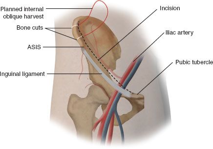
FIGURE 22.2 Exposure of the vascular pedicle can be accomplished with an incision along the iliac crest axis.
Incise the skin, and if raising a perforator flap, identify the perforator before exposing the external oblique muscle with its fibers running in a line from the costal margin to the pubic tubercle. At this point, dissect out the perforator vessel to where it should enter the DCIA and vein just anterior to the ASIS. Next incise the external oblique muscle about 20 mm above the iliac crest from the iliacs to the posterior osteotomy. This plane between the external and internal oblique is easy to separate as there is only a thin layer of adipose tissue. It is best to have access to the internal oblique muscle as far as the costal margin posterosuperiorly and the rectus sheath anteriorly. Next, outline the muscle to be harvested (Fig. 22.3
Stay updated, free articles. Join our Telegram channel

Full access? Get Clinical Tree


