The Pupils and Accommodation


The Pupils and Accommodation


Thomas L. Slamovits

Joyce N. Mbekeani

Joel S. Glaser


He sustained them in a desert land,

In an empty howling wasteland.

He encompassed them and raised them up,

Protecting them like the pupil* of His eye.

Deuteronomy 32:10

The pupil is a central aperture through which light is transmitted onto the eye’s special sensory apparatus, the retina. It serves as a kinetic indicator of both retinal and ocular motor function. The pupil size and reactivity depends on antagonistic influences of the parasympathetic and sympathetic autonomic transmissions supplying the sphincter pupillae and ciliary body and dilator pupillae. Although the neural mechanisms controlling the pupil size and reactivity are highly complex, they may be sampled and evaluated by simple clinical procedures. This chapter will describe the normal pupil structure, physiology, and the pathophysiology of pupillary anomalies commonly observed in clinical practice. For more encyclopedic information about the pupil, we recommend Irene Loewenfeld’s scholarly text, The Pupil: Anatomy, Physiology and Clinical Applications.1


ANATOMIC CONSIDERATIONS

The pupil is a mobile opening within the iris diaphragm, the most anterior projection of the uveal tract. Lying slightly inferonasal to the central iris, the pupil is surrounded by a thick frill or collarette of deeply pigmented tissue that rests on the anterior lens capsule. The iris consists of four parts: (1) anterior condensation of fibroblasts, melanocytes, and collagen fibrils; (2) loose fibrocollagenous stroma containing the muscles, blood vessels, nerves, fibroblasts, macrophages, and melanocytes; (3) dilator muscle; and (4) pigmentary epithelium. Mechanically, the diameter of the pupil is determined by the antagonistic actions of the iris sphincter and dilator muscles, with the dilator fibers playing the minor role. The sphincter muscle consists of circumferential smooth muscle fibers that lie in bundles parallel to the pupillary margin within the superficial iris stroma. The band measures 1 mm in width and occupies an area about 2 to 4 mm from the pupil. Although connected by gap and tight junctions, each group is innervated and functions separately. Thus, rather than retracting toward one quadrant when severed or ruptured, the sphincter continues to function except in the altered segment. The sphincter can be seen in light or atrophic irides and with infrared transillumination. Therefore, with prudence, pupillary reactions may be evaluated even in the presence of iris atrophy, traumatic rupture of the sphincter, and congenital or surgical coloboma.

The dilator muscle lies deep in the iris stroma anterior to the pigmented epithelium. The radially arranged fibers insert at the iris root at the ciliary body and extend anteriorly to the peripheral margins of the pupillary sphincter. The ciliary muscle consists of a circular ring of smooth muscle within the ciliary body, a triangular anterior extension of the uvea, between the ora serrata and the iris root. Composed of three muscle groups—longitudinal, meridional, and circular—it functions as one unit. Ciliary muscle contraction results in increased lens convexity, and anterior rotation of the iris-lens diaphragm, and its actions constitute the chief component of accommodative near synkinesis.

Pupillomotor nerves enter the orbit through the superior orbital fissure and annulus of Zinn. Preganglionic parasympathetic pupillomotor fibers travel with the inferior division of the oculomotor nerve and synapse in the ciliary ganglion. Several postganglionic short ciliary nerves pierce the posterior sclera around the optic nerve. They course anteriorly on the inner surface of the sclera to innervate the ciliary muscle and sphincter
pupillae. Cholinergic discharges from their muscarinic end plates result in miosis and accommodation. The dilator muscle is innervated by third-order, postganglionic sympathetics originating from the superior cervical ganglion. These nerve fibers surround the internal carotid artery as a plexus in the cavernous sinus from where they emerge to enter the orbit with the nasociliary branch of the ophthalmic nerve, a division of the trigeminal nerve. The nasociliary nerve gives off several long ciliary nerve branches that bypass the ciliary ganglion and travel with the short ciliary nerves before supplying the dilator pupillae. Their adrenergic discharges result in mydriasis. Both autonomic groups pass through the ciliary ganglion, a small body of variable size and shape that lies within orbital fatty tissue, posterolateral to the optic nerve and medial to the lateral rectus muscle. Only the cell bodies of postganglionic parasympathetics lie within this ganglion; vasomotor and sensory sympathetic fibers from the nasociliary nerve traverse the ganglion to emerge in the short posterior ciliary nerves without synapsing.


PUPIL FUNCTIONS

The pupil may be considered to have three major optical functions:


  • to regulate the amount of light reaching the retina;


  • to diminish the chromatic and spherical aberrations produced by the peripheral imperfections of the optical system of the cornea and lens; and


  • to increase the depth of field (analogous to the f-stop setting of a camera).

Pupillary function depends on the integrity of the iris and the structures along the course of the pupillomotor pathway (Fig. 15.1). These include (1) retinal receptors; (2) ganglion cell axons in the optic nerve, optic chiasm, and optic tract (but not the lateral geniculate body); (3) brachium of the superior colliculus; (4) pretectal area of the mesencephalon and the interconnecting neurons to pupilloconstrictor motor cells in the oculomotor nuclear complex; (5) the efferent parasympathetic outflow to the pupillary sphincter and ciliary muscle; and (6) the efferent sympathetic pathway from the hypothalamus to the pupillary dilator muscle. The ocular sympathetics may influence pupillary size directly by dilator muscle innervation or indirectly by central inhibition of the oculomotor parasympathetics (Edinger-Westphal nucleus [EWN]).2


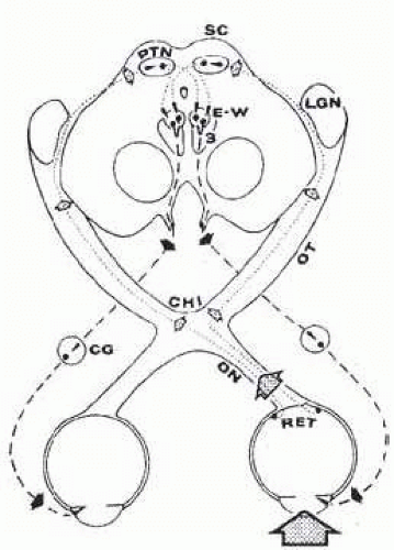
FIG. 15.1 Pupillary light reflex. Light in left eye (dotted arrow) stimulates retina (RET), whose afferent axons (fine dashed lines) ascend optic nerve (ON), decussate at chiasm (CHI), and terminate in pretectal nuclear complex (PTN). Lateral geniculate nucleus (LGN) is bypassed by these pupillomotor fibers. The PTN is connected by crossed and uncrossed intercalated neurons to both Edinger-Westphal parasympathetic motor nuclei (E-W), which comprise the dorsal aspect of the oculomotor nuclear complex.3 Preganglionic parasympathetic fibers (heavy dashed lines) leave ventral aspect of midbrain in the substance of the third cranial nerves. After synapsing in the ciliary ganglia (CG), the postganglionic fibers innervate the pupillary sphincter muscles. Note that uniocular light stimulus evokes bilateral and symmetric pupillary constriction. Brain stem diagram represents section through level rostral to superior colliculi (SC).


PUPIL SIZE AND REACTIVITY

As pupillary size increases, so does chromatic and spherical aberration. As pupillary size decreases, light diffraction at the pupil edge becomes a more significant factor in reducing image quality; this generally outweighs any benefit of miosis-induced increase in focal depth. In their experiments of optical line-spread function, Campbell and Gubisch4 found the optimal pupil diameter to be 2.4 mm;
scatter and focusing defects have an increasing effect with larger pupils. In addition to autonomic control, the size of the pupils is influenced by the physical integrity of the iris, intensity of retinal illumination, the near-effort reflex, the state of retinal light adaptation, and supranuclear influences from the frontal and occipital cortex above the pretectal area and from the reticular formation of the brain stem below. At a given moment, any or all of the aforementioned factors variably influence pupillary size and reactivity. Thus in the awake state, the pupil is in constant motion, a condition of physiologic unrest called hippus. Although it is described in diverse conditions ranging from encephalitis to schizophrenia and from cataracts to hemorrhoids, this incessant change in pupil size is of no pathologic significance.5

Age affects both pupillary size and reactivity6, 7 and the near response.8 Reduced scotopic pupillary diameter is only one of the factors contributing to higher light thresholds in older patients. The pupil of the neonate is miotic but increases in size during the first decade of life; it peaks in the second decade then steadily becomes smaller9 (Fig. 15.2). Loewenfeld has suggested three reasons for small pediatric pupils: (1) the small-sized pediatric eyes; limiting the size of all structures of the eye, (2) immature peripheral adrenergic neural transmission that limits pupillary dilation even to strong sympathomimetic mydriatics, and (3) immature brains with inadequate pupillomotor responses to arousal and central sympathetic inhibition of the EWN.9 Age-related miosis may result from decreased local sympathetic stimulation and central EWN stimulation.


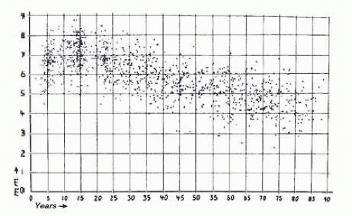
FIG. 15.2 Pupillary size in darkness of 1,263 subjects chosen at random; average pupil size was used ([R+ L]/2). Abscissa shows horizontal diameters in millimeters, ordinate shows subjects’ age in years. Note the wide scatter but obvious age trend. See also Figure 15.3 (top curve). (Reprinted with permission from Loewenfeld IE. Pupillary changes related to age. In: Thompson HS, Daroff RB, Frisen L, et al, eds. Topics in Neuro-ophthalmology. Baltimore, MD: Williams & Wilkins; 1979:129.)

Pupil reactivity, at least to “long” (3-second) light flashes, also seems related to age; the range of amplitude of the light reflex declines with increasing age (Fig. 15.3). Decreased central sympathetic inhibition to the EWN has been implicated as the mechanism for age-related diminished scotopic pupil size.2 Iris color, on the other hand, may affect constriction amplitude and velocity but not pupillary size.3

The correct estimation of pupillary diameter is relevant not only to neuro-ophthalmologists, neurologists, and neurosurgeons but also to refractive surgeons because larger scotopic pupils correlate with an increased incidence of postsurgical glare, halos, and reduced night vision. This is more common when the scotopic diameter exceeds the ablation zone. Indeed, cataract surgeons inserting new aspheric and multifocal lenses, likewise, may induce aberrations if pupil size is not taken into account. Rulers or the Rosenbaum card of semicircular templates are typically used to measure pupil size and should be sufficient in most clinical settings, but they suffer from imprecision, poor reproducibility, and observer bias. Measurement of pupillary size by computerized infrared video pupillography remains the gold standard against
which newer devices are compared. It affords more accurate measurements and documentation of the pupil diameter and reactivity. Numerous pupillometry devices are commercially available for specific uses and are less cumbersome than the original research tools. They have variable sources of illumination, spatial and temporal resolution, can be monocular or binocular, and have additional features such as detection of sleep waves and eye movements (Fig. 15.4). A practical clinically useful pupillometer will need to satisfy requirements such as portability, simplicity of use, reproducibility of measurements, technician-independence or automaticity, variable illuminations, and the capacity to reliably negate influences from accommodation, convergence, and consensual response. A study comparing the readily available techniques found that digital photography and the handheld Colvard monocular pupillometer (Oasis Medical Inc.) compared most favorably to infrared pupillographic pupil size measurements when compared with rulers and templates.10


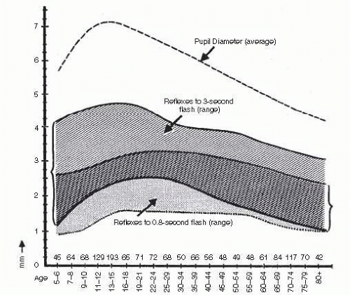
FIG. 15.3 Normal ranges of light reflex amplitude for long and for short flashes. Shaded area (left bracket) is normal range for 3-second flashes; stippled area is normal range for 0.8-second flashes. The numbers above the abscissa indicate the number of subjects per age group. Note the early peak, followed by decline with age for reactions to long light flashes. In contrast, reflexes elicited by short flashes show relatively flat age curve. (Reprinted with permission from Loewenfeld IE. Pupillary changes related to age. In: Thompson HS, Daroff RB, Frisen L, et al, eds. Topics in Neuro-ophthalmology. Baltimore, MD: Williams & Wilkins; 1979:137.)


PUPILLARY REFLEXES

Table 15-1 outlines the normal physiologic reflexes of the pupil. The pupillary light reflex remains the most researched and by far best understood of all pupillary reflexes. Different stimulating conditions or events result in either miosis or mydriasis and are mediated by the autonomic nervous system or by local neurotransmitters. Darkness, like light, is a stimulating event but results from an “off-response,” typically leading to sympathetic innervation of the dilator pupillae and central inhibition of the EWN. Drowsiness results in fluctuations between miosis and mydriasis and typical pupillographic wave patterns that can be used to assess states of wakefulness.11


Light Reflex Pathway

The pupillary light reflex pathway may be functionally considered a three-neuron arc (see Fig. 15.1): the afferent neurons from retinal ganglion cells (RGCs) to the pretectal area; an intercalated neuron from the pretectal complex to the parasympathetic motor pool (EWN) of the oculomotor nuclear complex; and the efferent parasympathetic outflow with the oculomotor nerve to the ciliary ganglion and from there to the pupillary sphincter.


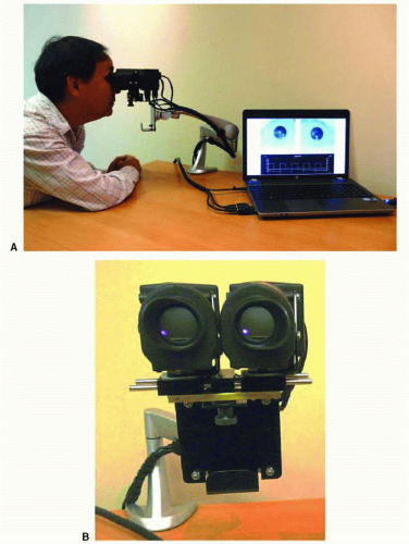
FIG. 15.4 Pupillometer (A) with close-up front view (B). In the past, most computerized pupillometers were designed primarily for clinical research. More patient-friendly pupil testing devices are now becoming available. Such instruments would optimally be compact, binocular (allowing both direct and consensual pupil responses to be recorded for each light stimulus), have a built-in light stimulus and should be easy to use in testing, with little setup time. Chromatic light stimuli (e.g., red and blue) might be advantageous to use in order to differentiate photoreceptor disorders from inner retina and optic nerve disorders. These devices are more suitable for routine clinical use to assess afferent input from the retina and optic nerve as well as efferent output abnormalities, manifesting as anisocoria. These pupillometers might be used as a screening test for a RAPD and anisocoria. The instrument shown above uses a compact optical “head” on an articulating arm that consists of two miniature video cameras and a bank of light-emitting diodes (LEDs) for each eye for light stimuli. The LEDs consist of an array of red, green, blue, and white lights that can be independently turned on over a 5-log unit range of intensities. A diffuser is used so that each light stimulus covers at least 20° of visual field and with scatter of light, even a greater area at brighter light intensities. The instrument can be run by a portable or desktop computer with software that allows one to custom-configure different clinical tests or use those provided by the company (Neuroptics, Inc.). Software will automatically analyze recorded pupil movements and will provide a report. (Courtesy Dr. Randy Kardon.)


TABLE 15-1 Outline of Physiologic Reflexes of the Pupil: the Stimuli, Effects, and Proposed Autonomic Mediation


Reflex


Stimulus


Pupil Response


Neurologic Control


Light


Light flash


Miosis and consensual


Parasympathetic EWN


Near


Blurred near image


Miosis and convergence and accommodation


Pathway unknown; occipital integration; efferent same as light reflex


Darkness


Darkness


Mydriasis


Direct sympathetic stimulation; central parasympathetic (EWN) inhibition


Psychosensory


Arousal, stress, anger, and pain


Mydriasis


Direct sympathetic stimulation; central parasympathetic (EWN) inhibition


Lid closure


Forcible opening of closed lids, blinking


Miosis


Light-induced miosis after brief darkness mydriasis (EWN)


Drowsiness


Fluctuations in wakefulness


Slow, high-amplitude oscillations


Fluctuations in sympathetic and parasympathetic input


Sleep/coma


Low cortical/reticular activity


Miosis


Decreased peripheral sympathetic activity and central EWN inhibition


Direct mechanical or chemical irritation, e.g., cataract surgery


Sensory sympathetic stimulation


Miosis


Unknown local neurotransmitter(s)


Considerable evidence exists that the visual cells of the retina (i.e., the rods and cones) also serve as light receptors, controlling pupillomotor activity. For example, pupillomotor light thresholds follow the same shifts in spectral sensitivity as visual thresholds, depending on the state of light adaptation of the retina (Purkinje shift). Pupillomotor sensitivity of the retina also frequently parallels visual form sensitivity, which is highest at the fovea and lowest in the periphery. Therefore, we may consider two intimately related systems emanating from the retina: the retinogeniculate pathway for visual perception and the retinomesencephalic for pupillomotor control.

Recently, a special group of RGCs has been discovered that transmit information to the pupillomotor pretectal area, quite distinct from the ganglion cells that transmit visual information to the lateral geniculate nuclei.12, 13, 14 and 15 Complementary to nonimage retinomesencephalic rods and cones, this small population of RGCs expresses melanopsin, a unique vitamin A-based visual pigment. In addition to transmitting impulses from rods and cones to which they are connected, these special RGCs themselves are also intrinsically photosensitive (ipRGCs) and peak at 480 nm (blue light). The axons project via the retinomesencephalic tract to the pretectal midbrain and via the retinohypothalamic tract to suprachiasmal nucleus of the hypothalamus, providing the main route for nonimage vision such as the pupillary light reflex and the circadian rhythm, respectively. Their existence explains the phenomenon of patients with blindness from photoreceptor degeneration exhibiting normal circadian rhythms and brisk light reflexes,12, 16, 17 whereas those with comparable optic nerve disease, presumably with destruction of ipRGCs, exhibit abnormal light reflexes. Indeed, experimental selective destruction of ipRGCs in mice has been shown to result in retention of formed vision but defects in the light reflex,18 providing further evidence to the division of light perception and pattern vision pathways. Like rods and cones, ipRGCs respond to both light and dark adaption.19 How melanopsin phototransduction produces physiologic responses or indeed what other vision-related functions ipRGCs may play is unknown and the subject of ongoing research.

The optic nerves transmit populations of both image and nonimage RGC axons. At the optic chiasm, slightly more than one-half of afferent axons cross to the opposite optic tract, where they are mixed with noncrossing axons from the contralateral optic nerve. The ratio of crossed to uncrossed fibers is approximately 53:47.20 From the chiasmal level posteriorly, afferent visual and pupillomotor information from either eye divides into crossed fibers
(from nasal retinal receptors of the contralateral eye) and uncrossed fibers (from temporal retinal receptors of the ipsilateral eye). In the posterior aspect of the optic tract (pregeniculate), the retinomesencephalic pupillomotor branches of the afferent axons gain the pretectal nuclear area by transversing the brachium of the superior colliculus into the rostral midbrain. Intercalated neurons interconnect from the preolivary nucleus of the posterior commissure and lentiform nucleus21 to the EWN by crossing dorsal to the aqueduct in the posterior commissure and by coursing ventrally in the periaqueductal gray matter. This simplistic anatomic approach belies the true complexity of the neuroanatomy and neurophysiology of the pretectal nuclear complex. The reader is referred to articles by Smith et al.,21 Breen et al.,22 Carpenter and Pierson,23 Benevento et al.,24 Burde,25 and Loewenfeld’s1 textbook.


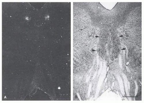
FIG. 15.5 A: Transneuronal autoradiographic label in the Edinger-Westphal nuclei seems bilaterally adjacent to the midline, ventral to the cerebral aqueduct. B: The label seen in (A) corresponds on each side to the fairly distinct cell group (thin arrows), the lateral visceral cell column of the Edinger-Westphal nucleus, shown in a Nissl-counterstained section. The somatic subnuclei of the oculomotor complex (thick arrows) contain larger nuclei. Fascicles from the oculomotor complex are seen streaming inferiorly toward the interpeduncular fossa. Scale bar = 1 mm. (Reprinted with permission from Kourouvan HD, Horton JC. Transneuronal retinal input to the primate Edinger-Westphal nucleus. J Comp Neurol. 1997;381:68.)

The organization of the oculomotor nuclear complex in the mesencephalon (midbrain) depicted by Warwick26 in 1953 and Jampel and Mindel27 in 1967 was modified by Burde and Loewy28 and Burde,25 recognizing two divisions: the anterior median nucleus rostrally and accessory cell columns caudally. The anteromedian nucleus is the source of special visceral efferent motor axons to the iris sphincter and ciliary musculature. This cell mass may be subdivided further into a rostral, caudal, and midportion associated with accommodation, pupil constriction, and both accommodation and constriction, respectively. Somatic subnuclei of the oculomotor nerve lie lateral and caudal to the visceral nucleus. Direct input to the iris sphincter and ciliary body may also be provided by the nucleus of Perlia.21, 28 The pretectal olivary nuclei receive direct retinal input and, in turn, provide direct input to the EWN. The precise location of the EWN in humans or indeed whether it is solitary or bilateral is the subject of ongoing research. First identified by Ludwig Edinger (1885) and further defined by Carl Friedrich Otto Westphall (1887) as parallel cellular columns ventral to the sylvian aqueduct, our understanding of this nucleus remains elementary. Kourouyan and Horton29 injected tritiated H-proline into macaque monkey eyes and found the primary pretectal retinal projections terminating in the olivary nuclei, ipsilaterally and contralaterally. The label for the EWN was found bilaterally in the midbrain, ventral to the cerebral aqueduct, in the central gray matter and in well-defined columns, corresponding to an area termed the lateral visceral column (Fig. 15.5).
Using multiple markers, including wheat germ agglutinin, May and Fratkin30 have further isolated the EWN in the macaque monkey and deduced that it is composed of a solitary column of cells. Increasingly sophisticated studies such as functional magnetic resonance imaging (MRI), positron emission tomography, and in situ hybridization should help to elucidate the detailed architecture and physiology of this nucleus.

According to the degeneration studies by Warwick,31 the ciliary ganglion contains more cells for innervation of the ciliary muscles than for innervation of the iris sphincter (about 30:1). Presumably, that same ratio occurs in the EWN, although the presence of diffuse projections may argue against it.

The efferent pupillomotor fibers join the trunk of the oculomotor nerve, adjacent to the inferior rectus and inferior oblique fasciculus32 and exit ventrally as one unit, coursing through the red nucleus and corticospinal tracts. These fibers travel within the oculomotor nerve in the interpeduncular space, then course above the cerebral tentorium and cross between the posterior cerebral and superior cerebellar arteries, lateral to the posterior communication artery before entering the cavernous sinus. According to Kerr and Hollowell,33 the pupillomotor fibers are located superficially in the nerve, lying just internal to the epineurium. It is believed that this superficial position makes the pupillomotor fibers especially vulnerable to direct compression, such as that from temporal lobe uncal herniation, increased intracranial pressure, or aneurysm of the posterior communicating and internal carotid arteries. In a more rostral part of its projections and in more anterior segments (e.g., the cavernous sinus), however, pupillomotor fibers may be spared preferentially even in the presence of total oculomotor palsy. It is likely that the involvement or “sparing” of the pupil sphincter reflects the nature and acuteness of the injury rather than merely the portion of the third nerve that is compromised.34

At about the level of the superior orbital fissure, the oculomotor nerve divides into superior and inferior divisions, with parasympathetic fibers traveling in the inferior division to the ciliary ganglion via the branch to the inferior oblique muscle. Although the ciliary ganglion contains afferent sensory fibers (nasociliary nerve) and sympathetic fibers to the vessels of the globe and dilator of the iris, only the parasympathetic fibers synapse here. The parasympathetic postganglionic fibers then pass to the globe via the short ciliary nerves.

The weight of anatomic evidence supports the view that the parasympathetic pupillomotor fibers synapse in the ciliary ganglion.31, 35 Parasympathetic denervation hypersensitivity does not occur solely with postsynaptic lesions. Preciliary ganglionic hypersensitivity to lowconcentration methacholine and pilocarpine has been reported, with complete36, 37 and 38 and (inferior) division oculomotor palsies.39

The pretectal pupilloregulator mechanism is subject to a variety of supranuclear influences, which may be summarized as follows:


  • Excitatory: retinomesencephalic (light stimulus) and occipitomesencephalic (near reflex) and


  • Inhibitory: corticomesencephalic and hypothalamomesencephalic pathways and the ascending reticulomesencephalic system.

During sleep and obtunded states, these supranuclear inhibitory influences are diminished, with resultant miotic but reactive pupils. Arousal results in pupil dilatation partly because of the return of supranuclear sympathetic inhibition. Sympathectomy does not eliminate this dilatation.


Oculosympathetic Pathways

Sympathetic outflow to the iris dilator muscles begins in the posterolateral area of the hypothalamus and descends uncrossed through the tegmentum of the midbrain and pons (Fig. 15.6). At the level of the medulla, the sympathetics lie laterally, where they may be affected in lateral medullary plate infarction (i.e., Wallenberg syndrome). The descending fibers, considered first-order preganglionic neurons, terminate in the intermediolateral cell column at the C8 or T2 cord level (the ciliospinal center of Budge). Second-order preganglionic fibers exit the cord primarily with the first ventral thoracic root (T1), but some pupillomotor sympathetics exit also with C8-T2. Via the white rami communicantes, the fibers enter the paravertebral sympathetic chain, which is closely related to the pleura of the lung apex. At this location, the sympathetics may be affected by apical lesions (e.g., the Pancoast syndrome; see discussion of Horner syndrome).

The fibers detour with the ansa subclavia around the subclavian arteries, ascend without synapsing through the inferior and middle cervical ganglia, and finally synapse in the superior cervical ganglion near the bifurcation of the common carotid artery at the base of the skull. Thirdorder postganglionic oculosympathetic fibers ascend the internal carotid to enter the skull, whereas fibers for sweat and piloerection of the face follow the external carotid and its branches.


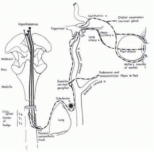
FIG. 15.6 Ocular sympathetic pathways. Hypothalamic sympathetic fibers comprise a polysynaptic system as they descend to the ciliospinal center. This intra-axial tract is functionally considered the “first-order neuron.” The second-order neuron takes a circuitous course through the posterosuperior aspect of the chest and ascends in the neck in relationship to the carotid system. Third-order neurons originate in the superior cervical ganglion and are distributed to the face with branches of the external carotid artery and to the orbit via the ophthalmic artery and ophthalmic division1 of the trigeminal nerve.

The intracranial sympathetics to the eye follow a circuitous course that includes


  • fibers to the tympanic plexus of the middle ear and petrous bone,


  • fibers temporarily joining the path of the intracavernous abducens nerve before anastomosing with the first division of the trigeminal nerve,


  • anastomoses with the ophthalmic-trigeminal nerve (the primary pupillomotor pathway via the nasociliary nerve), and


  • fibers surrounding the ophthalmic artery and ocular motor nerves at the level of the cavernous sinus.

Postganglionic sympathetic fibers:


  • orbital vasomotor,


  • pupillary dilators,


  • smooth muscles of the upper and lower lids (Müller muscle),


  • the lacrimal gland, and


  • trophic fibers to uveal melanophores.

Sympathetic fibers to the dilator muscle enter the globe with the long posterior ciliary nerve without traversing the ciliary ganglion. Vasomotor sympathetics, on the other hand, pass through the ciliary ganglion without synapsing and travel with the short posterior ciliary nerves with parasympathetic fibers.


Near Reflex and Accommodation

With accommodative effort, caused either by a blurred retinal image or conscious visual fixation on a near object
of regard, a “near synkinesis” is evoked that includes (1) increased accommodation of the lens, (2) convergence of the visual axes of the eyes, and (3) pupillary constriction. Many theories have been proposed to explain how accommodation enables near vision but von Helmholtz theory remains the most widely accepted.40 He proposed that contraction of the ciliary muscles cause the zonules to relax resulting in a more globular convex lens and anterior rotation of the lens-iris diaphragm. This change in architecture alters the focal point of the lens, allowing the eye to focus clearly on near objects.

The neural mechanisms of this motor triad are not as well understood as the pathways for pupillary light reactions or the saccadic and pursuit ocular motor systems. Awareness of decreased object distance probably evokes accommodative effort originating in frontal centers; blurred retinal images are sensed in the occipital cortex and corrected via occipitotectal tracts. Jampel41 obtained increased bilateral accommodation, convergence, and usually miosis by unilateral stimulation of the peristriate cortex (area 19) in primates. A group of midbrain cells subserving convergence has been identified in the monkey.42 The anteromedian nucleus in the midbrain rostrally and the EWN caudally have been mapped stereotactically,27 with the rostral portion concerned with accommodation, the caudal portion with pupillary constriction, and the middle segment with accommodation and constriction. Single-cell studies of the EWN of macaque monkeys have further differentiated cells for accommodation from those subserving convergence.43 Nervous supply for accommodation and pupillary constriction pass through the efferent preganglionic parasympathetic fibers, and convergence results from bilateral stimulation of the medial recti subnuclei of the oculomotor nerve.

The final pathway for pupil constriction, whether evoked by light or accommodative effort, consists of the oculomotor nerve, ciliary ganglion, and short posterior ciliary nerves. The ratio of ciliary ganglion cells innervating the ciliary muscle to cells innervating the iris sphincter is about 30:1, with a corresponding disparity in muscle bulk. Although tightly synchronized, accommodation, convergence, and miosis do not share the same efferent pathway. Indeed, each component of the synkinetic triad may be abolished independently. Accommodation and convergence may be suspended by substituting plus lenses and base-out prisms, without eliminating pupillary constriction. Likewise, accommodative convergence is still possible following atropine-induced cycloplegia and pupillary dilatation.

Pupillary constriction evoked by the near reflex is not as easy to evaluate as the light reaction. Accommodative vergence is under voluntary control, and the success of this maneuver is very much dependent on the patient’s cooperation and capacity to converge. In the elderly, convergence is diminished and the near reflex is especially difficult to test. An accommodative target such as a near card is useful for maintaining near fixation. Vision itself is not a prerequisite for the near response, which can be tested in the blind by proprioceptive “fixation” of the patient’s own fingertips. Because near synkinesis deficit rarely occurs independent of pupillary light reflex disruption, if pupillary reactions are brisk to light stimulus, the near reactions need not be examined. However, the student must learn this examination technique and become acquainted with the limits of normality. The light and near efforts are additive; that is, even with the eye brightly illuminated, further pupillary constriction is observed when gaze is shifted from distance to near. Therefore, when testing the light reflex, gaze (accommodation) should be controlled steadily by fixation on a distant target. If the pupil fails to react to light, the eye may be illuminated fully while the near reflex is examined. Light-near dissociation manifests as a poor a pupillary light response but a normal near response. The clinical picture probably results from a selective disruption of the rostral retinomesencephalic fibers to EWN that serve the light reflex with preservation of the caudal accommodative fibers.


THE PATIENT WITH ABNORMAL PUPILS

In office practice, patients present with relatively few isolated “pupil” problems, including pharmacologic accidents, sympathetic paresis (Horner syndrome), pupillary light-near dissociation, and essential anisocoria (Table 15-2). It is extremely unlikely that a patient with oculomotor paresis resulting from a posterior communicating aneurysm or other basal tumor will present solely with an abnormal pupil and no ocular motor or sensory disturbances. The reader is referred to the section on oculomotor nerve palsies to locate a more detailed discussion of pupillary findings with posterior communicating artery aneurysms. Direct trauma to the anterior ocular segment or surgery, local disease of the iris (e.g., cyst, melanoma, rubeosis, sphincter rupture, iritis), and angleclosure glaucoma are slit-lamp diagnoses that need not be discussed here, other than to point out that such local iris lesions have been misinterpreted as neurologic deficits.


Relative Afferent Pupillary Defect

The relative afferent pupillary defect (RAPD) is a clinical manifestation of asymmetric afferent pupillomotor input.
When there is a significant unilateral or asymmetric afferent visual pathway disruption caused by optic nerve or widespread retinal disease, the pupils show a subnormal response to light stimulation of the eye with the greater field or (generally) acuity loss. The pupils have a more extensive constriction response with light stimulation of the normal or less involved eye. It is this combination of subnormal direct pupillary light response and a normal indirect (consensual) response when the opposite eye is illuminated that constitutes the RAPD.


TABLE 15-2 Characteristics of Pupils Encountered in Neuroophthalmology


General Characteristics


Responses to Light and Near Stimuli


Room Condition in Which Anisocoria is Greater


Response to Mydriatics


Response to Miotics


Response to Pharmacologic Agents


Essential anisocoria


Round, regular


Both brisk


No change


Dilates


Constricts


Normal and rarely needed


Horner syndrome


Small, round, unilateral


Both brisk


Darkness


Dilates


Constricts


Cocaine 4%, poor dilation


Paredrine 1%, no dilation if third-order neuron damage


Tonic pupil syndrome (Holmes-Adie syndrome)


Usually largera in bright light; sector pupil palsy, vermiform movement


Unilateral or, less often, bilateral


Absent to light, tonic to near; tonic redilation


Light


Dilates


Constricts


Pilocarpine 0.1% or 0.125% constricts; mecholy 2.5% constricts


Argyll Robertson pupils


Small, irregular, bilateral


Poor to light, better to near


No change


Poor


Constricts


Midbrain pupils


Mid-dilated; may be oval; bilateral


Poor to light, better to near (or fixed to both)


No change


Dilates


Constricts


Pharmacologically dilated pupils


Very large,b round, unilateral


Fixedc


Light


Noc


Pilocarpine 1% will not constrict


Oculomotor palsy (nonvascular)


Mid-dilated (6-7 mm), unilateral (rarely bilateral)


Fixed


Light


Dilates


Constricts


a Tonic pupil may appear smaller after prolonged near effort or in dim illumination; affected pupil is initially large but with passing time gradually becomes smaller.


b Atropinized pupils have diameters of 8 to 9 mm. No tonic, midbrain, or oculomotor palsy pupil even if this large.


c Pupils may be weakly reactive, depending on interim after instillation.


The RAPD can be demonstrated clinically by the alternate cover test, also known as the Marcus Gunn pupillary test, as described by Kestenbaum44 or by the swinging flashlight test of Levatin45 (Fig. 15.7). The swinging flashlight test compares pupillary responses to brief bursts of light stimulation and is more sensitive than the Marcus Gunn test, which detects pupillary dilatation after prolonged light exposure.46, 47

The swinging flashlight test is best performed in a dimly lit room, using a bright light, such as a muscle light
or penlight. During the test, the patient must look at a distant fixation target to avoid accommodative miosis.


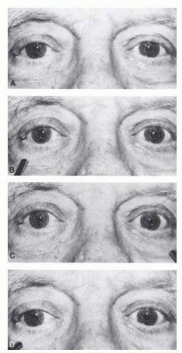
FIG. 15.7 Swinging flashlight test for afferent pupil defect. The patient is a 72-year-old man with right visual loss due to ischemic optic neuropathy. A: Pupils are equal in dim light. B: Illumination of right eye results in modest bilateral constriction. C: When the light swings to the left, there is more extensive constriction in both pupils. D: When the light swings back to the right, both pupils dilate.

The test light is shone directly into the visual axis to first illuminate one pupil and then the other. The alternating or swinging light should pause 3 to 5 seconds in each eye, and this maneuver should be repeated several times. As a rule, the pupils are round and practically equal in diameter (see “Essential Anisocoria” section) and briskly and symmetrically reactive to light stimuli. After an initial, prompt pupil constriction, a slight “release” dilation generally occurs. For example, in the presence of a right afferent defect (see Fig. 15.7), the following is observed with the swinging flashlight test: the pupillary diameters are equal and slightly larger bilaterally when the right eye is stimulated and bilaterally smaller when the normal left eye is illuminated. If only the illuminated pupil is observed—the other pupil being hidden in darkness— the following is seen: the normal left pupil constricts promptly on illumination; as the light is moved rapidly to the right, the right pupil is seen to dilate or “escape.” As the light moves again to the left, the left pupil again constricts briskly.

Afferent pupillary defects may be quantified conveniently by the use of neutral density filters placed before the normal eye. While performing the swinging light test, the neutral density filter is increased until the pupil responses are “balanced.” The neutral density value at which a positive (asymmetric) swinging light test is neutralized is read in log units.48 Previously used to assess the depth of amblyopia, the Sbisa bar (Bagolini filter bar) has also been found to be of comparable use in quantifying RAPD.49 Whether a dim, bright, or brilliant light is best-suited for pupil light-reaction testing is somewhat controversial,50 but an indirect ophthalmoscope light set at 6 V may be used as a handy “standardized” light source. Thompson and Jiang51 stress the importance of avoiding asymmetric retinal bleach, by maintaining a rhythmic “equal time” alternation of the light from one eye to the other and by not swinging the light too many times between the eyes. Finberg and Thompson48 and Thompson et al.52 provide detailed guidelines for proper performance of the pupillary examination and assessment of the RAPD.

Computerized infrared pupillography may allow the most reliable standardization of the swinging flashlight test.46 Personal computer-based infrared video pupillography has been developed for clinical use when detection and specific quantification is required. The proposed advantages to the swinging light test include the capacity to perform binocular tests, compensating for natural differences in midbrain decussation of the interneurons, asymmetric supranuclear influences on the EWN, and differences in efferent innervation of the pupils. Furthermore, the RAPD may be recorded at different light intensities independent of motion, pattern, and color, and free of observer bias.53, 54 Pupillography also measures response latency and cycling time, which can be delayed in diseases of the afferent and efferent visual pathway and amblyopia.55

Quantification of RAPD may be used to gauge the degree to which the anterior visual pathway disruption
has occurred or as a prognostic indicator for final visual recovery. In a small series, Alford et al.56 found that patients with traumatic optic neuropathy and an initial RAPD of log 2.1 or greater showed minimal visual recovery even after megadose intravenous steroid therapy. Those with RAPD measurements less then log 2.1 recovered to visions of 20/30 or better. Younis and Eggenberger examined 72 patients with unilateral or asymmetric demyelinating optic neuropathy and found significant linear correlation between retinal nerve fiber thickness, measured with ocular coherence tomography (OCT) and RAPD, measured in log units. These findings indicate that the log unit value of RAPD may be used to predict the extent of retinal nerve fiber layer (RNFL) loss in this type of neuropathy.57

An afferent pupillary defect may be assessed even if one of the pupils is unreactive because of mydriatics, miotics, oculomotor palsy, trauma (Fig. 15.8), or synechia formation. In such cases, when performing the swinging flashlight test, the direct and consensual responses of the single reactive pupil must be compared. The reactive pupil’s direct light response reflects the afferent function of the ipsilateral eye; its consensual response reflects the afferent function of the contralateral eye.


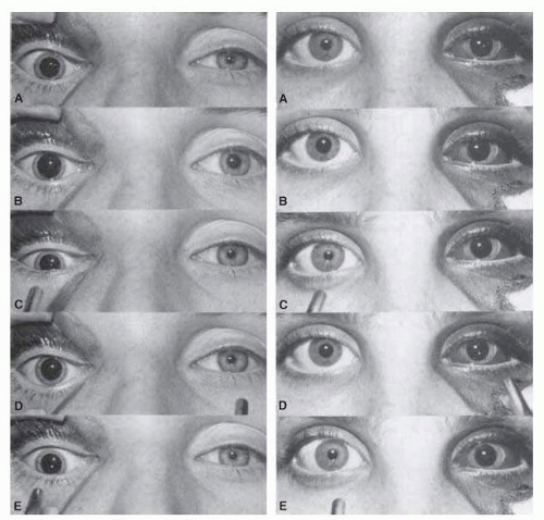
FIG. 15.8 Swinging flashlight test in two patients with mydriasis on side of orbital trauma. Pupils in bright (A) and dim (B) room lighting reveal normal responses on the uninjured side and minimal, if any, response on the affected side. As the flashlight is swung from right (C) to left (D) and again to right (E), one can observe the normally reactive pupil. The patient on the left has no afferent pupillary defect, whereas the patient on the right has a left RAPD.

Even severe unilateral visual loss due to retinal or optic nerve diseases associated with an afferent pupillary defect is not of itself a cause of anisocoria, despite past statements to the contrary (see Behr sign in the following text). If a patient with an RAPD also shows anisocoria, the pupillary inequality must be treated as a separate finding. The RAPD most typically provides objective evidence of optic nerve disease that is either unilateral or asymmetric, with more profound visual involvement on the side of the RAPD. In such cases, the RAPD is not specific and may
reflect optic neuropathy due to demyelination, ischemia, compression, or asymmetric glaucoma.

Because slightly more fibers cross than remain uncrossed at the level of the chiasm, RAPDs may also be seen with optic tract lesions, where greater visual field loss occurs in one eye. An obvious example would be an optic tract lesion with a complete homonymous hemianopia. In such a case, a RAPD would be expected in the eye with the temporal visual field loss (i.e., the eye contralateral to the side of the optic tract lesion).58 The same kind of RAPD can present without any associated visual field defect if there is a contralateral lesion affecting the pupillomotor fibers between the optic tract and pretectal region. The eye with the RAPD should have normal visual acuity, color vision, and visual field, and no other (occult) cause for the pupillary defect (i.e., no amblyopia, glaucoma, past optic neuritis). Ellis59 has reported an afferent pupillary defect contralateral to a pineal region tumor, suggesting that this was due to the involvement of afferent pupillary fibers between the optic tract and pretectal nucleus. Johnson and Bell60 have documented an RAPD in a pretectal lesion due to a pineal gland mixed-cell tumor and Kawasaki et al.61 have described four patients with dorsal midbrain lesions and normal visual fields who exhibited contralateral clinical and pupillographic RAPD. As with optic tract lesions, the midbrain RAPD is attributed to asymmetric nasal-temporal pupillomotor input. Unilateral disruption of homonymous paired fibers in the optic tract or midbrain result in a contralateral RAPD.61 Reported cases of isolated superior oblique palsy associated with RAPD and preserved vision similarly represent terminal retinotectal pupillomotor disruptions in the midbrain.62, 63

Previous descriptions of an RAPD associated with anisocoria, with the larger pupil opposite the side of the optic tract lesion (Behr sign of optic tract disease), have not been supported by subsequent reports. Loewenfeld1 has argued that reports of this phenomenon resulted from observation errors or misinterpretation. Because all afferent optic fibers synapse in intercalated interneurons that project to either side of the EWN, pupillary innervation and, thus, responses are more or less symmetrical.1 Therefore, unless another cause for anisocoria exists, anisocoria does not usually result from lesions causing RAPD.

RAPD most typically is an indicator of optic nerve disease. Retinopathy, maculopathy, or amblyopia can also lead to RAPD. However, even with very poor acuity and field depression, the latter diseases usually cause much less obvious RAPD than that found with optic neuropathy. An extensive retinal detachment, such as two detached quadrants, should cause an obvious RAPD, as do arterial occlusions. With unilateral or markedly asymmetric retinitis pigmentosa, one should see an RAPD; however, usually the disease process is quite symmetric, and thus an RAPD is typically not present.64 Furthermore, the preferential loss of photoreceptors in retinitis pigmentosa, as opposed to ipRGC results in brisk symmetrical pupillary light reflexes and a normal circadian rhythm (see section on Pupil Light Reflex). RAPD in general is proportional to the extent of visual field loss, and the size of the visual field defect is more closely correlated with the extent of the RAPD than is visual acuity loss.65, 66 As a rule, strictly macular disease leads to a much less profound afferent pupillary defect than the bulk of diseases affecting the optic nerve.67 Patients with central choroidopathy, for example, show either a small or no RAPD.68 Furthermore, the RAPD is much more likely to persist with resolved optic neuritis than resolved central serous choroidopathy.69 Patients have also been reported to have an RAPD70 with central retinal vein occlusion, the RAPD being more obvious (generally >1 log unit) in the ischemic than in the nonischemic variety, where most cases measure less than 0.3 log units. Thus, the RAPD may be helpful in distinguishing between ischemic and nonischemic central retinal vein occlusions.

RAPD may be observed in strabismic or refractive amblyopia71, 72 but these are small, measuring 0.3 log units or less and do not correlate with visual acuity.71 If a very obvious RAPD is seen in cases of amblyopia, advanced cataracts, retinopathy, or maculopathy, additional tests are recommended to rule out the possibility of a superimposed occult optic neuropathy that would better explain the pupillary findings. Occasionally, a small RAPD of neutral density filter of 0.3 log unit or less is detected in patients with other ocular pathology. These findings may be assumed to be benign if there are no etiologic clues from the history or clinical examination.73, 74 Asymmetric connections from the afferent visual pathways to the pretectal midbrain between the two eyes may partially explain the presence of RAPD in apparently normal patients. Wilhelm et al.75 obtained clinical and pupillographic measurements of 102 normal subjects and documented RAPD in 15% but none exceeded 0.39 log units. Followup with lack of progressive visual changes on subsequent examinations should provide further corroboration that small isolated RAPD are indeed “benign.” Interesting but rarely encountered in the usual clinical setting are RAPDs induced by contralateral monocular occlusion or resulting from dense contralateral cataracts.76, 77 and 78


Essential Anisocoria

All pupils are not created equal. In fact, benign pupillary inequality exists in practically all individuals, but not
necessarily all of the time. Using photographic techniques, Lam et al.79 determined pupil size in 128 healthy individuals, measuring greatest pupil diameter twice each day for 5 consecutive days. Forty-one percent showed anisocoria of 0.4 mm or greater at one time or another and 80% showed anisocoria of 0.2 mm or greater at some time. A majority of the subjects, therefore, had anisocoria of some degree at one time or another (Fig. 15.9). Anisocoria is as common in men as in women, in the morning as in the afternoon, in dark as in light irides, and in the young as in the aged (although Loewenfeld80 suggested that anisocoria is more common in the elderly). In a study of healthy neonates, anisocoria of 0.5 mm or more was found in about 20% of infants, but none had anisocoria greater than 1 mm.81 The physician can detect regularly a pupil difference of as little as 0.2 mm in diameter, given that clinical judgment of inequality is based more on impressions of pupil area than on measured diameter. Because the percentage difference in area is greater for smaller than for larger pupils, for any given diameter difference, anisocoria is detected more easily in smaller pupil pairs. The prevalence of anisocoria decreases in bright conditions when measured as a difference in pupil diameter but not when it is assessed as a ratio of pupil areas.82

Essential or “central” anisocoria should be identified easily because the pupillary light and near reactions are normal, eyelid positions are normal, and eye movements are full. The relative difference between pupil diameters is constant under various levels of illumination. With sympathetic disruption or Horner syndrome, the anisocoria is enhanced in dim lighting. Conversely, in parasympathetic disruption, the anisocoria is enhanced in bright illumination. Incidental ptosis due to traumatic or senile levator aponeurosis dehiscence and weakening, when ipsilateral to the smaller pupil of essential anisocoria, may be confused with a true Horner syndrome.83 When the distinction between true and pseudo-Horner syndrome is difficult by simple inspection alone, pharmacologic testing usually resolves the dilemma.


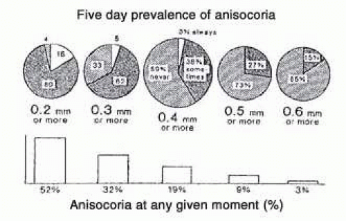
FIG. 15.9 Prevalence of simple anisocoria. (From Thompson HS. The pupil. In: Lessell S, van Dalen JTW, eds. Current Neuroophthalmology. Chicago, IL: Year Book Medical Publishers; 1989:214.)

Some individuals with long-standing anisocoria may suddenly discover the condition while shaving or applying makeup or may have it called to their attention over the breakfast table. In such situations, inspection of previous photographs is an invaluable aid in determining the nature and duration of pupil anomalies. A hand magnifying glass, a trial frame lens, an indirect ophthalmoscope lens, or even the high magnification of the slit-lamp beam84 may prove useful for examining pupil details in snapshot or portrait-quality photographs (Fig. 15.10).


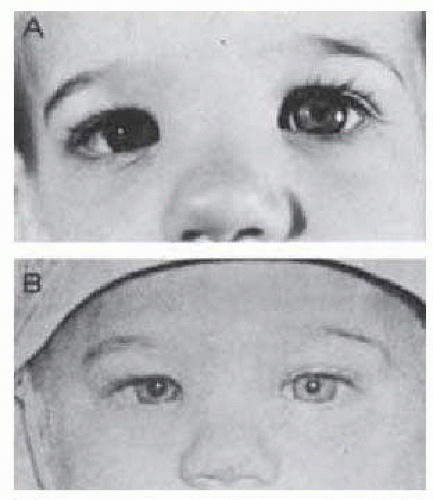
FIG. 15.10 A: Anisocoria noted at age 14 months in otherwise healthy infant. B: Photograph at age 6 months confirms chronicity and benign nature of finding. There was no iris heterochromia.

The evaluation of anisocoria is facilitated by the presence of the following associated signs (see Table 15-2): mild upper ptosis as well as lower lid elevation on the side of a relatively miotic pupil (Horner syndrome), a dilated pupil fixed to light but with very slow constriction on prolonged near fixation (tonic pupil), and small irregular pupils that react better to near than to light Argyll Robertson (AR) pupil. Table 15-3 outlines the conditions commonly associated with anisocoria and how alterations in illumination
affect the pupil size disparity. Generally, if anisocoria is greatest in the dark, the abnormal pupil is the miotic one. Conversely, if anisocoria is greatest in the light, the abnormal pupil is the mydriatic one. Because the etiology of anisocoria may range from abnormal iris structure to disruption of pupillomotor pathway and supranuclear integrity, it is imperative to take a comprehensive history and evaluate the patient for all levels of disruption.


TABLE 15-3 Causes of Anisocoria and Changes with Illumination


Anisocoria: Light = Dark


Anisocoria: Greater in Dark (Small Pupils)


Anisocoria: Greater in Light (Large Pupils)


Physiologic


Physiologic


Sphincter damage/traumatic mydriasis


Nonreactive unequal pupils (bilateral iritis, ischemia, atrophy)


Horner syndrome


Oculomotor nerve palsy


Old Adie pupil


Holmes-Adie pupil


Pharmacologic miosis


Pharmacologic mydriasis


Argyll Robertson pupil


Angle-closure glaucoma


Iritis (posterior synechiae)


Ocular ischemia


The history should include onset and duration, association with pain, ocular redness, ptosis, and whether the patient has noticed visual disturbances such as diplopia, decreased accommodation, glare, and halos. An intercurrent or past history of head trauma, surgery, malignancy, diabetes mellitus, syphilis, and neurologic, autoimmune, and cardiovascular disease as well as a social history of smoking, narcotic drug abuse, or occupational exposure (such as, e.g., exposure of gardeners to insecticides or of hospital workers to atropine) will help to focus the clinical exam and investigations. Table 15-4 details the examination of a patient with anisocoria. Pupillography may be employed for accuracy and documentation and old photographs examined to determine the duration and progression of anisocoria. The clinical findings will determine the need, if any, for ancillary investigations such as complete blood count (CBC) and erythrocyte sedimentation rate (ESR), blood chemistry, HbA1c, Venereal Disease Research Laboratory (VDRL) and FTA-ABS, CXR, computed tomography (CT) scan, and MRI/MRA.


TABLE 15-4 Clinical Approach to the Patient with Anisocoria


  1. Examination


    • VA (near and distant)


    • Patient to focus on distant object


      • Examine and measure pupils under normal room light


      • Examine and measure pupils in dark and bright light conditions


    • Determine whether anisocoria is greater in bright light or in the dark


    • Evaluate pupillary response to near target to rule out light/near disparity


      • Use accommodative target NOT light


    • Check lid position for ptosis


    • Check for globe retraction and enophthalmos


    • EOM—look for limitations


    • Slit-lamp examination


      • Segmental paralysis of iris ± wriggling and undulating movements


      • Signs of iritis


      • Iris atrophy, holes, sphincter tears


      • Signs of ocular ischemia/iris neovascularization


    • Measure intraocular pressure (IOP)


    • Undilated funduscopy (defer dilation)


  2. Determine whether the anisocoria is greater in dark or in bright light


    • Anisocoria greater in bright light


      • Larger pupil is the abnormal pupil


    • Anisocoria greater in the dark


      • Smaller pupil is the abnormal pupil


  3. Pharmacologic testing for suspected Adie or Horner syndrome


EOM, extraocular movements; IOP, intraocular pressure.


Light-Near Dissociation

With exception of errors in testing techniques or poor patient cooperation, there is no pathologic situation in which pupillary light reflex is normal while the near response is defective. Therefore, if the pupils respond briskly to light, the near response need not be examined. Lack of direct and near responses, in the absence of ocular motility disturbances, should raise the question of bilateral pharmacologic pupillary dilation or local ocular disease, such as sphincter trauma or synechiae. Rarely such findings represent congenital mydriasis.85 Paradoxical pupillary responses (pupillary constriction induced by darkness) have
been reported in association with concomitant stationary night blindness and congenital achromatopsia and subsequently with other retinopathies and optic neuropathies.86

The distinctive clinical syndromes demonstrating pupillary light-near dissociation include the Holmes-Adie tonic pupil syndrome, Argyll Robertson syndrome, and Parinaud dorsal midbrain syndrome. The tonic pupil is characterized by light-near dissociation and supersensitivity to dilute parasympathomimetic agents. It is distinguished from the AR pupil by certain characteristic features, including a larger pupil (see “Tonic Pupil [Holmes-Adie] Syndrome” section). The dorsal midbrain syndrome produces light-near dissociation in association with moderate mydriasis and a constellation of signs, including vertical ocular motility deficits and accommodative defects (see “Dorsal Midbrain [Parinaud] Syndrome” section). Other causes of light-near dissociation include significant visual loss, diabetes mellitus, and aberrant oculomotor nerve regeneration.

Any eye with a severe visual deficit due to retinal or optic nerve disease, with significant field loss, has a diminished ipsilateral pupillary response to light (RAPD) but an intact near reflex. For example, an eye blind from glaucoma demonstrates a light-near dissociation. Patients with profound bilateral visual loss caused by anterior visual pathway disease have bilaterally poor pupillary light responses but intact accommodative responses. Therefore, patients with bilateral end-stage glaucoma, total retinal detachments, or blindness resulting from optic nerve/chiasmal injuries should not be misconstrued as having dorsal midbrain disease on the basis of mid-dilated pupils and light-near dissociation alone because these findings may result solely from bilateral blindness. On occasion, patients with long-standing type I diabetes demonstrate moderate symmetric mydriasis with light-near dissociation that is clearly disproportionate to any concurrent retinopathy. Indeed, some patients with type I and type II diabetes may exhibit findings consistent with tonic pupils resulting from widespread autonomic dysfunction87 or local nerve damage from diode laser photocoagulation.88 Diabetic patients may also have typical AR pupils, including miosis.89, 90

When aberrant regeneration follows oculomotor nerve palsies, fibers originally associated especially with the medial rectus (although other oculomotor fibers are occasionally responsible) may anomalously innervate the pupillary sphincter as well. Frequently, aberrant-regeneration results from chronic oculomotor nerve compression, typically from meningiomas and aneurysms,91 as opposed to acute ischemic insults. In such cases, the light-paretic pupil can constrict when the medial rectus muscle acts, either in convergence or with conjugate lateral gaze. Therefore, a light-near dissociation (actually, a gaze-evoked) pupillary dissociation is observed. Unlike the AR pupil, the aberrant-regeneration pupil is large rather than miotic and is accompanied by other signs of third-nerve palsy, some paretic and some with “misdirection” features.92

Infrared videographic transillumination readily demonstrates denervated and aberrantly reinnervated sphincter segments.93 Pupillary changes seen on a slit-lamp examination may provide the early evidence of aberrant oculomotor regeneration; segmental pupillary sphincter contractions may be seen with attempted eye movements in the field of action of oculomotor innervated muscles.94 In more obvious cases, the pupillary constriction may be grossly visualized with attempted efforts by any of the muscles normally innervated by the oculomotor nerve.92 Ohno and Mukumo95 report anomalous pupillary innervation by the oculomotor nerve in 6 of 10 patients with aberrant oculomotor regeneration; pupillary constriction was seen most commonly with downgaze or adduction and less frequently with upgaze. In two patients, pupillary dilation was noted on abduction. Spiegel and Kardon96 evaluated 24 patients with aberrant oculomotor regeneration. Clinically, involvement of the eyelid and pupil was evident more commonly than that of ocular motility. Pupil signs of aberrant behavior, such as gaze-evoked anisocoria, were most noticeable in dim illumination.


Tonic Pupil (Holmes-Adie) Syndrome

One of the commonest causes of isolated internal ophthalmoplegia is the tonic pupil. It typically presents with mydriasis, light-near dissociation, and denervation supersensitivity to dilute cholinergic agonists. First described in 1931, in two separate reports, it was recognized to occur with other neurologic anomalies and subsequently named Holmes-Adie syndrome (after G.M. Holmes and W.J. Adie). Holmes-Adie syndrome has an annual incidence of 4.7 per 100,000.97 Pupillotonia is usually unilateral, but bilateral cases do occur in which the eyes are involved either simultaneously or sequentially (Fig. 15.11). Unilateral cases become bilateral at a rate of 4% per year.98 Although all ages and both sexes are affected, there is an unexplained predilection for women in the third to fifth decades.

Typically, the involved pupil is larger than its fellow. Because both constriction and dilation are defective, however, the tonic pupil may appear smaller in dim illumination because the normal pupil is free to dilate widely. Thus, the diameter of the normal pupil may be smaller (in bright illumination) or larger (in dim illumination) than
the tonic pupil. With the passage of time, the anisocoria becomes less marked as the initially larger tonic pupil gradually becomes less dilated and eventually even miotic over the years93, 99 (Fig. 15.11).


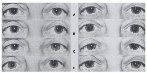
FIG. 15.11 A: A 52-year-old woman with right tonic pupil (left); 1 year later, involvement of the left pupil developed as well (right). Pupils are shown in bright (A) and dim (B) room lighting after near-convergence attempt (C), and in dim room lighting after instillation of 0.125% pilocarpine in both eyes (D). Note that the right pupil is smaller 1 year later (A) (right) than when it was “fresh” (A) (left). With time, the right pupil has also become more responsive to near-accommodation effort (C) (left and right). Parasympathetic hypersensitivity is seen in the right pupil (D) (left) and 1 year later bilaterally (D) (right).

As a rule, the tonic pupil is grossly defective in its reaction to light stimulus but may show a minimal degree of contraction. On slit-lamp examination, however, irregular spontaneous low-amplitude movements may be observed. Segmental sector contractions may be seen in the portions of the sphincter that are still either not denervated or reinnervated. Also, areas of sector palsy of the iris sphincter are seen. The combination of segmental contractions and areas of sphincter palsy exhibit the characteristic wormlike vermiform movements. With prolonged accommodative effort, the pupil slowly constricts, usually not extensively. When near effort is relaxed, the dilation movement is also gradual, requiring minutes or hours for redilation. Therefore, with accommodative effort and before redilations are complete, the tonic pupil can be relatively miotic with respect to its normal opposite. These pupillary kinetics constitute the “tonic pupil,” and pharmacologic testing (Fig. 15.11) is additive rather than diagnostic. Disruption of the final postganglionic nerve supply to the iris sphincter accounts for denervation supersensitivity, with pupillary constriction produced by dilute parasympathomimetic agents and is the basis for pharmacologic testing for the tonic pupil.

Supersensitivity to the parasympathomimetic drug methacholine 2.5%, with pupillary constriction, is a good but not foolproof test. Pilley and Thompson100 suggest the use of 0.125% pilocarpine as a substitute for methacholine. Actually, pilocarpine is, in some ways, the better drug because of its ready availability and stronger miotic action; supersensitivity is demonstrable in a greater proportion of patients with tonic pupils. Because it produces a more visible degree of anisocoria, a negative test is more valid with pilocarpine than with methacholine. Some authors have suggested the use of weaker solutions of pilocarpine, such as 0.0625% to 0.0313%.101, 102 These lower concentrations insignificantly constrict normal pupils and more reliably distinguish normal pupils from tonic pupils with parasympathetic supersensitivity. Weak-concentration arecoline, a pilocarpine-like drug, has also been used; its advantage, if any, may be that it brings about maximal miosis more quickly.103 Pupillary parasympathetic supersensitivity may occur with ganglionic lesions as well as with preciliary and postciliary ganglionic lesions.36, 37, 38 and 39, 63

Only gold members can continue reading. Log In or Register to continue

Stay updated, free articles. Join our Telegram channel

Jul 10, 2016 | Posted by in OPHTHALMOLOGY | Comments Off on The Pupils and Accommodation

Full access? Get Clinical Tree

Get Clinical Tree app for offline access